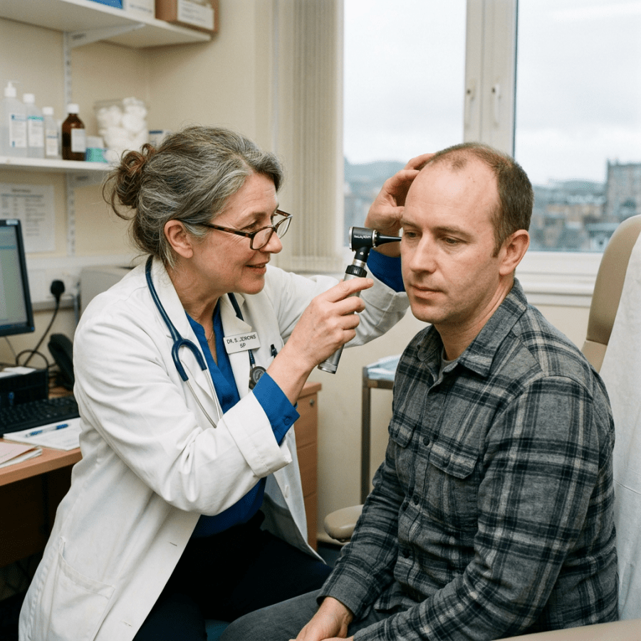
Clinician examining a seated patient’s ear with an otoscope

Ear Wax (Cerumen): What to Know About This Annoyance

shutterstock_147242300Ear wax, also called cerumen is a water resistant coating in the ear canal that protects the skin of the canal from water damage, infection, trauma and debris.  If it accumulates it is usually not noticeable, but can lead to hearing loss as well as pain/infection.  Normally it is supposed to migrate from near the ear drum along the canal and fall out of the ear. Cerumen is a mixture made up of secretions of both sebaceous (oil), dried skin cells, and sometimes hair.  It is identified in the ear canal in your doctor’s office with an instrument called an otoscope.  It’s appearance and texture vary widely – sometimes it has the appearance and texture of a liquid, and other times it looks like clay or rock. Causes of ear wax accumulation:  Narrowing of the ear canal can make it difficult for the ear wax to fall out of the ear. Skin disorders such as eczema can cause excessive cerumen.   Also as people age, they produce less fluid in the cerumen and it gets harder migrates slower out of the ear canal.  Epithelial migration can also occur as a result of using cotton swabs in the ears.  Q-tips tend to push ear wax deeper into the ear canal and over time can cause complete blockage of the ear canal in some people.  Earing aids, ear plugs and swim molds also after prolonged use can contribute to ceumen accumulation. Overproduction:  Some people produce more cerumen than others and this overcomes the ear canal’s ability to eliminate it. Symptoms of ear wax accumulation:  Hearing loss, earache, ear fullness, itchiness, dizziness, and/or ear ringing. Removal methods:  Ear wax is usually removed with one of three popular methods –medication to break down the wax, irrigation and manual removal with a small ear spoon to scoop it out. How to prevent cerumen accumulation:  Many people have difficulty preventing ear wax accumulation, especially if they have predisposing conditions such as a narrow ear canal, eczema or produce more was than average.  For many patients, topical drops such as mineral oil soaked on a cotton ball and left in the ear for about 10-20 minutes once a week while also not using a hearing aid overnight (if applicable) is helpful. Routine cleaning of the ears by a healthcare provider every 6-12 months is also helpful to many people.  If the wax becomes very difficult to remove, an Ear/Nose/Throat doctor can use a ear microscope and special instruments to help remove it more easily.   I hope that you have found this information useful.  Wishing you the best of health,

Scott Rennie, D.O.
Board Certified in Obesity Medicine and Family Medicine

This blog is for educational purposes only and does not constitute individual medical advice. Always consult your own physician before making changes to your health, medications, or treatment plan.

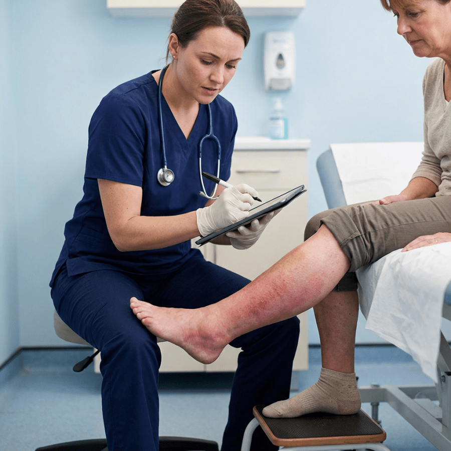
Nurse documenting examination of patient's reddened lower leg

Cellulitis – a soft tissue/skin infection – is it MRSA?

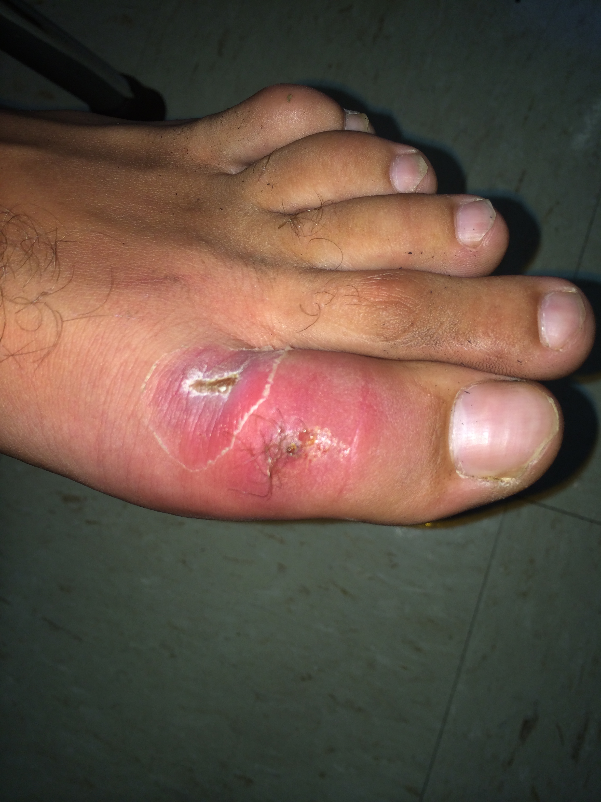
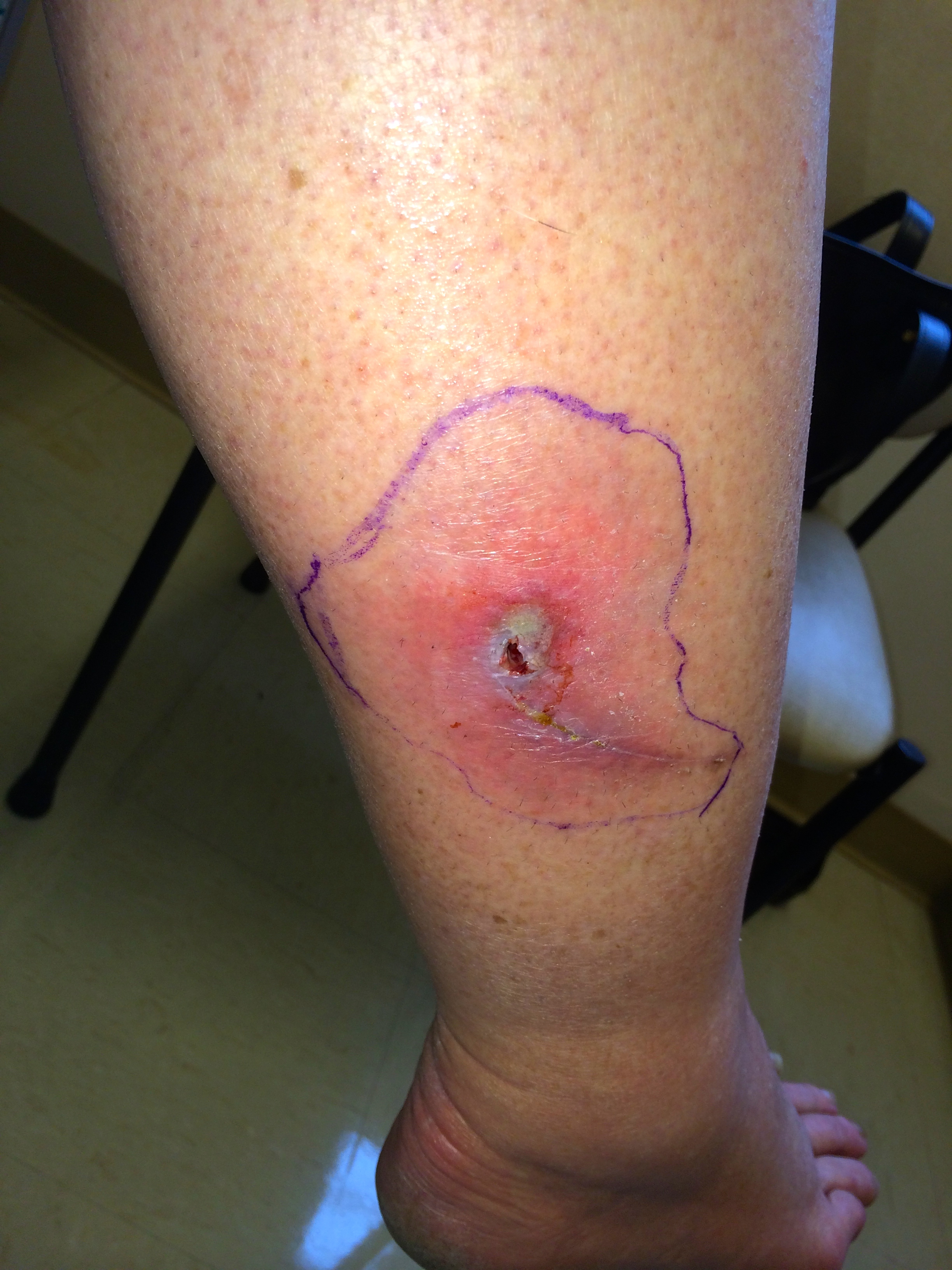
IMG_5864Cellulitis is an infection of the skin and soft tissues.  It is common and most often it’s caused by bacteria that normally live on the skin.  These bacteria don’t normally cause any problems unless the skin is scratched, torn or punctured.  The most common bacteria that cause cellulitis are streptococci “Strep” or staphylococci “Staph.” Most of the time cellulitis is mild and heals with the use of antibiotics, however it can become severe and cause an infection that spreads throughout the body if left untreated.  It can also lead to deep infections called abscesses. Risk factors: 1)   Chronic skin condition such as eczema or psoriasis 2)   Accumulation of fluid (edema) due to poor circulation possibly from heart failure, liver disease, or removal of lymph nodes 3)   Recent injury, wound, cut or laceration to the skin 4)   Current skin infection such as athlete’s foot or impetigo 5)   Being overweight Symptoms:  Pain, tenderness, increased warmth, redness and swelling in a distinct area of skin.  The skin can be smooth and shiny in this area.  Fever and chills are not usually present. Other skin infections:  different kinds of infections that can be confused with cellulitis include an abscess or boil.  These are different because they are usually bumpy, raised and filled with pus.  Abscesses are usually caused by a staph bacteria which may be MRSA.  Cellulitis is more commonly caused by “strep” bacteria. Treatment:  We commonly use antibiotics and also treat the underlying problem that caused the skin infection (such as athlete’s food or increased edema, eczema or psoriasis) if present. Keeping the area elevated to reduce inflammation is also helpful.  It is important to keep the infected area clean and dry.  Showering or bathing can be done normally, but pat the area dry with a clean towel afterwards to dry the area completely.  You can also use a bandage or gauze to protect the skin if needed. Antibiotics are often used and will be tailored to the individual patient.  The patient’s specific area of infection, medical history, history of allergies and examination will be important in determining the proper antibiotics to use.  Sometimes the antibiotics will need to be given by IV in more serious cases of cellulitis. Treatment time:  In most cases, the swelling, warmth and redness should improve within 1-3 days after starting the antibiotics.  We often use a Sharpie marker and draw a circle around the area of redness so that we can examine the red area over a few days to see if it’s increasing, decreasing or staying the same.  Symptoms of redness and swelling can persist for up to 2 weeks but it should be gradually decreasing.  If it’s not improving, you should call or return to your health care provider for re-evaluation.   I hope that you have found this information useful.  Wishing you the best of health,

Cellulitis of the lower leg often starts at a crack or a small cut on the foot, and with diabetic neuropathy that break can go unnoticed until the whole leg is red. It is the reason I keep after people about the diabetic foot check and what to look for.

Updated for 2026: Pus or No Pus Decides the Antibiotic

The most useful thing I can add to this post is a distinction that was fuzzy in 2012 and is now the thing the whole treatment decision turns on. Is there pus, or is there not. If there is an abscess, a pocket of pus you can feel giving under your finger, that is usually staph, MRSA is on the table, and it needs drainage plus an antibiotic that covers MRSA. I have written about that separately. If it is plain cellulitis, meaning spreading red, warm, tender skin with no drainable pocket, that is usually strep, and a cephalosporin like cephalexin is the right drug. Adding MRSA coverage on top does not help, and this has been tested directly. Moran and colleagues randomized patients with uncomplicated cellulitis to cephalexin plus trimethoprim-sulfamethoxazole or cephalexin alone. Cure was 83.5 percent against 85.5 percent (1). No benefit, two drugs, more side effects. So if you have been handed two antibiotics for a red leg with no abscess, that is worth a conversation. The 2014 infectious disease guidelines remain the operative document here and have not been superseded (2).

Draw a Line Around It

This is the single most useful thing you can do, and it is more valuable to me on a video visit than almost anything else. Take a pen and trace the edge of the redness. Write the time next to it. Then photograph it. When we talk tomorrow, or if you end up in an urgent care instead, that line answers the only question that really matters, which is whether this is advancing, holding, or retreating. Redness that has crossed well past the line in a few hours is a different problem from redness that has not moved. Without the line, everyone is guessing from memory, and memory is bad at this.

What I Can and Cannot Tell From a Screen

I can see color, extent, and whether there is an obvious pocket. I can ask about fever, chills, how fast it came on, whether there was a break in the skin, and how your blood sugars have been. What I cannot do is feel it. Warmth and firmness are things I am taking your word for, and the difference between cellulitis and something deeper and far more dangerous is partly a matter of how the tissue feels and how much pain there is relative to how it looks. That last one matters. Pain out of proportion to the appearance is a red flag I take seriously, and it is a reason for me to send you in rather than treat from here. Good photographs help more than people expect. Natural light, the same angle each time, and something for scale.

When To Be Seen, Today

Fever or chills. Redness advancing visibly past your marked line. Pain that seems far worse than the skin looks, or skin that is dusky, blistering, or numb. Cellulitis on the face. Any of this in someone diabetic, immunosuppressed, or with poor circulation. And no improvement after forty eight hours on the right antibiotic. The rapidly advancing, severely painful ones are surgical emergencies, not prescriptions, and they are the reason I would rather over-refer than under-refer on this particular condition.

The Bottom Line

No pus means a cephalosporin, and adding a second antibiotic for MRSA does not improve anything. Pus means drainage. Draw a line around the redness and note the time, because that line is the best data anyone will have.

Sources

1. Moran GJ, et al. Effect of Cephalexin Plus Trimethoprim-Sulfamethoxazole vs Cephalexin Alone on Clinical Cure of Uncomplicated Cellulitis. JAMA. 2017;317(20):2088-2096. https://jamanetwork.com/journals/jama/fullarticle/2627970 2. Stevens DL, et al. Practice Guidelines for the Diagnosis and Management of Skin and Soft Tissue Infections: 2014 Update. Clin Infect Dis. 2014;59(2):e10-e52. https://www.idsociety.org/practice-guideline/skin-and-soft-tissue-infections/

Related Reading

Abscesses and MRSA: What To Do About a Skin Infection Am I Truly Allergic to Penicillin? When to Use Telemedicine vs. Urgent Care: How I Spot the Sick One Newly Diagnosed With Type 2 Diabetes: What You Should Know Deep Vein Thrombosis (DVT): Blood Clot in the Leg Explained Ringworm, Athlete’s Foot and Jock Itch and Fungal Nail Infections – “There Are Fungus Among Us” Scott Rennie, D.O.

Board Certified in Obesity Medicine and Family Medicine

This blog is for educational purposes only and does not constitute individual medical advice. Always consult your own physician before making changes to your health, medications, or treatment plan.

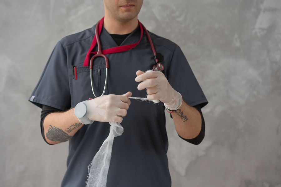
A clinician in scrubs and gloves preparing a length of sterile gauze for a dressing.

Abscesses – What to do about MRSA (Methicillin-resistant Staphylococcus aureus)

IMG_5872“Staph” infections are most often caused by the organism Staphylococcus aureus, a bacterium that is carried on the skin of about 30% of healthy individuals.  Most of the time, these bacteria do not cause any symptoms. They just hang out on the skin because the skin is a natural layer of defense against infections.  If the skin is damaged, even from a small scratch however, Staph can cause anything from a mild skin infection to a severe, life-threatening illness, especially in young children or older adults. The difference between normal Staph infections and MRSA Infections:  Staph infections in the past were treated with antibiotics such as penicillin.  Some strains of Staph have unfortunately become resistant to penicillin and related antibiotics.  These strains of MRSA cannot be cured with traditional penicillin-related medications.  MRSA is now found in up to 70% of people in the community with a diagnosed staph infection. The spread of MRSA:  Many people become “colonized” with MRSA and that means that they carry the bacteria on their skin or in their nose.  It doesn’t cause any problems unless there is an opening in the skin from a scratch or cut.  You can become colonized with MRSA bacteria in several ways: 1)   Touching the skin of someone else who has been colonized with MRSA 2)   By touching or inhaling small droplets from breathing, coughing or sneezing 3)   By touching a contaminated surface such as a phone headset, countertop or door handle Community MRSA:  You can more easily pick up MRSA by: 1)   Being overweight or obese 2)   Shaving body hair 3)   Sharing personal items or equipment that is not cleaned 4)   Skin trauma such as turf burns, cuts or sores 5)   Physical contact with someone with a draining cut or sore that has MRSA Symptoms:  A skin infection is the most common symptom and it may be mistaken for a spider bite.  A raised, red lump that is tender or cluster of  “pimples,” or large tender lump that drains pus may also be present.  If the bacteria enter the bloodstream, it is possible to develop an infection in areas other than the skin.  A staph infection can occur in a heart valve, inside a bone, a joint or in an implanted device such as an IV line, pacemaker or replacement joint.  Symptoms of these types may include fever, fatigue as well as swelling in the infected area. Diagnosis:  To actually diagnose a true MRSA infection, a culture of the infected area must be performed.  A small sample of bacteria or pus collected from the skin or bloodstream is collected and allowed to grow in an environment that the bacteria thrive in, and then we test these bacteria for resistance to certain antibiotics, including penicillin. Treatment:  in patients with community acquired MRSA we use antibiotics that may be different than patients who develop MRSA in the hospital.  This is because community acquired MRSA is usually sensitive to antibiotics such as Bactrim, clindamycin, or doxycycline.  MRSA that is acquired in the hospital often has to be treated with antibiotics given through an IV such as Vancomycin. A surgical procedure called an I&D (incision and drainage) can be done to drain the pus and heal the infection. If you’ve had an incision and drainage done at your doctor’s office:  After having a surgical procedure done at your doctors office, the wound is often packed with sterile gauze to allow the wound to heal from the bottom upwards.  This way, the wound doesn’t close up at the top and leave a space underneath the top of the skin where bacteria can start growing and cause an infection.  This type of wound needs special attention and observation each day. Basics of wound care after an I&D (incision and drainage): 1)   Keep the bandage/wound clean and dry. 2)   Only remove the bandage to clean around the wound and follow the advice of your medical provider on how to do this. 3)   The packing gauze should be removed little by little until the wound heals completely.  The decision on how much to remove (if any) should be made by a medical provider but this can be explained to the patient on an individual basis (ie. Wounds heal differently depending on the situation). 4)   You may need to wear a splint to decrease movement to the area and allow wound healing. 5)   Antibiotics and pain medication may be prescribed. Prevention:  The CDC has made several recommendations about how to prevent and control MRSA in our communities: 1)   Wash hands with soap and water paying special attention to the fingernails, wrists and between the fingers. 2)   If a sink is not available, alcohol-based hand sanitizers are a good alternative. 3)   Cover all cuts and scrapes and keep them clean until healed. 4)   Don’t touch wounds or bandages from other people 5)   Avoid sharing towels, razors, clothing, uniforms, towels, brushes, combs, make-up and towels. 6)   Athletes should shower after every sports activity and use soap and clean towels. 7)   Exercise equipment at sports clubs and schools should be wiped down with an alcohol-based solution after using it. Care for family members:  Our guidelines do not recommend that family members with MRSA be treated with antibiotics.  Proper preventative measures as described above, should be used. Should I be tested?:  Experts do not recommend widespread testing for MRSA because of the small risk of infection.  Currently only 4 out of 10,000 people in our communities develop a MRSA infection per year.   I hope that you have found this information useful.  Wishing you the best of health,
Recurrent boils and abscesses are one of the reasons I end up asking about someone’s blood sugar, since diabetes both invites skin infections and slows the healing after they are drained. If that is where you are, start with what a new diagnosis of type 2 diabetes actually means. Most of my writing now is obesity and metabolic medicine, including what Wegovy and Zepbound cost without insurance.

Updated for 2026: What Has Changed Since I Wrote This

I first published this in 2012. Most of it held up. One part did not, and the change matters enough that I would rather correct it here than leave it sitting. Back then the teaching was that a small abscess, once drained, did not need antibiotics. Drainage was the treatment. Antibiotics were for people who looked sick, ran a fever, or had redness spreading past the lump. That is what I wrote. It is what most of us were taught. Two randomized trials changed it. In 2016, Talan and colleagues published a placebo-controlled trial in the New England Journal of Medicine. Everyone got drainage. Half then got trimethoprim-sulfamethoxazole and half got placebo. Cure was 92.9 percent with the antibiotic against 85.7 percent with placebo (1). The following year Daum and colleagues ran a similar trial in smaller abscesses, adults and children together, comparing clindamycin, TMP-SMX, and placebo after drainage. Cure came in at 83.1 percent, 81.7 percent, and 68.9 percent. Clindamycin had fewer recurrences, 6.8 percent against 13.5 percent, and more diarrhea (2). Drainage still does the heavy lifting. But adding an antibiotic afterward buys a real, measurable improvement, and I now recommend it for most drained abscesses instead of holding it back for the ones who look ill. One limit worth knowing, because it cuts the other way. The benefit is specific to pus. In 2017 Moran and colleagues tested cephalexin plus TMP-SMX against cephalexin alone in uncomplicated cellulitis with no abscess. No difference, 83.5 percent against 85.5 percent (3). No pocket of pus, no reason to add MRSA coverage.

What This Actually Means on a Video Visit

I practice entirely by video, so let me be straight about what I can and cannot do with an abscess on a screen. I can look at it. I can ask you to press on it and tell me whether it feels like a firm knot or a soft pocket that gives under your finger. I can ask how fast it came up, whether you have had fever, how your blood sugars have been running. What I cannot do is drain it. If there is pus under there, somebody has to open it, and that somebody has to be in the room with you. So here is the honest version. Send me a photo of a red, painful, fluctuant lump and I am going to tell you it needs to be drained, and help you work out where to go. Once it has been drained, the antibiotic afterward is something I can handle from here without any trouble.

When You Need To Be Seen Today

Fever or chills. Redness spreading well past the edge of the lump. An abscess on your face, particularly near your eyes or nose. Poorly controlled diabetes, or a suppressed immune system. No improvement forty eight hours after drainage and antibiotics. Any of those and you belong in front of someone in person, not on a screen (4).

The Bottom Line

Cutting it open is still what fixes an abscess. The 2012 version of this post told you that was usually enough by itself. The evidence since then says an antibiotic afterward makes a real difference, and that is how I practice now.

Sources

1. Talan DA, et al. Trimethoprim-Sulfamethoxazole versus Placebo for Uncomplicated Skin Abscess. N Engl J Med. 2016;374(9):823-832. https://pmc.ncbi.nlm.nih.gov/articles/PMC4851110/ 2. Daum RS, et al. A Placebo-Controlled Trial of Antibiotics for Smaller Skin Abscesses. N Engl J Med. 2017. https://pmc.ncbi.nlm.nih.gov/articles/PMC6886470/ 3. Moran GJ, et al. Effect of Cephalexin Plus Trimethoprim-Sulfamethoxazole vs Cephalexin Alone on Clinical Cure of Uncomplicated Cellulitis. JAMA. 2017;317(20):2088-2096. https://jamanetwork.com/journals/jama/fullarticle/2627970 4. Stevens DL, et al. Practice Guidelines for the Diagnosis and Management of Skin and Soft Tissue Infections: 2014 Update. Clin Infect Dis. 2014;59(2):e10-e52. https://www.idsociety.org/practice-guideline/skin-and-soft-tissue-infections/

Related Reading

When to Use Telemedicine vs. Urgent Care: How I Spot the Sick One Allergic Antibiotic Drug Reactions: Am I Truly Allergic to Penicillin? Cellulitis – a soft tissue/skin infection – is it MRSA? What is Hidradenitis suppurativa? Pilonidal Cysts – A pain in the rear Epidermal Inclusion Cysts Explained: What Are They? Scott Rennie, D.O.

Board Certified in Obesity Medicine and Family Medicine

This blog is for educational purposes only and does not constitute individual medical advice. Always consult your own physician before making changes to your health, medications, or treatment plan.

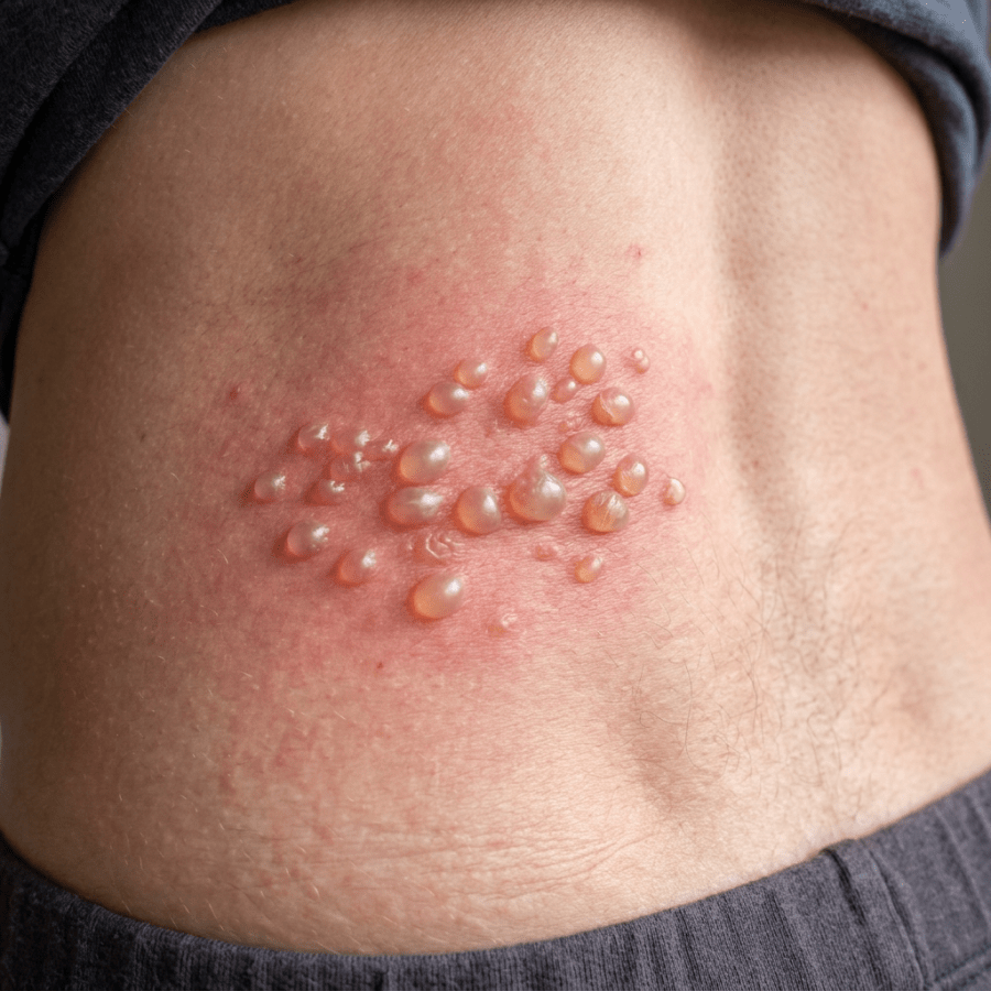
Clustered fluid-filled blisters on reddened skin of the torso

Shingles – “You mean I have Herpes?”

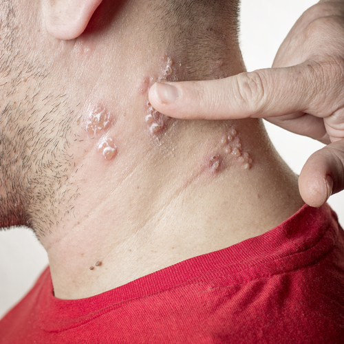
shutterstock_134601161Shingles, also called Herpes zoster is a painful rash caused by the same virus that causes chickenpox.  The term “shingles” comes from the latin word “cingulum” which means belt or girdle; the rash usually appears in a band or belt-like pattern. Singles can occur in people of all ages, but is more common in adults over age 50.  It can also be more common in those people with a weakened immune system. Cause:  After chickenpox goes away, the virus retreats to cells in the nervous system and hides out there quietly for many years.  Later in life, the varicella zoster virus can become active again and cause shingles.  There are many different types of herpes virus and shingles/chickenpox are a different form of herpes than the type that cause cold sores or genital herpes.   You are not at risk for getting cold sores or genital herpes by being around someone who has shingles or chickenpox. Risk of getting shingles:  About 20% of people will get shingles at some point in their life.  Some people actually get the recurrence of shingles multiple times.  Although it only occurs in people who have had chicken pox in the past, sometimes the chickenpox is mild enough that you might not even be aware that you have had it. Weakened immune system:  Certain people are more at risk for developing shingles if their immune system is compromised: 1)   Chemotherapy treatment for cancer 2)   Cancer itself can lower the immune system 3)   HIV 4)   Medications that suppress the immune system – such as after an organ transplantation or medications used to treat rheumatoid arthritis Symptoms:  Parasthesias are unusual sensations that may occur before any rash is noted.  It’s described as a tingling, itching or burning in the area of skin on one side of the body.  Some individuals develop a fever or headache, but many do not.  Within a day or two a rash of blisters occur on one side of the body in a band-like pattern.  The rash may occur on the chest, upper or lower back or even on the face.  If it occurs on the face near the eye, it can permanently affect the vision, so we will often have those patients be seen by an eye doctor. Shingles pain can be mild, moderate or severe and is usually described as a stabbing or burning pain.  The pain usually stays in the area of the rash and can interfere with sleep and daily activities.  Older adults typically have worse pain than younger patients. After 3-4 days the blisters usually become open sores or ulcers.  Rarely, the patient can develop a secondary bacterial infection in the area of the shingles rash.  We treat any underlying bacterial infection with antibiotics.  The sores then crust over and are not infectious after 7-10 days and the rash gradually resolves within 3-4 weeks.  Scarring or skin changes can occur.  Most of the time, no chronic problems occur after someone has a shingles outbreak. Can I catch it from someone else?   You can’t catch shingles from someone else but you can become infected with the varicella zoster virus.  If you’ve never had chickenpox or the vaccine, you can develop it after being in contact with someone who has shingles.  Even inhaling virus particles that are in the air can spread the infection.  If you have had chickenpox or the vaccine, being near someone with shingles will not cause shingles or chickenpox. Possible complications of shingles:  Possible complications of a shingles infection can occur: 1)   Pain called postherpetic neuralgia is the most common complication.  Is is usually described as a burning pain that affects 10-15% of patients after the rash has resolved. 2)   Skin infection from a bacteria can delay healing 3)   Eye complications can occur if the virus is on the face near the eyes Treatment:  There are multiple medications that can be used to treat shingles.  We try to keep the skin clean and dry to decrease the chance of developing a bacterial infection.  Some options might include: 1)   Antivirals such as acyclovir, famciclovir or valacyclovir are most effective when started within 3 days after the rash appears.  They stop the virus from multiplying and speed the healing of skin lesions.  This decreases the amount of pain the patient experiences 2)   Pain medication such as Ibuprofen, Aleve or prescription pain medication may be prescribed Return to work:  If the blisters are on the face, it is best to remain off work until the area has crusted over which is usually 7-10 days.  If the blisters can be covered, you can return to work after you are feeling well.  If you work in a healthcare facility, please consult your healthcare provider. Prevention:  There is a vaccine to help reduce the risk of shingles.  If a shingles infection occurs after the vaccine, it is usually less severe and there is less chances of developing post-herpetic neuralgia.  We recommend the vaccine for adults over age 60.  Even if you are unsure if you had chickenpox, we recommend the vaccine.  We don’t give the vaccine to pregnant women, or those with a weakened immune system.   I hope that you have found this information useful.  Wishing you the best of health,
When the pain outlasts the rash, gabapentin is one of the first drugs offered for postherpetic neuralgia, so it is worth reading where the gabapentin and dementia risk question stands before you start it.

Updated for 2026: The Vaccine in This Post No Longer Exists

That is not an exaggeration. When I wrote this in 2012, the shingles vaccine was Zostavax, a single live vaccine for adults sixty and over. Zostavax was discontinued in the United States on November 18, 2020 (1). You cannot get it. If this post sent you looking for it, that is on me, and here is what replaced it. Shingrix is now the only zoster vaccine available here. It is not live, it is given as two doses two to six months apart, and it is recommended for immunocompetent adults fifty and older (2). Two things people get wrong about it. You still need it if you have already had shingles. Having had it once does not protect you, and plenty of people get it twice. You also still need it if you had Zostavax years ago. Shingrix is recommended regardless (2).

If Your Immune System Is Suppressed

This is the group that had nothing in 2012, and it is the change that matters most. Because Zostavax was a live vaccine, immunocompromised patients could not have it, and they are precisely the people at highest risk. Shingrix is not live. In October 2021 the recommendation was extended to immunocompromised adults nineteen and older (2). The protection is real, though lower than in healthy adults, and it varies by situation. In people who had received their own stem cell transplant it was 68.2 percent. In people with blood cancers, 87.2 percent. In people on immunosuppressants for immune mediated disease, 90.5 percent (2). If you are on a biologic, on chemotherapy, or post transplant and nobody has raised this with you, raise it yourself. The interval can be shortened to one to two months if you need protection sooner. One technical point worth knowing: if the second dose lands less than four weeks after the first it does not count and has to be repeated. If more than six months go by, you do not start over, you just get the second dose (2).

Treatment, and the Seventy Two Hour Window

This part has not changed and it is still the thing that decides how your next few months go. Acyclovir 800 milligrams five times daily, valacyclovir 1,000 milligrams three times daily, or famciclovir 500 milligrams three times daily, all for seven days, and started within seventy two hours of the rash appearing (3). Seventy two hours. That is the whole game. Antivirals started inside that window shorten the illness and reduce the chance of the nerve pain that follows. Started late, they do much less. This is one of the better arguments for a same day video visit. You do not need anyone to touch you to start this, you need somebody to look at the rash quickly and act.

The Pain That Comes After

Postherpetic neuralgia is the reason shingles has a reputation. For what actually helps, the numbers are worth seeing side by side, expressed as how many people need treating for one to benefit. Tricyclics like nortriptyline do best on paper, around three, though roughly a quarter of people stop them over side effects. Pregabalin up to 600 milligrams a day comes in around four. Gabapentin, at a genuinely therapeutic 1,800 to 3,600 milligrams a day, around eight. The clinician applied capsaicin 8 percent patch, around seven. The lidocaine 5 percent patch has a very good safety profile and thinner evidence behind it (3). The gabapentin number is the one I would flag. It works, but only if it is pushed to a real dose. A great deal of gabapentin gets prescribed at 300 milligrams at bedtime, never titrated, and then written off as ineffective. That is an under dosing problem, not a drug problem.

When This Needs More Than a Video Visit

Shingles on the forehead, around the eye, or on the tip of the nose. That last one, the Hutchinson sign, predicts eye involvement, and this needs an ophthalmologist the same day, not tomorrow. A rash crossing more than about three dermatomes or scattered widely, especially if your immune system is suppressed, because that can mean the virus has spread internally. Facial droop, hearing change, or vertigo with a rash in or around the ear, which suggests Ramsay Hunt syndrome. Confusion, a stiff neck, or any neurologic change.

The Bottom Line

If you are fifty or over, or nineteen and over with a suppressed immune system, get Shingrix, two doses, even if you have had shingles before and even if you had the old vaccine. If a rash starts, be seen within seventy two hours. And if you end up on gabapentin for the nerve pain afterward, make sure somebody actually titrates it.

Sources

1. CDC. Zostavax discontinued in the United States as of November 18, 2020. https://archive.cdc.gov/www_cdc_gov/vaccines/vpd/shingles/public/zostavax/index.html 2. Anderson TC, et al. Use of Recombinant Zoster Vaccine in Immunocompromised Adults Aged 19 Years and Older: ACIP Recommendations, United States, 2022. MMWR. 2022;71(3):80-84. https://pmc.ncbi.nlm.nih.gov/articles/PMC8774159/ 3. Herpes Zoster and Postherpetic Neuralgia: Prevention and Management. American Family Physician. November 15, 2017. https://www.aafp.org/pubs/afp/issues/2017/1115/p656.html

Related Reading

When to Use Telemedicine vs. Urgent Care: How I Spot the Sick One Am I Truly Allergic to Penicillin? All about Cold Sores (Oral Herpes) The Viral Rash – Exanthem How to Manage Chronic Pain Without Relying on Medication Scott Rennie, D.O.

Board Certified in Obesity Medicine and Family Medicine

This blog is for educational purposes only and does not constitute individual medical advice. Always consult your own physician before making changes to your health, medications, or treatment plan.

Swimmer’s Ear (Otitis Externa): How to Spot and Treat It

Otitis externa, commonly known as “Swimmer’s Ear,” is a frequent condition we see in the clinic—especially in the warmer months or among frequent swimmers. It occurs when the delicate skin lining the ear canal becomes inflamed, often due to moisture, infection, or irritation.

What Causes It?

Swimmer’s ear can result from bacterial or fungal infections, but it can also be triggered by allergies, eczema, or psoriasis. The nickname “Swimmer’s Ear” comes from its strong association with prolonged water exposure, which creates an ideal environment for bacteria to grow.

How It Differs from a Middle Ear Infection

Many people use the term “ear infection” to refer to otitis media, which affects the middle ear behind the eardrum. Otitis externa, on the other hand, affects the outer ear canal—and the treatment and causes are different.

Risk Factors

Several things can increase your risk of developing otitis externa:

  • Over-cleaning the ears, which strips away protective earwax and may scratch the canal.

  • Swimming, especially in untreated water, can trap moisture in the ear.

  • Hearing aids, ear plugs, or in-ear headphones that block airflow and may trap moisture.

  • Skin trauma, such as from Q-tips or fingernails, can create entry points for infection.

Symptoms

Look out for:

  • Ear pain, especially when tugging on the outer ear.

  • Itching deep in the ear canal.

  • Fluid drainage, including pus or clear fluid.

  • Swelling or muffled hearing—the canal may swell shut in more severe cases.

Diagnosis

Your healthcare provider will examine the ear using an otoscope—a small lighted tool that allows them to see signs of redness, swelling, or discharge in the ear canal.

Treatment

The main goals of treatment are to:

  1. Clear the infection.

  2. Reduce inflammation and pain.

  3. Prevent recurrence.

Depending on the case, we may flush the ear canal gently with a mix of water and hydrogen peroxide to remove debris and bacteria. Most patients will be prescribed antibiotic ear drops, often with a small amount of steroid to reduce inflammation.

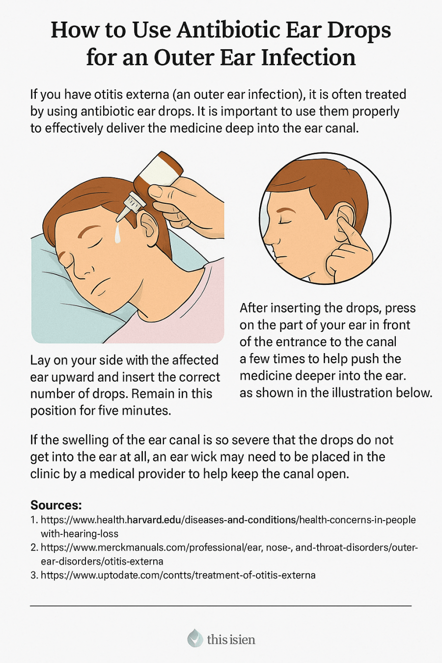
How to Use Ear Drops Properly

Correct technique matters. Follow these steps to make sure the medication gets deep into the canal:

  1. Lie on your side with the affected ear facing up, or tilt your head.

  2. Place the prescribed number of drops into the ear canal.

  3. Stay in that position for 20 minutes, or use a small cotton ball at the ear opening to help hold the drops in.

  4. Finish the full course of treatment, even if you feel better in a few days.

  5. Call your provider if symptoms persist after 36–48 hours.

If the ear canal is too swollen for drops to enter, this may require an in-person visit for placement of a wick—a small sponge that helps carry the medication deeper into the canal.

Pain Management

Most cases respond well to ibuprofen or naproxen (Aleve). Prescription pain relievers are rarely needed.

Protect the Ear During Treatment

  • Avoid getting the ear wet. During showers, cover the ear with a dry cotton ball.

  • Do not swim for 7–10 days after starting treatment.

  • Avoid inserting hearing aids, earbuds, or Q-tips until fully healed.

Prevention Tips

To reduce the risk of future infections:

  • Never insert Q-tips, fingers, or towels deep into the ear canal.

  • Dry your ears after swimming by shaking them out or using a blow dryer on low, held at least 12 inches away.

  • Use over-the-counter drying drops after swimming.

  • Swim with earplugs designed to keep water out of the canal.


Scott Rennie, D.O.
Board Certified in Obesity Medicine and Family Medicine

This blog is for educational purposes only and does not constitute individual medical advice. Always consult your own physician before making changes to your health, medications, or treatment plan.

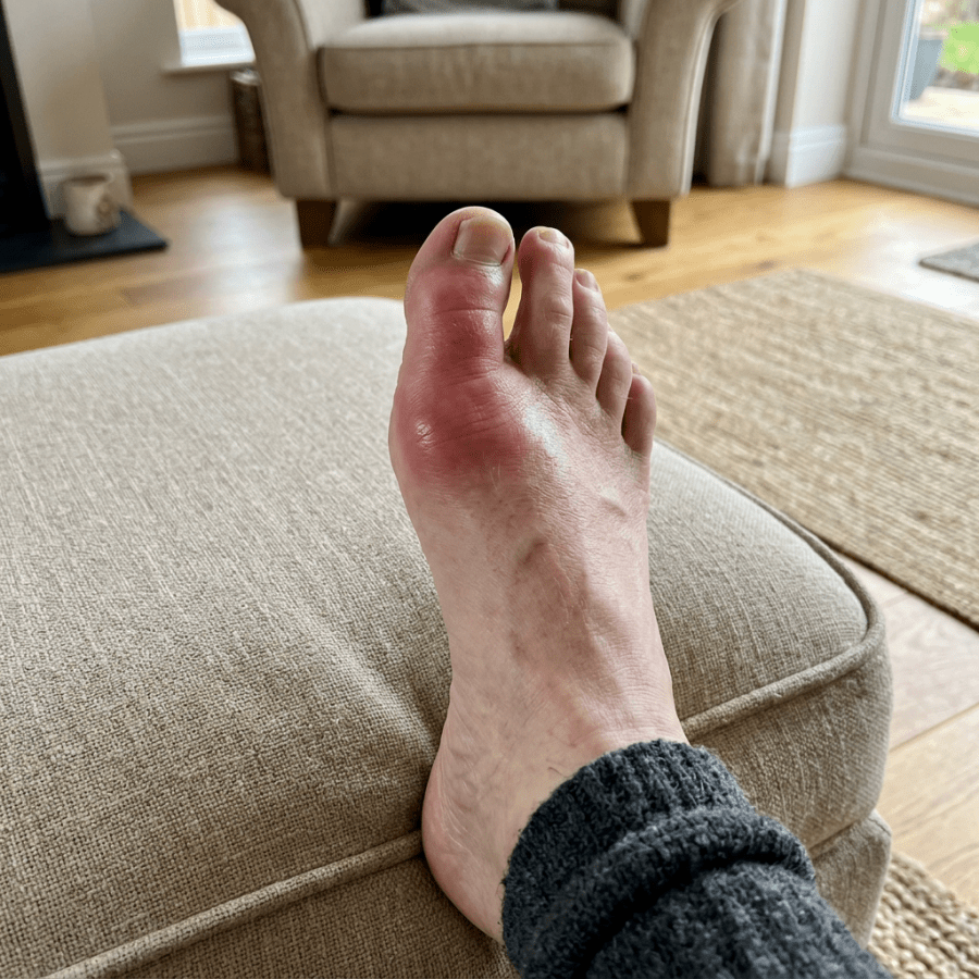
Swollen, reddened big toe resting on a beige ottoman

Diagnosed With Gout? What It Means and How to Prevent Flares

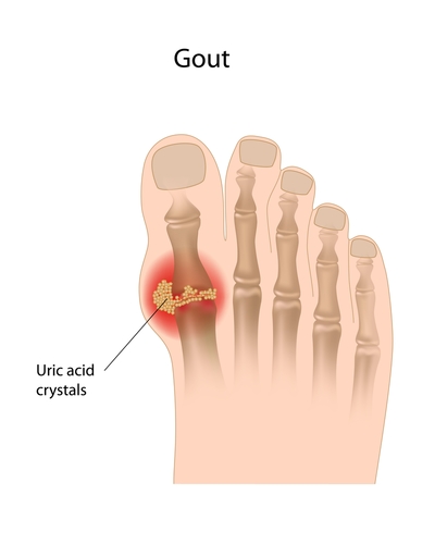
shutterstock_114066385At least once a week, I have patient who presents to the clinic due to pain from gout.  It is a painful condition that develops in some patients who have elevated levels of uric acid in their blood (hyperuricemia).  Up to 66% of patients who have elevated uric acid levels never develop symptoms.  We’re not sure why some people develop symptoms and others don’t have symptoms at all. Uric acid crystals most commonly affect one or multiple joints in the body, but may also cause uric acid crystals to develop within the kidney or urinary tract.  A stone within the kidney may cause impaired kidney function or pain.  Only 15% of people with gout develop kidney stones due to uric acid. Gout risk factors:  Gout is estimated to affect about 2% of the population in the USA.  It is most common in men between 30-45 and women between 55-70.  There is an increased risk of gout with the following: 1)   Taking medications that affect uric acid levels, such as diuretics 2)   Obesity 3)   Ingesting large amounts of meat or seafood 4)   High blood pressure 5)   Consuming high quantities of alcohol 6)   Overeating 7)   Recent surgery or injury Symptoms: 1)   Sudden severe joint pain (usually one joint), sometimes with redness and swelling 2)   Pain and inflammation are worst within several hours and improve over a few days to weeks completely even if untreated Gout phases: 1)   Acute gouty arthritis:  attacks usually involve the big toe or knee which become painful and inflamed 2)   Intercritical period:  The time between attacks.  Usually a second attack occurs within 2 years.  If uric acid levels are uncreated over several years the time between attacks may shorten or the symptoms may become more severe when attacks occur. 3)   Chronic tophaceous gout:  Large numbers of uric acid crystals may collect in joints, bones or cartilage and cause a nodule called a tophus to form.  This tophus is not usually tender, but cause erosion of bone and permanent deformity.  The tophi when the occur on the knuckles can cause an unfortunate cosmetic problem. Diagnosis:  Gout may be suspected if the person has the sudden onset of joint pain in the commonly affected joints such as the big toe.  If the patient has a period when there are no symptoms, and then symptoms return this is very typical of the disease.  The best way to diagnose gout is to examine fluid from the affected joint to look for uric acid crystals.  A medical provider uses a needle and syringe to withdraw a small sample of synovial fluid from the joint and this fluid is sent to the lab for microscopic analysis.  Other criteria for diagnosis gout may be: 1)   Blood testing showing high uric acid levels 2)   Rapid improvement of symptoms after the patient is treated with colchicine 3)   History of pain and inflammation involving one joint at a time, especially at the base of the large toe 4)   The symptoms totally go away between attacks Treatment:  The goals of gout treatment are to decrease the pain of a gout attack and also to reduce the frequency of their attacks.  Some of the treatment options are listed below: 1)   Nonsteroidal anti-inflammatory drugs (NSAIDs) reduce the swelling in the joint.  Examples of these are Ibuprofen or indomethacin.  They are most effective if started as soon as possible after the gout attack starts. 2)   Colchicine is a medication that may decrease the pain of a gout attack very rapidly. 3)   Steroids such as prednisone may be used if an NSAID is not used.  This type of medication can be injected directly into the affected joint or can be given as pills or intramuscular injection.  There is a risk of “rebound gout attack” as the steroids are stopped however, so usually steroids are slowly tapered over 7-10 days. Prevention therapy:  Some medications as well as dietary changes can be helpful to reduce the frequency of gout attacks. 1)   Uric acid lowering medications are used indefinitely because there is not a benefit from taking a break from the medications.  Examples of this are Probenacid, Losartan, and Allopurinol.  Rapid lowering of uric acid can cause a gout flare . 2)   Increased water intake – at least 2 liters/day are recommended Dietary changes:  Obesity is a major risk factor for gout so changing diet may decrease the frequency of gout attacks.  Current recommendations also include eating less of the following: 1)   Red meat 2)   Seafood 3)   Beer and hard alcohol (gin, vodka, etc.) 4)   Foods and drinks that contain high-fructose corn syrup (fond in some non-diet sodas for example) Some foods that may be good to eat or drink if you have gout may include: 1)   Low fat dairy 2)   Foods with complex carbohydrates such as whole grains, brown rice, oats and beans 3)   Vitamin C (500mg/day has been found to have a mild uric acid lowering affect). 4)   Coffee in moderation 5)   Wine in moderation – (1-2 5oz servings/day me be helpful) 6)   Pineapples – see my blog article about preventing gout by eating pineapples 7)   Drink plenty of water – twelve 8oz glasses of water during every day.   I hope that you have found this information useful.  Wishing you the best of health,
Related Reading

Can Eating Pineapple Help Treat Gout Pain Naturally? How to Manage Chronic Pain Without Relying on Medication All About Knee Pain: What Every Patient Should Know I Hurt My Finger or Toe: How Do I Know if It’s Fractured? How Do You Actually Lose Weight? A Doctor Explains

Scott Rennie, D.O.
Board Certified in Obesity Medicine and Family Medicine

This blog is for educational purposes only and does not constitute individual medical advice. Always consult your own physician before making changes to your health, medications, or treatment plan.

Woman using an inhaler while seated in a bright living room

Wheezing and Possible Asthma: What Should You Do Next?

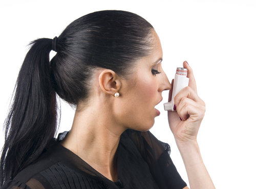
shutterstock_137805167Millions of people suffer from asthma worldwide.  The cause is due to narrowing of the airways (small tubes) in the lungs which is partially or completely reversible. Symptoms of asthma: 1)   Coughing 2)   Wheezing 3)   Chest tightness 4)   Shortness of breath Asthma symptoms tend to come and go and are affected by the amount of inflammation or narrowing of the airways are present.  Things that can cause the airways to narrow may include: 1)   Viruses that increase the production of mucus 2)   Allergens 3)   Exercise 4)   Certain foods 5)   Environmental conditions Asthma Triggers:  We call factors that set off or worsen asthma symptoms “triggers.”  One of our goals is to identify factors that cause asthma flare-ups so we can reduce or prevent asthma attacks.  Some of these asthma triggers in most patients are: 1)   Smoke or chemical irritants 2)   Allergens such as dust, pollen or pet fur 3)   Respiratory infections 4)   Emotional stress 5)   Physical activity 6)   Menstrual cycle in some women 7)   Certain medications such as aspirin, ibuprofen or Aleve have caused asthma symptoms in a small number of patients.  This is rare however. Categories of asthma:  We categorize asthma according to the severity and adjust the treatment plan depending on the patient’s age, level of control and severity of symptoms. 1)   Intermittent asthma:  These patients usually have symptoms that occur two or fever times per week, the asthma does not interfere with their daily activities and nighttime symptoms awaken the patient two or fever nights per month and oral steroid medication is needed no more than once per year to treat symptoms. 2)   Persistent asthma:  These patients have symptoms more frequently, and there may be days that activities are limited due to symptoms.  Symptoms tend to awaken to patient from sleep more often need to be treated more regularly than for intermittent asthma to keep symptoms from returning. Treatment:  Asthma treatments tend to be separated into two catergories: 1)   Short acting bronchodilator medications to treat an acute asthma flare.  These medicines work by temporarily relaxing the muscles around the narrowed airways.  Albuterol (also called Ventolin, Proventil and Proair) is the most commonly used inhaled bronchodilator used in the US There is no benefit to just using short-acting short acting bronchodilators on a regular basis.  If symptoms are occurring more than twice a week, other medications for asthma may be more helpful. 2)   Asthma controller medications:  For patients with more persistent symptoms, inhaled glucocorticoids, long acting bronchodilators, leukotriene modifiers, or oral glucocorticoids may be used. The benefit of regularly using a inhaled glucocorticoid medication is that it will reduce of frequency of symptoms so that the need for the short acting inhaled bronchodilator (albuterol) is not needed as often and improves quality of life and decreases serious attacks. Patients who present to the medical clinic or urgent care generally have progressed beyond treating a mild episode of asthma and usually require a medication in addition to a short acting bronchodilator such as albuterol.  We usually use an oral steroid medicine such as prednisone or dexamethasone. In patients who use an inhaler, it is important to understand how to use it properly.  Most inhalers work best if you hold the mouthpiece of the inhaler 1 to 2 inches in front of your mouth when you activate it.  If you close your mouth on the mouthpiece of the inhaler, less medicine generally reaches the lungs.  A spacer can be very helpful because it allows you to breathe in slowly and fully to inhale more of the asthma medication. Exercise induced asthma:  In patients who get asthma symptoms with exercise, we usually recommend an extra dose of albuterol before exercise to prevent the asthma symptoms.  Sometimes a leukotriene modifier (such as Singulair) or cromolyn may be used on a daily basis to prevent flares in these patients.   I hope that you have found this information useful.  Wishing you the best of health,
Scott Rennie, D.O.
Board Certified in Obesity Medicine and Family Medicine

This blog is for educational purposes only and does not constitute individual medical advice. Always consult your own physician before making changes to your health, medications, or treatment plan.

Woman washing her hands at a kitchen sink with irritated forearm

Skin burns – 1st degree, 2nd degree, 3rd degree and what to do about them

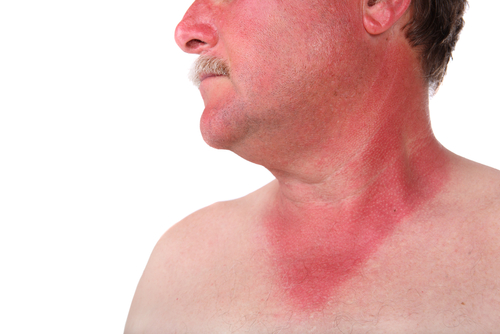
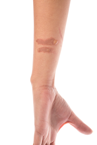
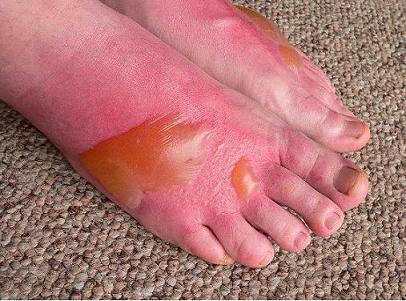
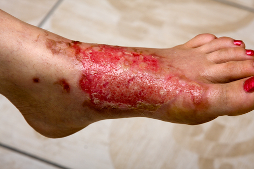
Patients frequently come into the medical clinic with burns.  Several possible sources of exposure can lead to skin burns including hot water or steam, hot objects or flames, chemical, electricity or exposure to the sun. Since moderate to severe burns can cause serious complications, it is important to recognize the signs of a serious skin burn so you can be evaluated by a healthcare provider. When to seek help: 1)    If the burn is on or near a joint such as the fingers, feet or hands 2)    If the burn is on the genital area 3)    If the burn goes all the way around (encircles) an arm, leg, foot, finger or your chest 4)    If the burn is larger than 3 inches or deep 5)    If the patient is younger than 5 years old or older than 70 years old 6)    If there is increasing pain, swelling, redness or pus-like discharge or fever > 100.4 degrees F. Burn classification:  The classification system that we used in the past was based on the thickness of the burn.  The new classification is as follows: 1)  Superficial burn or 1st degree involves only the top layer of skin.  They heal within 3-6 days.shutterstock_56132251 2)  Partial-thickness  or second degree burns involve the two top layers of skin.  These burns are painful with air movement or temperature changes and form blisters.  Partial thickness burns turn white when pressed.  These burns usually health within 7-21 days.  The skin that was burned may become darker or lighter color permanently in some patients. shutterstock_127655633 3)  Deep partial-thickness burns were previously called 3rd degree burns and are painful with deep pressure.  They form blisters and do not turn white with pressure.  These burns take longer than 21 days to heal and usually develop a scar that may be severe.  If the skin blisters immediately, it is at least a deep partial-thickness burn.  bb2_2 4)  Full Thickness Burns:  Extend through all layers of the skin and destroy it completely.  The burned area usually does not hurt and is waxy white to leathery gray in color and the skin is usually dry.  Full-thickness burns do not heal without surgery.  These burns used to be called 4th degree burns.  shutterstock_101309467 Treatment:  Most of the time small superficial or partial-thickness burns can be treated at home, but deeper wounds should be evaluated by a medical provider.  Treatment in the home: 1)   Clean the wound:  If clothing is stuck to the wound, seek care by a medical provider.  Gently wash the burned skin with soap and water.  Do not use alcohol, iodine or other cleansers. 2)   Cool the wound:  After cleaning, you may apply a cold compress or soak the skin in cool water for comfort and reduce pain.  Do not put ice directly on the wound itself however. Infection prevention:  Apply a small amount of antibiotic ointment such as bacitracin or silvadine cream may be applied.  A minor burn may be covered with a bandage or dressing.  If there are blisters, these should be covered with a clean bandage or dressing.  Do not pop the blisters with your fingernail as this may increase the risk of infection.  Leave the blisters alone or gentle pop them on the side of the blister sterile needle if they are large.  Use a non-stick dressing such as Vaseline impregnated gauze or Telfa dressing to cover the wound. If your tetanus has not been updated within 5 years, you need a tetanus booster if the burn is superficial partial-thickness or deeper. Burn Prevention: 1)   Keep matches, lit candles and lighters out of the reach of children 2)   Do not smoke when sleepy 3)   Use a cool-mist humidifier instead of a warm mist humidifier 4)   Apply sunscreen liberally to prevent sunburn and stay out of the sun when possible 5)   Be careful when placing a child in a car seat or stroller because the metal or vinyl can become hot 6)   Set your hot water thermostat to lower than 120 degrees F 7)   Keep children away from fireplaces, hot stoves and ovens 8)   Children’s sleepwear should be non-flammable.   I hope that you have found this information useful.  Wishing you the best of health,
A burn is one of the problems where a camera only tells you so much, and I have written about which problems a video visit can handle and which ones need to be seen in person. The rest of my work these days is obesity medicine, where the argument right now is whether Ozempic and Zepbound cause muscle loss.

Updated for 2026: The Cooling Advice Has Gotten More Specific

When I wrote this in 2012 the standard advice was to run cool water over a burn. That was right, but it was vague, and vague advice gets applied badly. Here is the current version. Cool running tap water, somewhere between 46 and 77 degrees Fahrenheit, for at least twenty minutes, started within the first half hour. There is evidence the window stays useful out to about three hours, so if an hour has already passed, still do it (1). Not ice. Ice constricts the blood supply to skin that is already injured and makes the wound deeper. I still hear ice recommended and it is worth saying plainly: skip it. Plain tap water is fine. It does not raise the infection risk compared with sterile solutions, and the twenty minutes of cooling matters far more than what the water came out of (1).

Silver Sulfadiazine Has Fallen Out of Favor

This is the part of the old post I would write differently today. For decades silver sulfadiazine cream was the default. It is no longer where the evidence points. There is reasonable concern that it impairs the cells doing the healing, slowing things down and possibly worsening scarring, and the trial support for it was never as strong as its popularity suggested (1). What has replaced it is less glamorous and works better. Either a topical antimicrobial under simple gauze changed every twelve to twenty four hours, or one of the modern occlusive dressings, silver foam, hydrocolloid, or silicone, left alone for as long as two weeks. The second approach hurts less and gets opened less often, which is why I favor it, particularly in children (1). Antibiotic pills are not part of routine burn care. An uncomplicated burn does not need them, and giving them anyway does not prevent infection. One thing that has not changed: tetanus. A partial or full thickness burn in someone whose shots are incomplete or unknown needs Td or Tdap, and immune globulin if the primary series was never finished (1).

What I Can and Cannot Do With a Burn Over Video

Burns are one of the harder things to assess on a screen, and I would rather tell you that than pretend otherwise. Depth is the problem. On camera I can usually tell superficial from something deeper, but the line between a deep partial thickness burn and a full thickness one is a judgment I would rather make with the wound in front of me. Good lighting and a close, steady photo help more than people expect. So does telling me exactly what caused it, how hot, and how long the contact lasted. What I can do well from here is talk you through the first twenty minutes while it is happening, sort out dressings and pain control, check your tetanus status, and tell you honestly when this is beyond what a camera can settle.

Burns That Need a Burn Center, Not a Video Visit

The American Burn Association keeps a referral list and it is worth knowing (2). Any full thickness burn. Partial thickness burns covering ten percent or more of the body. Any deep burn on the face, hands, feet, genitals, perineum, or across a major joint. Anything suggesting smoke inhalation, including facial flash burns or singed nasal hair. Chemical burns. All electrical burns, and high voltage or lightning injuries immediately. Pain you cannot get on top of. And essentially any significant burn in a child.

The Bottom Line

Twenty minutes of cool running water, measured, not guessed. No ice. Skip the silver sulfadiazine your medicine cabinet may still be holding. And know which burns are past the point where any of this applies.

Sources

1. Rice PL, Orgill DP. Outpatient Burn Care: Prevention and Treatment. American Family Physician. April 15, 2020. https://www.aafp.org/afp/2020/0415/p463 2. American Burn Association. Guidelines for Burn Patient Referral. https://ameriburn.org/burn-care-team/resources/guidelines-for-burn-patient-referral

Related Reading

When to Use Telemedicine vs. Urgent Care: How I Spot the Sick One Abscesses and MRSA: What To Do About a Skin Infection Basic Wound Care Tips for Non-Medical Professionals Cellulitis – a soft tissue/skin infection – is it MRSA? It’s Cloudy in Seattle: Can You Still Get a Sunburn? Frostbite and cold induced injuries Diabetic Foot Care: How to Check and Protect Your Feet Scott Rennie, D.O.

Board Certified in Obesity Medicine and Family Medicine

This blog is for educational purposes only and does not constitute individual medical advice. Always consult your own physician before making changes to your health, medications, or treatment plan.

Forearm with multiple raised red bumps and irritated skin

Hives – What am I allergic to?

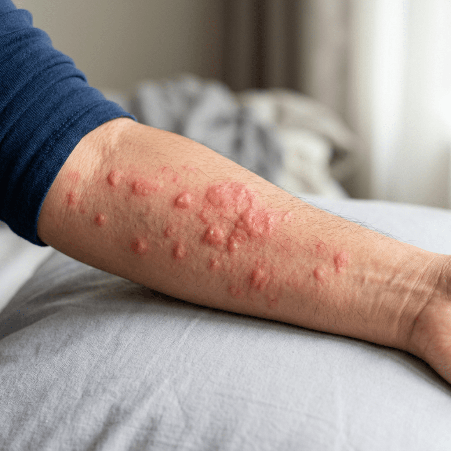
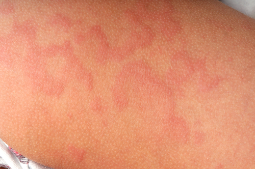
shutterstock_46021174The medical term for hives is urticaria and they are described as raised areas of skin that itch intensely and they are red and often have a pale color to the center.  They are common and occur in at least 20% of people at some point in their lives. Why hives develop:  This skin rash occurs as the body’s immune system is exposed to an allergen (such as a food or chemical) that it inappropriately believes could be harmful.    The body then responds by activating it’s army of immune cells in the skin to fight off the allergen.  When these immune cells (called mast cells) are activated, they release histamine that is a natural chemical that causes itching, redness and swelling of the skin.   In most cases, hives appear and can reappear suddenly – within several hours. Skin appearance:  Hives can occur anywhere on the body and actually appear and disappear and give the appearance that the rash is actually moving around the body.  They are raised areas of skin that itch and these red areas may enlarge and merge together.  The itching is usually the most bothersome symptom and it can be severe enough to interfere with work or sleep. Angioedema:  Some people who get hives also have puffiness of the face/lips, eyelids, mouth, hands, feet and genitalia.  Swelling usually affects one side of the body more than the other and may give the sensation of fullness or discomfort in the area of swelling. Anaphylaxis:  Hives can occur as part of a serious allergic reaction called Anaphylactic shock.  You should see a medical provider right away if you have symptoms along with hives such as: 1)   Trouble breathing 2)   Tightness in the throat 3)   Nausea/vomiting 4)   Cramping or abdominal pain 5)   Passing out 6)   Dizziness or light-headedness Types of hives:  Hives are usually classified based on how long they’ve been present.  Acute (brief) hives, vs. chronic (long-standing) or physical (triggered by a physical stimulation such as sun exposure or cold). Acute hives:  These are the type that we see most often in the medical clinic.  They usually don’t last beyond a few days to a week or two.  Triggers that can cause acute hives include: 1)   Infections:  Viral infections actually have been found to cause more than 80% of the cases of hives in children.  The hives usually last a week or two. 2)   Medications:  Many different kinds of drugs can cause hives including nonsterioidal anti-inflammatory drugs (NSAIDs) such as aspirin, ibuprofen or Aleve.  Antibiotics can also cause hives, as well as narcotic pain medication such as morphine or codeine. 3)   Insect stings:  Stings from bees, wasps, hornets or fire ants can cause hives around the sting.  If you get hives all over your body after a sting, this could be sign of anaphylaxis, and you should be seen by a medical provider as soon as possible. 4)   Food allergies:  food-associated hives usually occur within 30 minutes to an hour after eating the food.  Common foods which can cause hives include milk, eggs, peanuts or other nuts, soy, wheat, fish or shellfish. 5)   Physical contact:  After you touch a certain substance that you are allergic to, you may get hives.  Common substances that can cause hives are plants, raw fruits and vegetables, latex (found in balloons, certain gloves, condoms, etc.) Chronic hives:  Many people have hives that occur daily or almost every day.  We call hives that last longer than 6 weeks chronic hives.  Sometimes they can even last for years.  Hives are frustrating and can effect how you look but it is important to remember that hives are not contagious, they are rarely permanent, they are not life-threatening and symptoms are treatable in most people. For most people who get chronic hives, the cause is unknown.  Chronic hives can be a sign of other medical problems including thyroid or liver problems, chronic infection or lupus.  People with these problems usually have symptoms other than just hives however. Physical hives:  Hives can be triggered by exposure to cold (they often appear as the skin warms up), changes in body temperature or sweating, vibration, pressure, exercise, sunlight or water.  Some people can develop reddened, raised lines if the skin is stroked firmly or scratched.  This is called dermatographism. Diagnosis:  Most people do not need testing. The diagnosis can be made by the medical provider by taking a history and performing a physical examination.  Sometimes testing is done if symptoms are not resolving within six weeks.  Skin testing may be ordered by an allergist to test for food and medication allergies.  Blood tests can also be performed to look for other underlying medical problems.  A skin biopsy is a small sample of skin that is removed and examined under the microscope.  The biopsy can sometimes be helpful for patients who have chronic hives and it is usually performed by a dermatologist (skin doctor). Treatment: 1)  Avoid the cause/trigger.  Many times the cause in not found and the hives usually disappear over days or weeks anyway. 2)  Antihistamines – there are two types of antihistamines.  The older type such as Benadryl often work well, but can cause drowsiness, and are usually taken multiple times per day.  The newer types of antihistamines such as Zyrtec, Claritin or Allegra have fewer side effects are usually taken only once or twice a day. 3)  Oral steroids such as prednisone at a high dose are often used in combination with antihistamines.  We are careful with the use of steroid medications because long-term use can cause serious side effects if taken of long periods of time (months to years). 4)  Topical steroids or creams:  Sometimes topical creams may help with the itching, however we usually don’t use topical steroid creams on the hives because the rash may move from one area of the body to another rapidly.   I hope that you have found this information useful.  Wishing you the best of health,
Hives were everyday family medicine for me. These days I write more about metabolic and menopause care, including the FDA pulling the black box warning off menopause hormone therapy.

Related Reading

Hives and Angioedema: Signs of an Allergic Reaction Allergic Antibiotic Drug Reactions – Am I Truly Allergic to Penicillin? Doctor Said You Have Allergies? How to Know for Sure GLP-1 Skin and Nerve Pain: Ozempic, Wegovy, Mounjaro

Scott Rennie, D.O.
Board Certified in Obesity Medicine and Family Medicine

This blog is for educational purposes only and does not constitute individual medical advice. Always consult your own physician before making changes to your health, medications, or treatment plan.

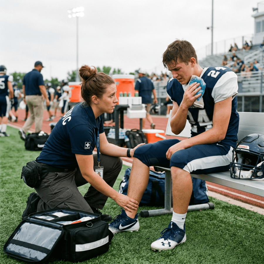
Sports trainer examining a seated football player on the sideline

Head Injury: Is It a Concussion or Something More Serious?

shutterstock_89651788 Head injuries are common all year long in the urgent care setting.  In the winter months, skiing or snowboarding is one of the leading causes of head injuries.  In the summer months, more sports injuries occur with the improved weather in the Seattle area. Most head injuries are not associated with brain injury or longer-term complications.  Rarely, however more significant injuries may develop which could be due to bleeding around the brain. Causes of head injuries:  Most of the time falls are the cause, but motor vehicle accidents, pedestrian or bicycle accidents and sports-related trauma also cause head injuries.  In the medical setting, we also have to be alert for signs of child abuse as well.  The risk of brain injury depend on the type of trauma and the age of the patient. Higher risk injuries: 1)   High speed motor vehicle accidents 2)   Fall from a great height 3)   Being hit by a high speed, a heavy or sharp object 4)   Inflicted injury such as abuse Symptoms of head injuries: 1)   Scalp swelling 2)   Loss of consciousness/passing out – Happens only 5% of the time and usually lasts less than 60 seconds 3)   Headache – Occurs in 20% of patients.  In children who are too young to speak, they may become irritable. 4)   Vomiting – occurs in about 10% of patients.  Children who have a head injury and vomit, do not necessarily have a serious brain injury. 5)   Seizures – Less than 1% of patients have a seizure right after a head injury.  A few of these patients will have a serious head injury.  We will usually do a CT scan of the head if  the patient has a seizure. 6)   Concussion – common symptoms include confusion or inability to remember events around the time of the injury, headache, vomiting, and dizziness. When to seek help: 1)   The patient has recurrent vomiting 2)   The patient has a seizure (convulsion) 3)   The patient loses consciousness after the injury 4)   Severe headache after the injury, or it worsens with time 5)   Head injury in a child with behavior change (lethargic, difficulty to wake, extremely irritable, other abnormal behaviors). 6)   Difficulty walking, is clumsy or has lack of coordination 7)   Slurred speech or confusion 8)   Dizziness that is not resolving 9)   Blood or watery fluid comes from ears 10)  The patient is a child less than 6 months of age 11)  There is a cut that will not stop bleeding after pressure is applied for 10 minutes 12)  The patient fell from a height greater than 5 feet, or was hit with a high speed object or with great force 13)  Patient’s friends/family are concerned about how the patient is acting When do I need a Cat Scan/CT Scan of the brain?  A CT scan is a special X-ray that expose children/adults to radiation and should be avoided if possible.  Sometimes, however a Cat scan will be recommended to diagnose more severe injuries.  Some possible symptoms which may prompt your medical provider to order a CT scan are: 1)   Prolonged loss of consciousness 2)   Persistent or severe memory loss/confusion 3)   Persistent vomiting 4)   Seizure 5)   Severe, persistent or worsening headache 6)   Suspicion of intentional injury (abuse) 7)   Behavioral changes (lethargy, decreased alertness, extreme irritability) 8)   Signs of skull fracture such as a bulging fontanel or skull deformity 9)   Abnormal neurological exam 10)  Severe scalp bruising or swelling in a very young child Why not an MRI?  We use CT scanning instead of an MRI to look for brain injury in most head injuries because it is available at most hospitals, and CT is relatively quick compared to MRI.  MRI requires patients – including children to be completely still for at least 30 minutes and that can be challenging. Head injury treatment at home: 1)   Rest – lie down or participate in quiet activities 2)   If the head is bleeding, clean the area with soap and water and apply pressure with gauze.  If bleeding does not stop, the child should be evaluated 3)   Tylenol may be given in most cases for pain/headache.  If the headache worsens, please have the child evaluated (see above). Monitoring after head injury:  The patient should be observed for signs of worsening injury.  Please call your healthcare provider if any of the following are noted: 1)   Vomiting more than once or vomiting continues for 4-6 hrs after the injury. 2)   Severe/worsening headache 3)   Becomes more drowsy or hard to wake up. 4)   Confused or not acting normally. 5)   Has trouble walking, talking or seeing 6)   Develops stiff neck. 7)   Has a seizure (convulsion) or any abnormal movements or behaviors. 8)   Cannot stop crying – children 9)   Has weakness or numbness involving one side of the body. Return to normal activities:  Patients who have sustained a concussion are at a risk for serious or even fatal complications of they have a second injury within a short time after the first injury – this is called second impact syndrome.  It important not to participate in high impact sports or risky activities for 6 weeks. Post-concussion syndrome:  Sometimes the patient who has sustained a head injury may develop a group of symptoms in the first few days after the injury called “post concussion syndrome.”  These symptoms can include headaches, anxiety, irritability, dizziness, or impaired memory or concentration.  In 85-90% of patients, this resolves within a few weeks-few months after the injury.  There isn’t any specific treatment for post-concussion syndrome. Head injury prevention: 1)   Wear a bicycle helmet when riding bikes, skating , sledding or participating in activities where you may hit your head 2)   Install car seats/booster seats correctly.  At least a booster seat is needed until the child is at least 4 feet 9” tall.  Individual states may have additional regulations about the use of booster/car seats. 3)   Use gates on stairways to prevent injuries in infants/young children 4)   Install window guards on all windows above the first floor 5)   Do not use wheeled baby walkers 6)   Teach kids to safely cross the street.  Young children should never cross the street alone. 7)   Discuss sports safety with your healthcare provider.  Be sure that a child has appropriate protective equipment.   I hope that you have found this information useful.  Wishing you the best of health,
Scott Rennie, D.O.
Board Certified in Obesity Medicine and Family Medicine

This blog is for educational purposes only and does not constitute individual medical advice. Always consult your own physician before making changes to your health, medications, or treatment plan.