What Is MASLD? The New Name for Fatty Liver Disease

Liver disease is showing up more often, and it tracks closely with rising rates of obesity, diabetes, and metabolic syndrome. What many of us trained calling “fatty liver” or NAFLD has been renamed and reframed. The term now is MASLD, metabolic dysfunction-associated steatotic liver disease, and it reflects a better understanding of what actually drives the condition.

Why the change? NAFLD was a definition by exclusion. It told you the disease was not caused by alcohol without saying what it was. It also excluded patients with both alcohol and metabolic drivers, and the word “fatty” carried stigma that most patients felt immediately. In 2023 a multisociety Delphi process involving 236 panelists from 56 countries settled on the new nomenclature. Sixty-six percent of respondents found “fatty” stigmatizing and 61% said the same of “nonalcoholic.” The new definition requires at least one of five cardiometabolic risk factors, and it added MetALD for patients with metabolic dysfunction who also drink significantly (Rinella et al., J Hepatol, 2023).

MASLD is common. Roughly 30% of U.S. adults are affected. Among people with diabetes that figure climbs above 60%, and up to 15% carry advanced fibrosis (Le et al., Clin Mol Hepatol, 2022). Worldwide it is projected to overtake hepatitis C and alcohol as the leading cause of cirrhosis, hepatocellular carcinoma, and liver transplant.

The liver isn’t where most of these patients die. Cardiovascular disease is the leading cause of death in MASLD. The same inflammatory and metabolic pathways that damage the liver drive atherosclerosis. Diabetes worsens MASLD and MASLD worsens diabetes. The relationship runs in both directions.

One point matters more than any other: liver enzymes are a poor marker of severity. Normal ALT and AST are entirely compatible with advanced fibrosis. Fibrosis stage is what predicts progression, complications, and mortality. In a meta-analysis of 4,428 patients, all-cause mortality rose with each fibrosis stage, reaching a relative risk of 3.42 at stage 4 compared with stage 0, and liver-related mortality reached 11.13 (Taylor et al., Gastroenterology, 2020; Ekstedt et al., Hepatology, 2015). That is why guidelines now point everything at fibrosis assessment.

In primary care, FIB-4 is the practical first step. Age, AST, ALT, and platelet count. Under 1.3 suggests low risk and those patients can generally stay in primary care. Above 2.67 means high risk and warrants hepatology referral. Intermediate scores land in a gray zone that usually needs imaging such as FibroScan or a blood-based marker like the ELF test. FibroScan is fast and non-invasive but loses accuracy in patients with obesity, which is a real limitation given who has this disease. MR elastography is the most accurate option and the least available.

Treatment still starts with lifestyle. Weight loss of 5 to 10% improves steatosis and inflammation. The Mediterranean pattern is consistently associated with lower liver fat and better insulin sensitivity. Exercise at 150 minutes a week of moderate activity reduces liver fat even without weight loss, which is worth telling patients who are discouraged by the scale. Cutting sugar-sweetened beverages and limiting fructose is standard advice. Coffee earns its reputation here: a meta-analysis of observational studies found coffee consumption associated with 35% lower odds of significant fibrosis, with three or more cups a day the threshold most often cited, caffeinated or not (Hayat et al., Nutrients, 2021).

Medication options are expanding. Vitamin E has histologic benefit in non-diabetic patients with biopsy-proven MASH, though long-term safety concerns persist. Statins remain badly underused and are safe in MASLD, and they should be prescribed for cardiovascular risk reduction (Kargiotis et al., World J Gastroenterol, 2015). GLP-1 receptor agonists reduce liver fat and support weight loss.

In March 2024, resmetirom became the first FDA-approved drug for MASH with fibrosis. It is a liver-directed thyroid hormone receptor-beta agonist. In the phase 3 MAESTRO-NASH trial, MASH resolution without worsening fibrosis occurred in 25.9% of patients on 80 mg and 29.9% on 100 mg, against 9.7% on placebo, and both doses beat placebo on fibrosis improvement (Harrison et al., NEJM, 2024). It is approved for adults with non-cirrhotic MASH and stage F2 to F3 fibrosis. Those response rates are meaningful and they are also modest, and patients should hear both halves.

Endoscopic and surgical options matter too. Endoscopic sleeve gastroplasty and intragastric balloons reduce liver fat and improve fibrosis. Bariatric surgery remains among the most effective interventions available, with a systematic review and meta-analysis finding NASH resolution in roughly half of patients and fibrosis improvement in about a third (Lee et al., Clin Gastroenterol Hepatol, 2019).

MASLD management has moved well outside hepatology. It needs primary care, cardiology, endocrinology, nutrition, and gastroenterology working the same problem. Screen at-risk patients with FIB-4, particularly those with diabetes or obesity. Counsel on weight and diet. Prescribe statins when indicated. Refer for advanced assessment when fibrosis is suspected.

MASLD reframes liver disease as part of the broader cardiometabolic picture. Treating it means protecting the liver while cutting cardiovascular risk, improving glycemic control, and addressing systemic inflammation. That is where the impact lives.

Scott Rennie, D.O.

References:

1. Rinella ME, Lazarus JV, Ratziu V, et al. A multisociety Delphi consensus statement on new fatty liver disease nomenclature. J Hepatol. 2023;79(6):1542-1556. https://pubmed.ncbi.nlm.nih.gov/37364790/

2. Le MH, et al. Global incidence of non-alcoholic fatty liver disease. Clin Mol Hepatol. 2022;28(4):841-850. https://pubmed.ncbi.nlm.nih.gov/36117442/

3. Taylor RS, et al. Association Between Fibrosis Stage and Outcomes of Patients With Nonalcoholic Fatty Liver Disease: A Systematic Review and Meta-Analysis. Gastroenterology. 2020;158(6):1611-1625.e12. https://pubmed.ncbi.nlm.nih.gov/32027911/

4. Ekstedt M, et al. Fibrosis stage is the strongest predictor for disease-specific mortality in NAFLD after up to 33 years of follow-up. Hepatology. 2015;61(5):1547-1554. https://pubmed.ncbi.nlm.nih.gov/25125077/

5. Hayat U, et al. Effect of Coffee Consumption on Non-Alcoholic Fatty Liver Disease Incidence, Prevalence and Risk of Significant Liver Fibrosis: Systematic Review with Meta-Analysis of Observational Studies. Nutrients. 2021;13(9):3042. https://pubmed.ncbi.nlm.nih.gov/34578919/

6. Kargiotis K, et al. World J Gastroenterol. 2015;21(25):7860-7868.

7. Harrison SA, et al. A Phase 3, Randomized, Controlled Trial of Resmetirom in NASH with Liver Fibrosis. N Engl J Med. 2024;390(6):497-509. https://pubmed.ncbi.nlm.nih.gov/38324483/

8. Lee Y, et al. Complete Resolution of Nonalcoholic Fatty Liver Disease After Bariatric Surgery: A Systematic Review and Meta-analysis. Clin Gastroenterol Hepatol. 2019;17(6):1040-1060.e11. https://pubmed.ncbi.nlm.nih.gov/30326299/

Board Certified in Obesity Medicine and Family Medicine

This blog is for educational purposes only and does not constitute individual medical advice. Always consult your own physician before making changes to your health, medications, or treatment plan.
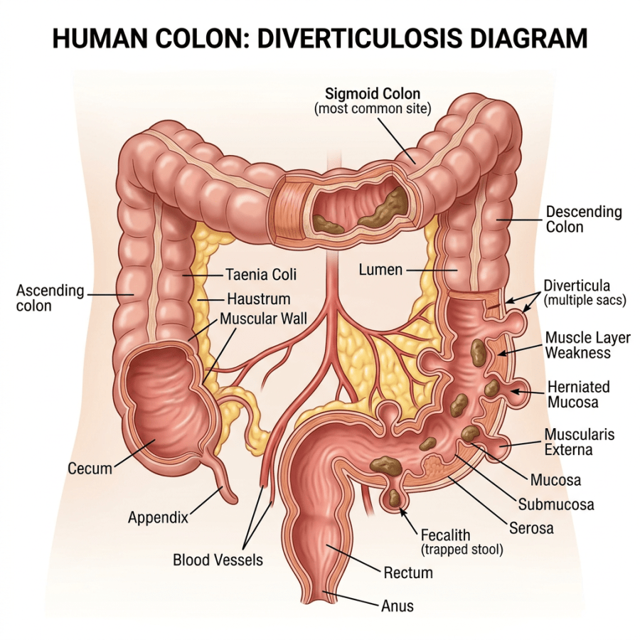
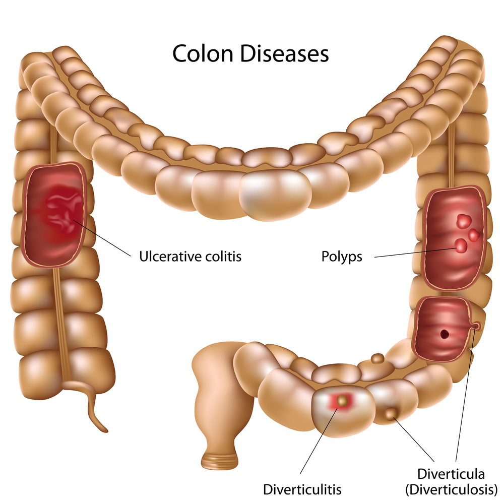
Diagram of the human colon highlighting diverticulosis features including diverticula, muscle layer weakness, herniated mucosa, and fecalith.

Diverticular Disease: Diverticulosis vs Diverticulitis Explained

shutterstock_75923548

When a patient came in with abdominal pain or blood in their stool, one of the conditions we considered was diverticulitis. You may not have heard much about diverticulitis, so this post will try to answer some common questions about diverticular disease.

A diverticulum is a small pouch-like structure that sometimes forms in the muscular wall of the colon. These little pouches often cause no pain, and we usually become aware of them only after a procedure such as a colonoscopy, flexible sigmoidoscopy, a barium enema or a CT scan.

What is diverticulosis? Diverticulosis simply means that diverticula are present. Most people have no symptoms and will remain free of symptoms throughout their lives. (Roughly 15 to 25 percent go on to develop diverticulitis, which is more severe.) I usually think of a diverticulum as a small area where the colon balloons out. These are potentially weak spots that might become inflamed or may rupture under pressure.

What is diverticulitis? Diverticulitis is inflammation of a diverticulum, which occurs when there is thinning or breakdown of the wall of the colon. It is more severe than diverticulosis because the pouches have become irritated, inflamed or stretched, either from pressure within the colon or from hard particles of stool lodged inside them. The diverticulum becomes painful and may rupture, meaning the wall of the colon breaks down and allows stool and bacteria to enter the abdomen. Diverticulitis may also cause bleeding within the colon, because blood vessels often run through the area where diverticula form and they can bleed when the area gets inflamed.

Symptoms of diverticulitis: The symptoms depend on how severe the inflammation inside the colon is. The most common symptom is pain in the left lower abdomen. Patients may also have blood in the stool, nausea, vomiting, constipation, or diarrhea.

Classification of diverticulitis: Simple diverticulitis is more common and usually responds well to medical treatment without surgery. Complicated diverticulitis, about 25 percent of cases, usually requires surgery to remove the section of colon that has become inflamed or ruptured.

Treatment: Diverticulosis itself usually needs no treatment, since diverticula that are not inflamed are generally painless and cause no problems. Many providers do recommend increasing dietary fiber to reduce the chance of constipation, since constipation raises pressure within the colon, which can in turn lead to diverticulitis. Increasing fruits, vegetables, hydration and fiber has not been proven to prevent diverticulitis, however. We used to tell patients with diverticulosis to avoid seeds and nuts, on the theory that they increased the risk of diverticulitis. Research has since shown that advice to be wrong.

Diverticulitis does require treatment, and what you receive depends on the severity of your symptoms. Mild abdominal pain from diverticulitis can usually be managed at home with a clear liquid diet and oral antibiotics. If a patient develops a fever greater than 100.4 degrees F, worsening or severe abdominal pain, or an inability to keep fluids down, hospital treatment is usually recommended.

Hospitalization: If you need to be in the hospital for diverticulitis, you usually will not be allowed to eat or drink until you start feeling better, and antibiotics and IV fluids are given. If an abscess forms, a surgeon may need to drain it by placing a tube through the abdominal wall.

Complications: Peritonitis is a generalized infection within the abdomen that occurs if the colon ruptures. An emergency operation is often required in these cases to remove the diseased section of colon and reconnect the unaffected ends. Sometimes those two steps are done at separate times, with the removal first, so the colon is allowed to heal before the pieces are reattached.

Surgical treatment: Patients who don’t respond well to medical treatment, or who have repeated attacks of diverticulitis, may benefit from surgery to remove the diseased section of colon.

Bleeding from diverticulitis: Most of the time the bleeding stops on its own, but sometimes a colonoscopy or another procedure is necessary to get it under control.

Scott Rennie, D.O.

Board Certified in Obesity Medicine and Family Medicine

This blog is for educational purposes only and does not constitute individual medical advice. Always consult your own physician before making changes to your health, medications, or treatment plan.

Blog: https://doctorrennie.wordpress.com

Abstract artistic illustration of the human digestive system with surrounding microbiome cells

Rectal Bleeding: What Are the Possible Causes of It?

Colon-Endoscopes
anat_colon_1.0

Photo credit: http://www.naturalhealingsolutionsllc.com/learn-about-colon-hydrotherapy.html

Photo credit: http://www.thenurseslockerroom.com/2013/03/sigmoidoscopy-screening-test-for.html

One of the more common problems that brought patients in to see me, believe it or not, was seeing blood in the toilet, on the stool, or on the toilet paper after a bowel movement. Since I had seen several patients about this, I thought I would write about some of the possible causes.

Healthcare providers take this seriously, because blood noticed after a bowel movement can sometimes be a sign of colon or rectal cancer. Fortunately, most causes of rectal bleeding are not cancer.

Causes of rectal bleeding:

  1. Hemorrhoids: Swollen blood vessels in the rectum or anus can cause itching, pain, and bleeding. Patients usually describe hemorrhoidal blood as bright red. It may coat the surface of the stool, drip into the toilet and turn the water red, or show up on the toilet paper. Hemorrhoids do not have to hurt. Painless rectal bleeding during a bowel movement is a common presentation.
  2. Anal fissure: A tear in the lining of the anus can bleed, and there may be pain with a bowel movement.
  3. Other causes include infection, colitis (which may be due to an autoimmune disease such as ulcerative colitis or Crohn’s disease), colon polyps, and colon cancer. If the bleeding comes from higher in the digestive tract, such as in or above the stomach, the blood may look dark black or tarry.

Diagnosis and testing: To find the cause of bloody stools, your provider may perform tests or refer you to a specialist. They will take into account the information you give them, your history, your symptoms, and your age.

1) Rectal exam: Your provider will usually examine the rectum and look for a source of bleeding such as a hemorrhoid or fissure. This may include a digital rectal exam, where the doctor inserts a gloved and lubricated finger into the rectum to feel for possible rectal cancers.

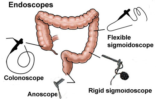
2) Anoscopy: Your doctor may use a small plastic device with an attached light to get a better look at the source of bleeding. Most of the time this is not painful, though it can be a bit uncomfortable, and it can be done in the office.

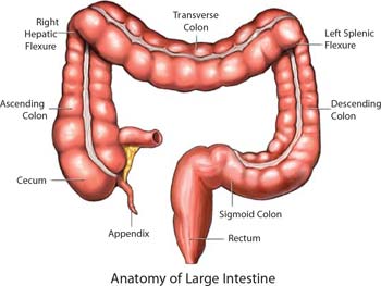
3) Sigmoidoscopy: This is usually done in an outpatient center, and the patient is usually not sedated. Sigmoidoscopes come in rigid and flexible forms, and the flexible one is used most often. A flexible tube, roughly 70 cm long and 1 cm wide, carrying a tiny video camera and a light, is inserted through the anus and gently into the colon while air is introduced to open the area and improve the view. A biopsy is often taken with a small tool. Sigmoidoscopy allows visualization of the anus, rectum, sigmoid colon and the top of the descending colon. It does not reach the entire colon, so it can miss cancers, polyps or bleeding sources in other areas.

4) Colonoscopy: Similar to sigmoidoscopy, but it allows the doctor to examine the entire colon with a longer flexible tube. The colonoscope, about 140 cm long, reaches everything the sigmoidoscope can and also allows visualization of the transverse colon, ascending colon and cecum. The patient is usually sedated.

When to seek help: It is impossible to know the cause of rectal bleeding without an examination. Everyone with rectal bleeding should talk to their healthcare provider about what evaluation they need. Even though there are common causes that are not cancerous, bleeding can be caused by cancer or by precancerous conditions.

Precancerous polyps may sit in the colon for years before turning cancerous, and they can be removed safely, which prevents them from ever becoming cancer. These polyps can produce symptoms very similar to an innocent hemorrhoid.

I’ve met patients who have ignored rectal bleeding for years because they thought it was because they had a hemorrhoid and it turned out to be cancer. With increasing age comes increasing risk of polyps and colon cancer.

Colon cancer screening: For patients at average risk, screening was started by checking the stool for small amounts of blood, which may be hidden, each year beginning at age 50. A first colonoscopy at age 50 was also recommended, unless other risk factors were present, and every 10 years thereafter unless a person was at increased risk based on family history or a previous diagnosis or biopsy result.

Update, 2026: The starting age has changed since this post was written. The American Cancer Society lowered it to 45 for people at average risk in 2018, the U.S. Preventive Services Task Force adopted 45 in 2021, and the ACS reaffirmed age 45 in its 2026 guideline update, with screening continuing through age 75 for those with a life expectancy over 10 years. If you are 45 or older and have not been screened, talk to your provider. See https://www.cancer.org/cancer/types/colon-rectal-cancer/detection-diagnosis-staging/acs-recommendations.html

People at increased or high risk:

If you are at increased or high risk of colorectal cancer, you should begin screening before the standard starting age, be screened more often, or both. The following raise your risk above average:

  1. A personal history of colorectal cancer or adenomatous polyps
  2. A personal history of inflammatory bowel disease (ulcerative colitis or Crohn’s)
  3. A strong family history of colorectal cancer or polyps
  4. A known family history of a hereditary colorectal cancer syndrome such as familial adenomatous polyposis (FAP) or hereditary non-polyposis colon cancer (HNPCC)

Scott Rennie, D.O.

Board Certified in Obesity Medicine and Family Medicine

This blog is for educational purposes only and does not constitute individual medical advice. Always consult your own physician before making changes to your health, medications, or treatment plan.
Blog: https://doctorrennie.wordpress.com

Celiac Disease and Gluten Sensitivity: Why Go Gluten Free?

One of the most common questions I hear from patients lately is about the gluten free diet. Many want to know if going gluten free can help with weight loss, low energy, bloating, constipation, or heartburn. The conversation around gluten is everywhere. Some of the information is accurate, but a lot of it is based on rumor or advertising.

So what exactly are Celiac disease, gluten intolerance, and gluten sensitivity?

Celiac disease is an autoimmune condition that affects about 1% of the population. When patients with Celiac disease eat gluten, their immune system reacts by damaging the small intestine. Gluten is a protein found in wheat, rye, and barley. For people with this condition, avoiding these foods is essential. Even small amounts can cause illness.

Gluten intolerance, sometimes called non-Celiac gluten sensitivity, is different. It may affect as many as 10% of people. Symptoms can be milder than Celiac disease and may involve more than just the gastrointestinal tract. Definitions of gluten intolerance have shifted over time as we learn more, but the general idea is that some people feel unwell after eating gluten even without the intestinal damage seen in Celiac disease.

The small intestine’s main job is absorbing nutrients from food. In Celiac disease, that lining gets damaged, which makes it harder for the body to take in vitamins and minerals. Symptoms can include diarrhea, abdominal pain, gas, weight loss, and deficiencies in iron or vitamins. Some patients develop related conditions, such as osteoporosis or thyroid disease. Celiac disease is also linked to dermatitis herpetiformis, a very itchy rash that can form on the elbows, knees, or back.

There’s even an increased risk of intestinal lymphoma in patients with untreated Celiac disease. While this cancer is rare, the risk decreases when gluten is eliminated from the diet.

So how do we test for Celiac disease?

The most definitive test is a biopsy of the small intestine, usually done during an endoscopy. Blood tests can also help, such as tissue transglutaminase antibody (TTG) or endomysial antibody (EMA). These are often used in a Celiac panel. Your doctor can help decide which test makes sense based on your symptoms.

There isn’t a test for gluten intolerance. If blood work and biopsy are normal but symptoms improve on a gluten free diet, that’s sometimes how the diagnosis is made.

Celiac disease appears to have a genetic component. It’s more common in patients with European ancestry, as well as in parts of the Middle East, North Africa, and South Asia. It is rare in populations from northern Asia and southern Africa.

Treatment is simple in theory but difficult in practice. The only effective treatment is to completely avoid gluten. About 70% of patients feel better within two weeks of eliminating gluten. Antibody levels in the blood often return to normal as well. But gluten is in many foods, and avoiding it requires major lifestyle changes. It’s not just bread and pasta. Gluten can hide in supplements, sauces, and processed foods.

A dietician who specializes in Celiac disease is often the best partner for patients making these changes. They can help identify hidden sources of gluten and ensure nutritional needs are still being met. That’s important because gluten free foods often contain less iron, vitamin D, and B vitamins. Some processed gluten free products add extra sugar or fat to make up for texture.

Patients sometimes ask if they should just “try going gluten free.” I usually recommend testing first if symptoms suggest Celiac disease. That way, you don’t lose the chance to confirm the diagnosis. If the tests are negative, you can still try eliminating gluten to see if you feel better, but do it with support from a healthcare provider or dietician.

Practical considerations matter. Many people with Celiac disease also need extra calcium and vitamin D, and sometimes blood tests for iron, folic acid, or B12. Osteoporosis can be a risk, so bone density testing may be appropriate. Dairy intolerance is also common until the intestine heals.

Some naturally gluten free foods include rice, corn, potatoes, beans, nuts, seeds, and soy. Foods to avoid are wheat, rye, barley, malt, brewer’s yeast, and most oats unless specifically labeled gluten free. Most wines are safe, but most beers are not.

Gluten intolerance and Celiac disease are real conditions, but gluten isn’t harmful for most people. It’s important to understand why a gluten free diet might be necessary and not assume it’s a guaranteed path to better health.

For more information:

This is for educational purposes only. If you think you may have Celiac disease or gluten intolerance, talk with your healthcare provider.

Scott Rennie, D.O.
Board Certified in Obesity Medicine and Family Medicine

This blog is for educational purposes only and does not constitute individual medical advice. Always consult your own physician before making changes to your health, medications, or treatment plan.
Reference: Ciclitira PJ, King AL, Fraser JS. AGA technical review on Celiac Sprue. Gastroenterology 2001;120:1526.

shutterstock_146760950

Clear water bottle on a wooden café table with coffee and menu

Traveler’s Diarrhea: What Every Traveler Should Know

shutterstock_158924261I give credit to Dr. Gene Allred for the information that I’ve learned and posted here.  He is an expert in Wilderness and Travel Medicine. I enjoy travelling, but I am always concerned the possibility of developing diarrhea when I go the backcountry or remote areas.  The majority of cases of traveler’s diarrhea (TD) occur in the first 2 weeks of travel. Symptoms:  The “typical” symptoms are either 4 loose stools in 24 hours or 3+ loose stools per day x 4-5 days plus at least one of the following:  nausea, vomiting, abdominal pain, fever (10%), blood in the stools (15%).  Symptoms can begin as early as 8-10 hours after exposure to contaminated food or water.  With persistent diarrhea (>14 days) we usually consider a parasitic infection with Giardia, E. histolytica, Crypto, or Cyclospora more likely. The majority of diarrheal diseases improve on their own and specific tests to identify the pathogen may not be necessary.  Treatment typically involves ensuring adequate fluid replacement, and empiric antibiotic treatment.  If there is fever along with abdominal pain and dysentery we will obtain a stool culture to look for Salmonella, Shigella and Campylobacter as well for ova and parasites. We used to think that water contaminated with bacteria was the way the infection was spread from person-to-person but we now know that the flies spread the disease very effectively.  The travel from fecal samples and then land on food that’s been sitting out. Diarrhea can be caused from viruses, bacteria or protozoan.  The most common pathogen that causes diarrhea in travellers is E-Coli.   Bacteria cause 50-75% of TD with E-Coli causing 25-40%, Campylobacter – 0-30%, Salmonella 0-15%, and Shigella 0-15%.  Protozoan cause 0-5% of TD, and viruses cause 0-20%. I try to take meticulous drinking water precautions (including ice) but it’s often very challenging.  When I go out in the backcountry or to remote areas I take along water purification tablets such as chlorine dioxide.  Boiling water for at least 1 minute can be helpful, as can chemical disinfection, filtration and use of UV devices.  Remember to cook your food well, and peel all your fruits and vegetables – it’s easy to remember but impossible to do.  As the saying goes “Boil it, cook it, peel it, or forget it!” Diagnosis:  I’m not sure if there is any hard and fast rule for diagnosing traveler’s diarrhea when out in the field without access to a laboratory, but if I’m with a group of people in a developing country and one of the group members has at least one loose stool, I treat them for traveler’s diarrhea.  In the clinic, we can check the stool cultures and for ova & parasites, but we usually only need to do this if there are symptoms of fever, bloody stools or persistent abdominal pain. Treatment:  There is increasing resistance of campylobacter to the fluroquinolone medications such as Cipro.  Azithromycin has been found to be a better choice – 1000mg taken daily for 2-3 days usually works well. Rifaximin is a newer antibiotic with few side effects and has a broad spectrum of coverage – the dose is 200mg three times a day for three days.   PeptoBismol often improves diarrhea and cramps (2 tablets every 30 minutes for up to 8 doses).   If there is no fever, vomiting or blood in the stool, loperamide can help reduce the frequency of diarrhea and can be invaluable for long bus rides. If there is persistent infection, even after treatment with antibiotics we think about the cause being a protozoa.  Giardia is a protozoan that can be detected by a immunoassay and causes diarrhea that lasts for 10 days or longer.  Nitazoxanide (Alinia) 500mg twice a day for three days or Tinidazole (Tiniba) 2 grams in a single dose for adults. Cryptosporidiosis can be a concern, however it is very sensitive to heat.  Even 65 degrees C will kill it, so just boil the water. In third world countries, we wary of swimming in fresh water or going barefooted because there are infectious agents that can get through the skin and cause serious infections (Schistosomiasis, Leptospirosis and Crytopsoridium). Prevention when travelling:  Here are a few of my recommendations about preventing traveler’s diarrhea with water precautions: 1)   Drink carbonated water from a sealed container – the carbonation process kills many pathogens – beer is also acceptable 2)   If non-carbonated, bring beverages to a boil – you can bring them just up to boiling temperature 3)   Avoid ice cubes in your beverages unless you prepared them If you are interested in learning more about wilderness medicine, a great resource for information is the wilderness medical society:  http://www.wms.org/   Stay safe, and happy travels,

Scott Rennie, D.O.
Board Certified in Obesity Medicine and Family Medicine

This blog is for educational purposes only and does not constitute individual medical advice. Always consult your own physician before making changes to your health, medications, or treatment plan.

Blue cushion on a wooden chair in front of a fireplace

Hemorrhoids Explained: Why They’re a Real Pain in the Rear

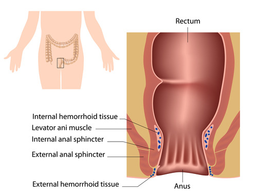
shutterstock_125891585Hemorrhoids are a common problem in both men and women who come to see their primary care or urgent care doctor.  Hemorrhoids are enlarged or swollen veins in the lower rectum.  They can cause pain, itching and rectal bleeding.  Sometimes you can feel or see hemorrhoids around the outside of the anus.  Other times they may be hidden from view inside the rectum (internal hemorrhoids). They are more common in older individuals and patients during and after pregnancy, those with diarrhea or constipation or in people who sit for prolonger periods of time. Symptoms: 1)   Anal pain or itching 2)   Tissue bulging around anus 3)   Painless rectal bleeding 4)   Difficulty cleaning or leakage of feces after bowl movement Diagnosis:  Your healthcare provider will examine the rectum and anus to inspect for hemorrhoids.  This exam may include inserting a gloved finger into the rectum.  The reason for the digital exam is to inspect for possible rectal cancers and also to collect any stool that’s present and test for blood.  If there is blood present, your healthcare provider may use a special tube called an anoscope, that is clear and allows visualization of the rectum/anus. Treatment:  If hemorrhoids are diagnosed, one of the most important steps is avoiding constipation because they can lead to rectal bleeding or tearing the anus (called a fissue).  Also, it is important to avoid pushing or straining during bowl movements because that can worsen existing hemorrhoids and risk causing development of additional hemorrhoids.  Reducing constipation can be done by: 1)   Increasing fiber – increasing fruits and vegetables contain fiber.  In addition fiber supplements such as Metamucil may be helpful. 2)   Laxatives – These are not “addictive” or increase your risk of constipation in the future. 3)   Warm sitz baths – soaking the rectal area in warm water for 10 minutes 3x/day improve blood flow by relaxing the internal anal sphincter. 4)   Topical treatments – Suppositories are creams with hydrocortisone may be helpful to relieve pain, itching and irritation. More invasive procedures: 1)   Rubber band ligation – a rubber band or ring is place around the base of the internal hemorrhoid which restricts the blood supply to that area.  This shrinks the hemorrhoid and reduces it. 2)   Laser surgery – uses a laser or infrared light to destroy internal hemorrhoids 3)   Sclerotherpay – a chemical solution is injected into the hemorrhoidal tissue causing it to break down and form a scar 4)   Surgery – hemorrhoidectomy is the treatment of choice for patients with large internal hemorrhoids. A gastroenterologist is a doctor who specializes in the gastrointestinal tract.  If you are looking for a gastroenterologist in your area, the American Gastroenterological Association has a locator:  https://secure.gastro.org/GILocator/locator.asp   I hope that you have found this information useful.  Wishing you the best of health,

Scott Rennie, D.O.
Board Certified in Obesity Medicine and Family Medicine

This blog is for educational purposes only and does not constitute individual medical advice. Always consult your own physician before making changes to your health, medications, or treatment plan.
Person holding abdomen, appearing in pain, wearing grey sweatshirt

What’s Causing My Abdominal Pain? Common Causes Explained

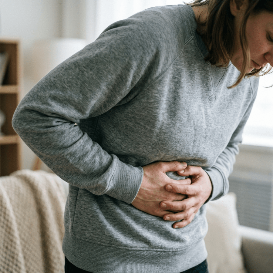
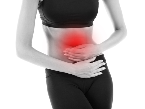
shutterstock_152569646Patients who present to the clinic or urgent care with abdominal pain can be some of the most challenging, because of the many different causes.  The job of your primary care or urgent care provider is to determine whether the pain is requiring immediate surgical evaluation. Medical providers must try to determine which patients can safely be observed and their symptoms treated and which patients require further investigation by a specialist such as a surgeon.  This is difficult because abdominal pain is often non-specific and presents with other symptoms very commonly. Triage:  We must urgently investigate abdominal pain in many patients.  Some patients require assessment of their airway, breathing and circulation followed by appropriate resuscitation.   Patients who may need surgery must be transferred to a facility where they can receive that care where appropriate nursing care, laboratory, surgical consultation, and radiology facilities are available. Patients who are having less severe pain or signs on exam may require consultation or referral for further management. Helpful information from patients: 1)   Time course of pain 2)   Location of pain 3)   Radiation of pain 4)   Factors that make the pain worse or better (such as foods or antacids) 5)   Associated symptoms including fever, chills, weight loss, nausea, vomiting, diarrhea, constipation, blood in the stools, jaundice, change in stool or urine color or diameter of the stools. 6)   Past medical history, including history of abdominal surgeries 7)   Family history of bowl disorders 8)   Alcohol intake 9)   Medications – including Tylenol, aspirin, and ibuprofen/aleve 10)  Menstral and contraceptive history in women Surgical abdomen:  Usually defined as a condition with rapidly worsening course without surgical intervention.  Obstructions of the intestines and peritonitis (inflammation/irritation of the inner wall of the abdomen that covers most abdominal organs) are reasons for referral to a surgeon. Sometimes tests will be ordered such as an abdominal radiograph,  CBC, comprehensive metabolic panel with liver enzymes, lipase, a urine analysis and pregnancy test (in women of childbearing potential). Other things we consider in determining the cause of the abdominal pain are the location of the pain and changes in where the pain radiates as well as how rapidly the pain gets worse. Some possible causes of abdominal pain are many – gallstones or gallbladder dysfunction, peptic ulcer, hiatal hernia, pneumonia, heart attack,  pancreatitis, heartburn, lactose intolerance, celiac sprue, pregnancy (including ectopic), endometriosis, sickle cell disease,  appendicitis, ovarian cyst or torsion, UTI, kidney stones, constipation, colitis, diverticulitis, pelvic inflammatory disease, gastroenteritis, intestinal ischemia (decreased blood flow to the intestines), diabetic ketoacidosis, kidney infection, abdominal aortic aneurism, or even trauma. Treatment:  Is tailored to address the cause of the pain.  If no cause can be found at your doctor’s office, the goal is to determine whether it is safe for you to go home with medications to help with the pain, and testing done as an outpatient or whether you need to be transferred to the hospital where further workup can be done immediately.   I hope that you have found this information useful.  Wishing you the best of health,

Scott Rennie, D.O.
Board Certified in Obesity Medicine and Family Medicine

This blog is for educational purposes only and does not constitute individual medical advice. Always consult your own physician before making changes to your health, medications, or treatment plan.

Orange Electrolyte+ drink bottle and bowl of crackers on a wooden tray on bed

Gastroenteritis: Nausea, Vomiting, Diarrhea and Dehydration

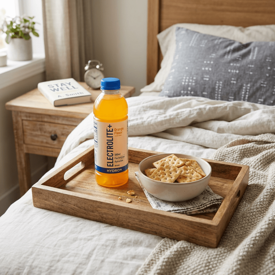
shutterstock_103672964Viral gastroenteritis is an illness that we often see in the urgent care and medical clinic in both adults and children.  It causes diarrhea and vomiting. How do I get infected?  If you touch an infected person or an object that has been touched by someone who is infected (that has the virus on it), you could become contaminated.  If you eat foods or drink liquids with the virus you may also become infected.  It is important for people with the virus to wash their hands. Symptoms: Possible symptoms include nausea, vomiting, diarrhea, fever, headache or muscle aches, belly pain or cramping and loss of appetite.  If your body loses too much water, you can become dehydrated.  Symptoms of dehydration include dark yellow urine, feeling thirsty, tired, dizzy or confused.  Dehydration that is severe can be life threatening.  Babies, infants, young children and the elderly are more likely to become dehydrated. When should I call a doctor or nurse?  If you or your family member has symptoms of dehydration (mentioned above), diarrhea or vomiting that lasts longer than 24 hours, vomiting blood or have bloody diarrhea, they haven’t had anything to drink in a few hours or been able to urinate in the past 6-8 hours during the day, if a baby or young child hasn’t had a wet diaper in 4-6 hrs. Diagnosis:  Most of the time the diagnosis can be made after obtaining a history and performing a physical exam, but sometimes your healthcare provider may obtain blood tests, urine tests or tests on a stool sample. Treatment of viral gastroenteritis:  For severe gastroenteritis with dehydration patients are sometimes treated with IV fluids (a thin tube that goes into the vein).  We do not give antibiotics for viral gastroenteritis because they don’t help cure a viral infection.  We can give medicine in the IV to help reduce the nausea as well.   Sometimes suppositories or oral medications for nausea can be helpful.  Small sips of clear fluids at home every 15 minutes can help prevent dehydration.  We generally try to avoid red colored fluids so that if it passes right through, we don’t confuse the red color for blood in the stool.  Once vomiting has stopped for 24 hours, starting small bites of crackers can be tried.  The diet usually advances to a “BRATTY” diet at this point – ie. “Bannanas, Rice, Apples, Tea, Toast and Yogurt” are all bland foods that are easy on the stomach. Prevention of viral gastroenteritis:  Wash your hands with soap after using the bathroom or change your child’s diaper and before you eat.  Avoid changing diapers near where you prepare food and make sure your baby gets the rotavirus vaccine.  Rotavirus is a common infection that causes severe nausea, vomiting and diarrhea in children. Dehydration:  This is a term that medical providers use to describe when the body loses too much water.  It can be mild or severe.  Usually mild dehydration doesn’t cause problems, however severe hydration is a medical emergency and can be life threatening.  You children and babies are more at risk for dehydration because they have a small body mass and less to lose to begin with. Causes of dehydration:  Vomiting, diarrhea, sweating/high fever, medicines called diuretics or water pills.  Also some people who have nausea or sore throat might not drink enough fluids. Symptoms of dehydration:  With mild dehydration, patients might not notice any symptoms, however as dehydration gets worse it can cause: 1)   Feeling thirsty 2)   Urinating less often, or having dark yellow or brown urine 3)   Dry mouth or cracked lips 4)   No tears when a child cries 5)   Feeling tired or confused 6)   Feeling light headed or dizzy 7)   Eyes look sunken in the face 8)   Babies can have a sunken gap between the bones in the babies skull. This soft spot can feel/look caved in. When to seek help:  Call your medical provider if your child has any symptoms of dehydration.  You should also call if the patient has diarrhea that lasts more than a few days, vomits blood or has bloody diarrhea, vomiting lasts more than 24 hours, urinating much more than usual, haven’t had anything to drink in many hours, hasn’t needed to urinate in the past 6-8 hours (in adults/older children) or hasn’t had a wet diaper in 4-6 hrs.   I hope that you have found this information useful.  Wishing you the best of health,

Scott Rennie, D.O.
Board Certified in Obesity Medicine and Family Medicine

This blog is for educational purposes only and does not constitute individual medical advice. Always consult your own physician before making changes to your health, medications, or treatment plan.

Breakfast table with quinoa bowl, porridge with berries, sliced avocado, whole grain toast, and fresh fruits

I think I may be constipated – How do I know?

shutterstock_136335587Constipation is a common condition that I see in the clinic.  It’s a topic that few people enjoy talking about but is very common.  In fact, almost everyone gets constipated at some time during his or her life.  Women and the elderly are more commonly affected.  Though not usually serious, constipation can be a concern. Constipation:  A bowl motility disorder that occurs when bowl movements become less frequent.  The normal time between bowl movements varies widely from person to person – some people having a bowl movement 3 times a day, and others only 1 to 2 times per week.  In general, going more than 3 days without a bowl movement can cause pain.  After 3 days, the stool becomes harder and more difficult to pass. Some signs that you may be constipated (2 or more during 3 months at least):  Straining during bowl movement more than 25% of the time, lumpy or hard stools more than 25% of the time, sensation of anorectal obstruction, sensation of incomplete evacuation more than 25% of the time, manual maneuvering required to defecate, three or fewer bowl movements in a week. Symptoms of constipation:  Infrequent bowel movements and/or difficulty passing stool, swollen abdomen or abdominal pain, vomiting. Some causes of constipation:  Medications (especially strong pain medications such as narcotics, antidepressants or iron pills), low fiber diet, dehydration, disruption of regular diet or routine (traveling), inadequate activity or exercise, eating large amounts of dairy products, stress, resisting the urge to have a bowl movement (sometimes the result of painful hemorrhoids), overuse of laxatives, hypothyroidisms, Parkinson’s disease or Multiple Sclerosis, antacid medications containing calcium or aluminum, depression, eating disorders, irritable bowl syndrome, pregnancy and colon cancer. Treatment options: Increase hydration – drink 1.5 – 2 quarts of water each day Add fruits and vegetables to your diet Eat prunes and/or bran cereal A mild stool softener or laxative may be used Medication choices: 1)   Bulk forming laxative:  Metamucil, Citrucel, Fibercon or Benefiber 2)   Surfactants – Lower the surface tension of stool, thereby allowing water to more easily enter the stool.  Docusate/Colace is an example 3)   Osmotic agents: Cause intestinal water secretion and thereby increase stool frequency.  Examples are Polyethylene glycol (PEG) which is also known as Miralax.  Excessive use of these agents may result in electrolyte and volume overload in patients with renal or cardiac dysfunction.  Lactulose is a synthetic disaccharide that is not metabolized in the intestine, thus water and electrolytes remain in the lumen of the intestine. Sorbitol is a less expensive alternative.   Saline laxatives such as milk of magnesia and magnesium citrate are poorly absorbed and act as hyperosmolar solutions. 4)   Stimulant laxatives:  Dulcolax, senna and sodium picosulfate primarily exert their effects via alteration of electrolyte transport by the intestinal mucosa. 5)   Suppositories:  For treatment of functional defecation disorder – glycerin or bisacodyl suppositories may help liquefy stool and thereby overcome the obstruction. Other treatments for Constipation:  Behavioral approaches (habit training has been especially successful for children), biofeedback – a behavioral approach that can be used to correct inappropriate contraction of the pelvic floor muscles and external anal sphincter during defecation in patients with functional defecation disorder. Warning about constipation:  Since constipation can be a sign of possible bowl obstruction or even cancer – I recommend calling your doctor if this is a new problem for you, if you have blood in your stool, you are losing weight even if you’re not dieting, you have severely painful bowl movements, your constipation has lasted more than two weeks.   I hope that you have found this information useful.  Wishing you the best of health,

Scott Rennie, D.O.
Board Certified in Obesity Medicine and Family Medicine

This blog is for educational purposes only and does not constitute individual medical advice. Always consult your own physician before making changes to your health, medications, or treatment plan.

Diagram of esophagus, lower esophageal sphincter, stomach, stomach acid, and acid reflux causing heartburn

Heartburn and GERD: Answers to Your Common Questions

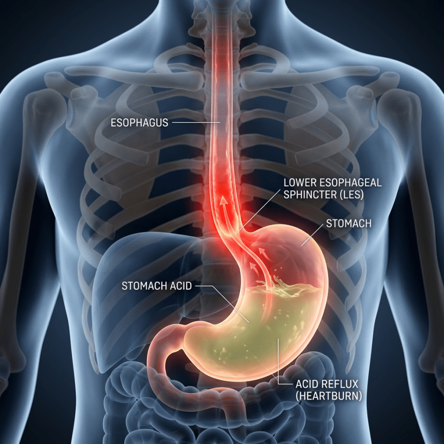
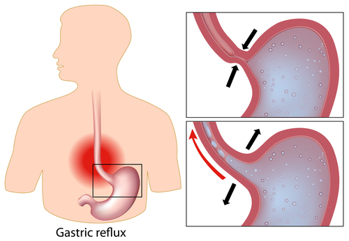
shutterstock_74586250I often get questions from patients asking how to treat their heartburn also called gastroesophageal reflux disease or GERD.  It is sometimes confusing to diagnose and can also cause dangerous complications if not treated. GERD is a common condition where acid contents from the stomach go backwards up the esophagus (food tube between your mouth and stomach).  This can irritate and inflame the esophagus causing a burning sensation and other problems.  There is a sphincter/valve at the top of the stomach that helps keep the stomach contents from going backwards, but if the spincter (called the lower esophageal sphincter or LES) doesn’t fully close, liquid contents of the stomach which contain acid can leak back into the esophagus. Risk factors:  Obesity, pregnancy, smoking, hiatal hernia (a problem where part of the stomach moves above the diaphragm), alcohol and medications.  The medication list is extensive and includes: 1)   Non-steroidal anti-inflammatory medications such as ibuprofen, aspirin or aleve 2)   Beta-blockers which are used for high blood pressure 3)   Bronchodilators such as prednisone used for asthma 4)   Calcium channel blockers used for high blood pressure 5)   Dopamine-active medications used for Parkinson’s disease 6)   Sedative medications which are used for insomnia/anxiety 7)   Tri-cyclic anti depressant medications – used for pain/IBS Symptoms of GERD:  Nausea after eating, hiccups, feeling that food is stuck behind breastbone, burning pain in the chest which is increased by bending, stooping, lying down or eating, usually worse at night and relieved by antacids. How do I find out if I have GERD/Heartburn?  There are many different tests that can be used to help diagnose esophageal reflux such as:  a trial of a medication for heartburn such as a GI cocktail (a mixture of Mylanta, viscous lidocaine and donnatol), esophagogastroduodenoscopy(EGD) – which is when a doctor inserts a camera that looks down the esophagus when you are under anesthesia, barium swallow, esophageal pH monitoring, and esophageal manometry.  Sometimes a stool blood test is used to diagnose bleeding that might be coming from the esophagus, stomach or intestines. Helicobacter Pylori, also known as H. pylori is a bacterium that is commonly found in the stomach.  About half of the world’s population has this bacteria and they have no symptoms and will never develop problems.  However, H. pylori can cause stomach ulcers, and much less commonly, stomach cancer.  We really don’t know why some people with H. pylori get these symptoms and others do not. Stomach ulcers can cause symptoms such as pain or discomfort in the upper abdomen, bloating, feel full after eating only a small amount of food, lack of appetite, nausea and vomiting, black or tar-colored stools,  and even low blood count and fatigue. Treatment:  Treatment is tailored toward the individual patient and might include some of the following options: 1)   Treat H. pylori with antibiotics if it is present 2)   Diet modification – avoid a core group of reflux-inducing foods (fatty foods, chocolate, peppermint, excessive alcohol) and spicy foods like citrus fruits, coffee, tomatoes/marinara sauce (found in spaghetti and pizza), garlic, onions, peppers, chili, carbonated beverages 3)   Stop smoking or chewing tobacco 4)   Reduce or stop medicines which can make GERD worse such as NSAIDs (aspirin, ibuprofen, Aleve, Excedrine etc. – see list above) 5)   Refrain from laying in a supine (on your back) after meals and avoid meals right before bedtime 6)   Lose weight – obesity is a risk factor for GERD, erosive esophagitis, and esophageal adenocarcinoma (cancer). 7)   Promotion of salivation by chewing gun, or oral lozenges might be helpful for mild heartburn as saliva neutralizes refluxed acid. 8)   Acid-suppressive medications:
  1. Antacids such as tums
  2. Prescription H2 (such as Zantac) blockers twice a day for mild GERD
  3. Prescription PPI (such as Prilosec) – for more severe GERD
9)    Acid barrier medications:  Sucralfate protects the stomach or ulcer from acid by acting as a barrier It is important to be monitored closely if you are taking medication for heartburn because some of the complications that come with heartburn/reflux and the possibly with the medications used to treat them might be:  Pneumonia, hypergastrinemia, atrophic gastritis, intestinal infections, vitamin B12 malabsorption, iron malabsorbtion, hip fracture and calcium malabsorption, magnesium absorption and drug interactions.   I hope that you have found this information useful.  Wishing you the best of health,

Scott Rennie, D.O.
Board Certified in Obesity Medicine and Family Medicine

This blog is for educational purposes only and does not constitute individual medical advice. Always consult your own physician before making changes to your health, medications, or treatment plan.