Photo credit: http://en.wikipedia.org/wiki/Sebaceous_cyst
Patients often came into the urgent care with a small skin lump that had become red or painful. Frequently, thinking back, they would recall feeling a small nodule under the skin in that same spot months or even years before it became swollen and red. The epidermal inclusion cyst is one of the most common skin cysts. It can occur anywhere on the body, though it is more common on the face and upper body. Most of the time these cysts cause no problems, but they can be cosmetically unwelcome.
Other names for epidermal cysts:
1) Epidermoid cyst
2) Sebaceous cyst
3) Keratin cyst
4) Epidermal inclusion cyst
5) Infundibular cyst
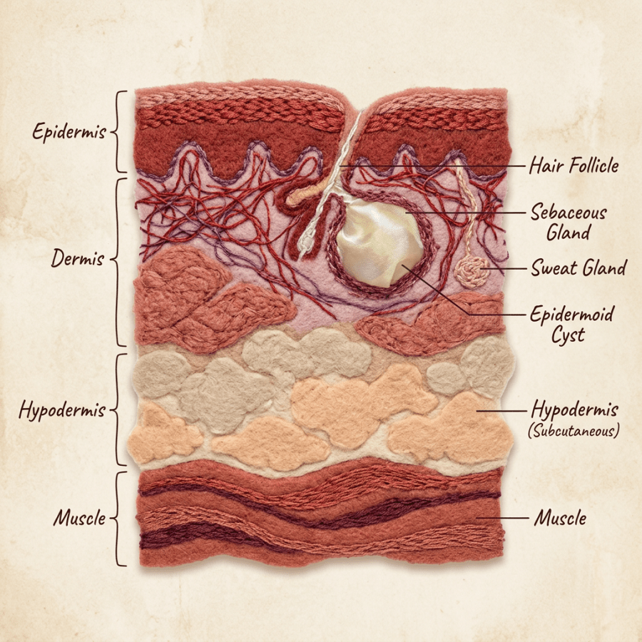
Appearance: Epidermal cysts have a wall made of skin cells from the outer layer of the skin, called the epidermis. The cyst wall is like a balloon that extends down into the second layer of skin, the dermis. That wall produces keratin, a protein found in skin and nails, which is usually white and either cheesy or firm in consistency. It is often foul smelling as well.
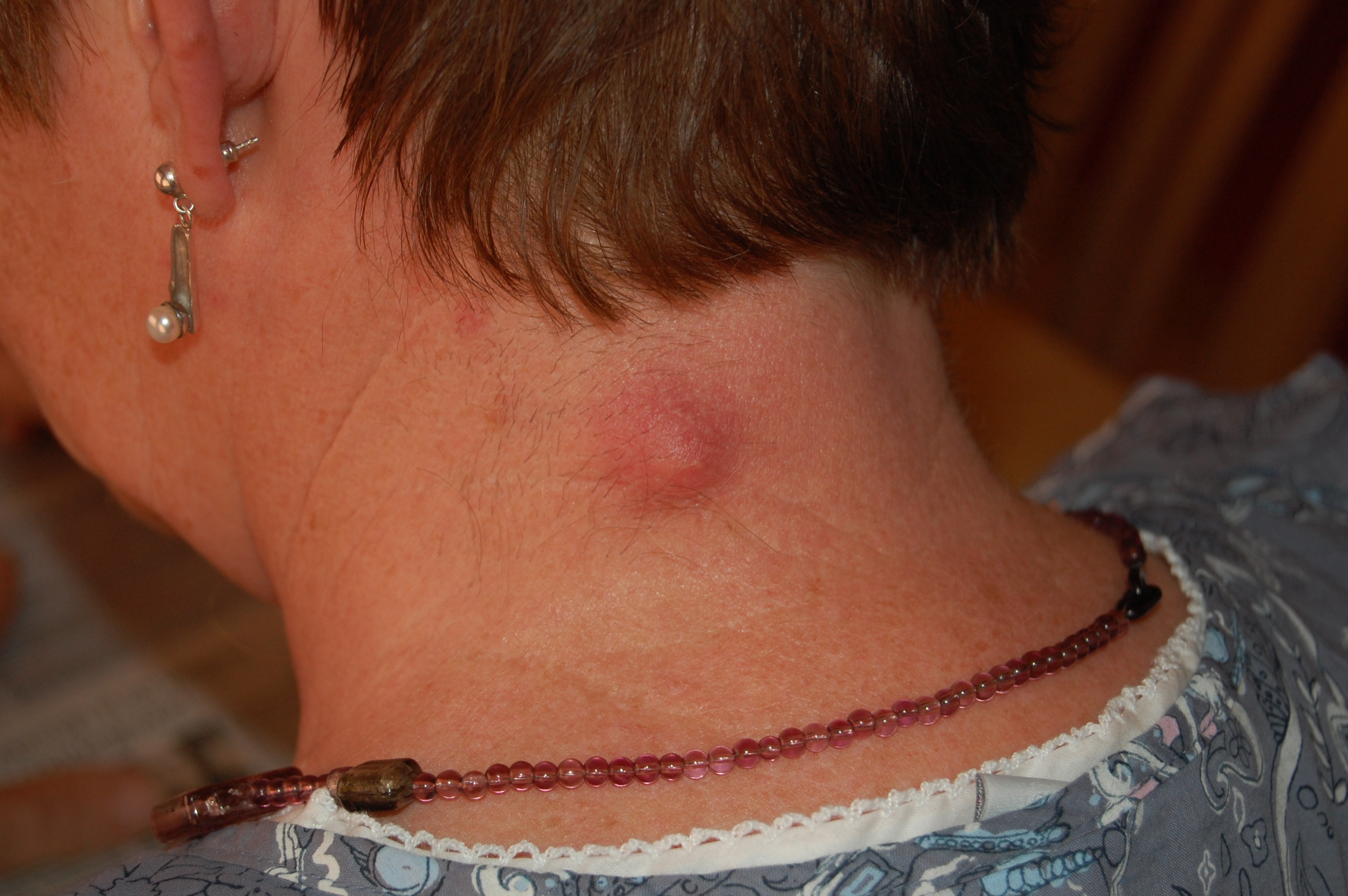
Cyst Rupture: If the cyst wall ruptures underneath the skin, usually from trauma or from bumping the area unintentionally, the keratin escapes into the surrounding tissue and is very irritating. The skin becomes red, swollen and painful. It’s best to see your doctor rather than trying to “pop” or drain the cyst yourself. Your doctor may recommend treating you with an oral antibiotic before opening the cyst if he or she thinks it has become infected.
How epidermal cysts are removed: If the cyst needs to come out, your doctor will try to remove the entire cyst including the wall. Remember, the cyst wall is what makes the keratin. If the wall is left behind under the skin, it may start producing keratin again and the cyst can come back.
Usually we make an incision over the cyst, separate the overlying skin from the cyst wall, and try to remove it in one piece. If the cyst has ruptured, which is most likely what brought the patient in, it is removed piecemeal, with an effort to get all of the keratin and all of the wall out. The doctor may irrigate the wound with sterile solution afterward. The skin is usually left open rather than stitched. The doctor may place a small piece of packing gauze under the skin where the cyst was, and then cover it with a bandage. This allows the wound to drain while it heals. The wound is usually examined by a medical provider every two to three days to check on healing, and part or all of the packing gauze is removed at those visits.
The reason your provider may pack the wound and ask you to come back, instead of simply putting stitches over it, is that these areas are sometimes contaminated with bacteria. If the skin is closed right away with sutures, the bacteria have a small sealed pocket in which to grow and form an abscess. Letting the wound heal from the inside out lowers that risk and often speeds healing.
When to have a cyst removed: If it’s small, painless, and not red or swollen, it probably doesn’t need to be removed. I might recommend removing a cyst if:
1) It keeps getting red and irritated or infected
2) It’s getting larger quickly
3) It’s in a place that rubs against clothing or jewelry and gets irritated
4) It becomes red, inflamed or painful
Board Certified in Obesity Medicine and Family Medicine
This blog is for educational purposes only and does not constitute individual medical advice. Always consult your own physician before making changes to your health, medications, or treatment plan.
 Doctor Rennie's Blog