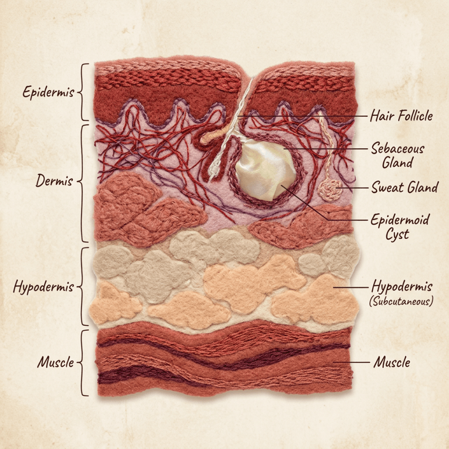
Cross-section of skin layers showing epidermis, dermis, hypodermis, muscle, hair follicle, sebaceous gland, sweat gland, and epidermoid cyst

Epidermal Inclusion Cysts Explained: What Are They?

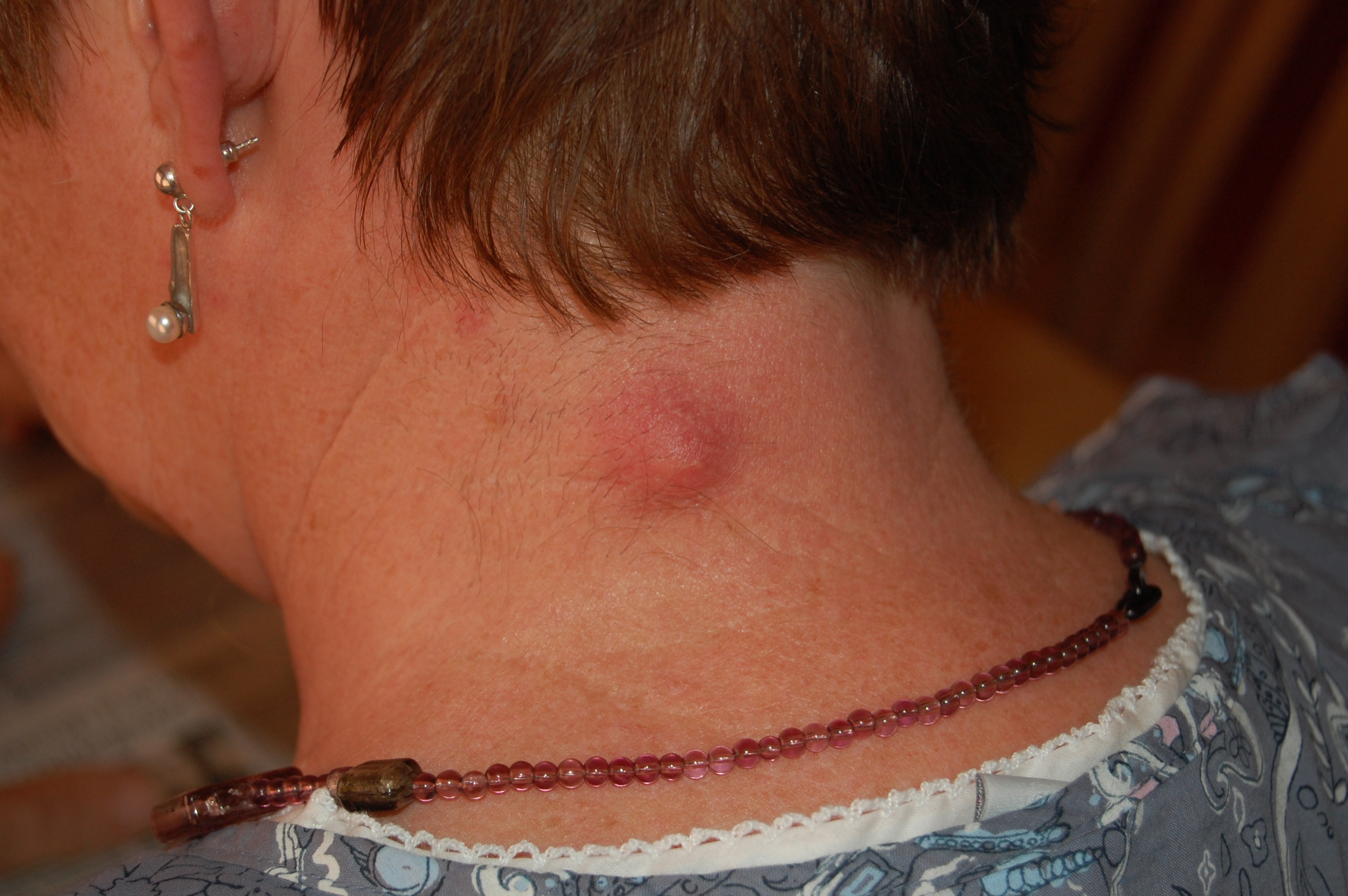
Inflamed_epidermal_inclusion_cyst

Photo credit: http://en.wikipedia.org/wiki/Sebaceous_cyst

Patients often came into the urgent care with a small skin lump that had become red or painful. Frequently, thinking back, they would recall feeling a small nodule under the skin in that same spot months or even years before it became swollen and red. The epidermal inclusion cyst is one of the most common skin cysts. It can occur anywhere on the body, though it is more common on the face and upper body. Most of the time these cysts cause no problems, but they can be cosmetically unwelcome.

Other names for epidermal cysts:

1) Epidermoid cyst

2) Sebaceous cyst

3) Keratin cyst

4) Epidermal inclusion cyst

5) Infundibular cyst

Appearance: Epidermal cysts have a wall made of skin cells from the outer layer of the skin, called the epidermis. The cyst wall is like a balloon that extends down into the second layer of skin, the dermis. That wall produces keratin, a protein found in skin and nails, which is usually white and either cheesy or firm in consistency. It is often foul smelling as well.

Cyst Rupture: If the cyst wall ruptures underneath the skin, usually from trauma or from bumping the area unintentionally, the keratin escapes into the surrounding tissue and is very irritating. The skin becomes red, swollen and painful. It’s best to see your doctor rather than trying to “pop” or drain the cyst yourself. Your doctor may recommend treating you with an oral antibiotic before opening the cyst if he or she thinks it has become infected.

How epidermal cysts are removed: If the cyst needs to come out, your doctor will try to remove the entire cyst including the wall. Remember, the cyst wall is what makes the keratin. If the wall is left behind under the skin, it may start producing keratin again and the cyst can come back.

Usually we make an incision over the cyst, separate the overlying skin from the cyst wall, and try to remove it in one piece. If the cyst has ruptured, which is most likely what brought the patient in, it is removed piecemeal, with an effort to get all of the keratin and all of the wall out. The doctor may irrigate the wound with sterile solution afterward. The skin is usually left open rather than stitched. The doctor may place a small piece of packing gauze under the skin where the cyst was, and then cover it with a bandage. This allows the wound to drain while it heals. The wound is usually examined by a medical provider every two to three days to check on healing, and part or all of the packing gauze is removed at those visits.

The reason your provider may pack the wound and ask you to come back, instead of simply putting stitches over it, is that these areas are sometimes contaminated with bacteria. If the skin is closed right away with sutures, the bacteria have a small sealed pocket in which to grow and form an abscess. Letting the wound heal from the inside out lowers that risk and often speeds healing.

When to have a cyst removed: If it’s small, painless, and not red or swollen, it probably doesn’t need to be removed. I might recommend removing a cyst if:

1) It keeps getting red and irritated or infected

2) It’s getting larger quickly

3) It’s in a place that rubs against clothing or jewelry and gets irritated

4) It becomes red, inflamed or painful

Scott Rennie, D.O.

Board Certified in Obesity Medicine and Family Medicine

This blog is for educational purposes only and does not constitute individual medical advice. Always consult your own physician before making changes to your health, medications, or treatment plan.

Blog: https://doctorrennie.wordpress.com

Bottle of scalp care shampoo labeled Renew & Refresh with hairbrush and towel

Seborrheic Dermatitis – Dandruff, Cradle Cap and Adult Seborrhea

??????????????????????????????????????????????????????????????????????
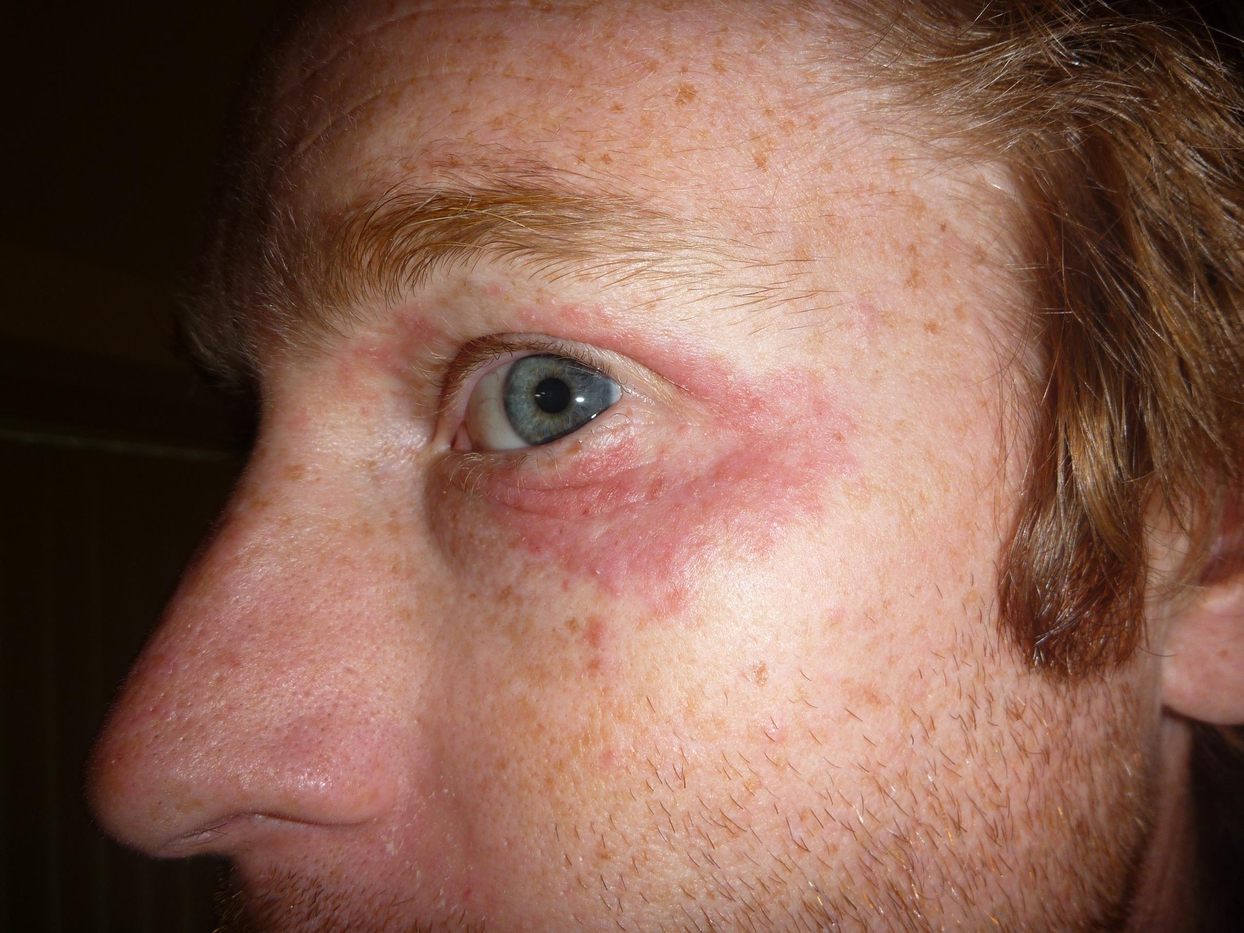
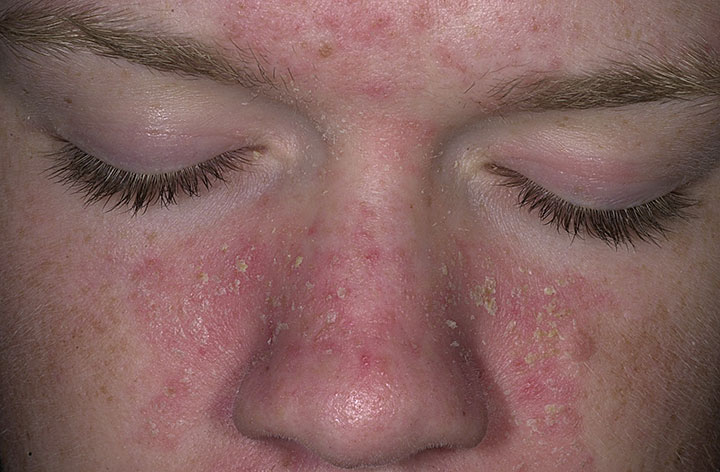
seborrheic-dermatitis-eyes
??????????????????????????????????????????????????????????????????????

Photo credit: http://hardinmd.lib.uiowa.edu/dermnet/seborrheicdermatitis22.html

Photo credit: http://siklusair.com/seborrheic-dermatitis-eyes

Photo credit: http://medicalcontent.hubpages.com/hub/Seborrheic-Dermatitis-Pictures-on-Face-Scalp-Hair-loss-Causes-Treatment

Seborrheic dermatitis is a common skin condition affecting both infants and adults. It causes redness, greasy scales, flaking along the eyebrows, and itching. It was one of the most common skin conditions I saw as an urgent care physician.

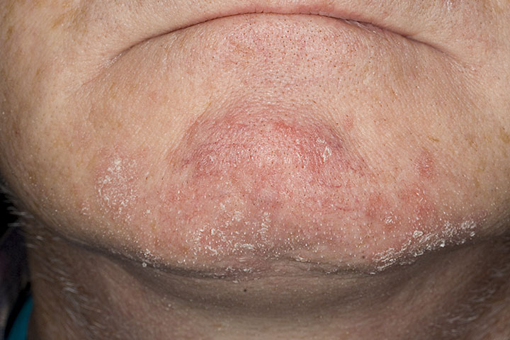
Seborrheic dermatitis, also called seborrhea, is an inflammation of the skin that usually occurs in areas with an abundance of oil glands: the scalp, face, around the eyes, ears, neck, and even the diaper area. In men it is more common where there is facial hair. Infants often have seborrhea on the scalp, where it is known as cradle cap. A mild form confined to the scalp, in children or adults, is what we call dandruff. Symptoms may include:

  1. Skin scales, white or yellow, usually oily or greasy
  2. Itching
  3. Mild redness
  4. Skin lesions or plaques

The exact cause is unknown, but there is evidence that it flares in times of stress, with hormonal changes, and during particular seasons, especially in extreme cold or warm weather. It is more common in people with weakened immune systems, in those with oily skin, and in certain neurological conditions such as Parkinson’s disease.

Diagnosis: Your healthcare provider can usually diagnose seborrheic dermatitis by examining you and taking a history. A biopsy, a sample of skin removed surgically and examined under a microscope, is rarely needed.

Treatment: Treatment is tailored to the individual patient and to which part of the body is affected. Options include:

  1. Medicated shampoos such as Neutrogena T-Gel or T-Sal, Head and Shoulders, or Nizoral. These usually contain salicylic acid, coal tar, zinc, selenium sulfide, ketoconazole, or resorcinol. Even if you do not have dandruff, these shampoos can be used on the face and other affected areas, and they work best left in place for five to ten minutes before rinsing.
  2. Topical steroid creams, lotions, foams or shampoos. Low, medium or even high potency steroids are sometimes prescribed to reduce inflammation, depending on severity and location. Lower potency preparations are used on the face, such as desonide 0.05% lotion.
  3. Antibacterial creams. Topical agents such as sodium sulfacetamide with sulfur are sometimes prescribed.
  4. Antifungal creams. Ketoconazole 2% cream and ciclopirox 1% cream are commonly used.
  5. Other anti-inflammatory medications such as pimecrolimus cream or tacrolimus ointment, sometimes prescribed for facial seborrhea.
  6. Dermatologists sometimes recommend a compounded mixture combining a steroid with an antifungal or antibacterial agent.

Cure: Unfortunately there is no cure for seborrheic dermatitis. It is a chronic, lifelong condition. It may disappear for months or years and then relapse. We focus on controlling it, usually by combining the treatments above with efforts to reduce known triggers such as emotional stress, extreme cold, and excess body weight.

Blog: https://doctorrennie.wordpress.com

This one is from 2014. I am board certified in obesity medicine and most of what I write now sits there, down to narrow questions like whether you can do intermittent fasting while you are on Ozempic or Wegovy.

Updated for 2026: A New Drug, and the Same Old Shampoo

Most of what I wrote here in 2014 still stands. Ketoconazole 2 percent or ciclopirox 1 percent shampoo, used a couple of times a week for at least a month, is still the backbone. A short run of a mild steroid for a flare. A calcineurin inhibitor like tacrolimus for the face, where I would rather not leave someone on steroids. What is new is roflumilast foam 0.3 percent, sold as Zoryve. It is a topical PDE4 inhibitor, approved in December 2023 for seborrheic dermatitis in anyone nine and older, applied once a day. It is the first genuinely different mechanism approved for this condition in more than twenty years (1). The trial numbers are good. In the phase 3 STRATUM study, 79.5 percent of patients on roflumilast reached clear or almost clear skin at eight weeks against 58 percent on vehicle, with about half completely clear. Itch improved within two days (2). My honest read: it works, and it is a real option when shampoo and a steroid have not been enough or when the face is involved and you want off steroids. It is also expensive, and for a lot of people ketoconazole shampoo bought over the counter handles the problem for a few dollars. I would still start there.

Cradle Cap Has Not Changed

Worth saying because parents ask constantly. It resolves on its own over weeks to months. Baby shampoo, soften the scale with mineral oil or petroleum jelly, lift what comes away easily, leave what does not. Do not use medicated dandruff shampoo on an infant. If it is genuinely inflamed and persistent, a low potency steroid or topical antifungal briefly, and that is a conversation to have rather than something to start yourself (1).

What a Video Visit Handles Well Here

This is a good fit for the format. Seborrheic dermatitis has a recognizable look and a recognizable distribution, scalp, eyebrows, the creases beside the nose, behind the ears, sometimes mid chest, and a photograph in decent light usually tells me what I need. The questions matter as much as the picture. How long, whether it comes and goes with stress or season, what you have already tried and for how long, and whether you have been using a steroid cream continuously, which causes its own problems on facial skin. Where I want a closer look is when the distribution is not typical, because psoriasis, tinea, and contact dermatitis all get called seborrhea and are treated differently.

When To Be Seen In Person

Seborrheic dermatitis that is unusually widespread or severe deserves a conversation about HIV testing, because that association is real and longstanding. Signs of bacterial infection on top, meaning spreading redness, warmth, pus, or fever. Failure to respond to standard treatment, which usually means the diagnosis is wrong rather than the drug. And in an infant, scale spreading well beyond the scalp along with poor feeding or poor weight gain needs a pediatrician.

The Bottom Line

Start cheap. Antifungal shampoo, used properly and for long enough, still fixes most of this. Roflumilast is a genuine addition for the cases it does not, particularly on the face. Cradle cap needs patience and mineral oil, not medicine.

Sources

1. Vidal SI, Menta N, Green L. Child and Adult Seborrheic Dermatitis: A Narrative Review of the Current Treatment Landscape. Dermatol Ther (Heidelb). 2025;15(3):599-613. https://pmc.ncbi.nlm.nih.gov/articles/PMC11909311/ 2. Blauvelt A, et al. Roflumilast foam 0.3% for adolescent and adult patients with seborrheic dermatitis: a randomized, double-blinded, vehicle-controlled, phase 3 trial. J Am Acad Dermatol. 2024;90(5):986-993. https://pubmed.ncbi.nlm.nih.gov/38253129/ 3. American Academy of Dermatology. Seborrheic dermatitis: Diagnosis and treatment. https://www.aad.org/public/diseases/a-z/seborrheic-dermatitis-treatment

Related Reading

Ringworm, Athlete’s Foot and Fungal Nail Infections When to Use Telemedicine vs. Urgent Care: How I Spot the Sick One Scott Rennie, D.O.

Board Certified in Obesity Medicine and Family Medicine

This blog is for educational purposes only and does not constitute individual medical advice. Always consult your own physician before making changes to your health, medications, or treatment plan.
Hospital bed with medical equipment and patient repositioning guide on wall

What is a Pressure Ulcer (AKA Pressure sore)?

shutterstock_89421025
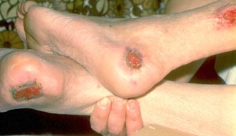
bed-sores

Photo credit: http://diseasespictures.com/bedsores/

A patient came in to see me today with a sore on his heel that’s been bothering him for the past few months. He’s diabetic and has lost feeling in the bottom of his feet. He’s had these pressure sores in the past but has trouble getting them to heal up.

Pressure sore: An area of skin damaged by pressure, such as sitting or lying in one position for a long period. They are also called bedsores. They are most common where bone sits near the surface of the skin: the hips, elbows, ankles, and back or buttocks. The skin and soft tissue become damaged because not enough oxygenated blood reaches the area to support healing, usually because the soft tissue is compressed between the surface and the hard bone beneath.

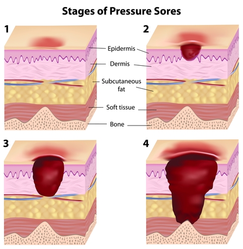
Appearance: The sores change in appearance depending on how long they have been present and how much damage has been done. At the start, the sore looks like a small red patch of skin. If it is not treated, the skin breaks down and a hole or crater forms, which is what we call an ulcer.

Stage 1: The skin is intact without ulceration, but when you press on it the color does not change the way it should. Healthy tissue is pink, and pressing on it with a finger blanches it, with the pinkness returning within a couple of seconds. That does not happen in damaged skin at this stage, and the area may look darkly pigmented.

Stage 2: There is an open, shallow ulcer with a red-pink base. Blisters may be present, either intact or ruptured.

Stage 3: Structures beneath the skin such as fat may be exposed, but at this stage you should not see bone, tendon or muscle.

Stage 4: Bone, tendon and muscle may be visible in the base of the ulcer.

People at risk: Some patients are far more likely than others to develop pressure sores.

  1. Patients who cannot move well because of a medical problem. They may sit or lie in one position for a long time and need help repositioning so the skin does not break down.
  2. Older people, who often move less and whose skin is thinner and more fragile.
  3. Patients with diabetes or nerve problems in their feet, who may not feel a pebble in a shoe or pressure building against the foot.
  4. Patients in the hospital or a nursing home, who are at especially high risk because several of these factors stack: increased age, reduced mobility, and other complicated medical problems.

Prevention: Several things lower the chance of developing pressure sores.

  1. Repositioning the patient’s body every two hours so no one area of skin stays crushed, pinched or under pressure.
  2. Putting pillows between the ankles and knees to reduce pressure on the skin over those bony areas.
  3. Raising the head of the bed when the patient is lying on their side to reduce pressure on the hip bone.
  4. Using special foam or soft mattresses that reduce pressure over the areas that bear the most load.

For patients in wheelchairs:

  1. Use a cushioned seat if possible to prevent pressure on the sacrum.
  2. Tilt forward or to the side every hour to release pressure on the seat.
  3. If the ankles or heels press against the chair, use foam padding to protect against sores.
  4. Check the skin regularly for signs of pressure or ulceration.

Treatment: Pressure sores are treated differently depending on the stage and how severe the skin damage is.

  1. If there is mild erythema, treatment is generally offloading the area by reducing the time it spends compressed, through repositioning and pillows to cushion it. We also use transparent films over the ulcers to protect them.
  2. In patients with diabetes, keeping blood sugars under good control is very important. Elevated blood sugars impede wound healing.
  3. If there is dead or dying skin or soft tissue, it often needs to be removed to help prevent infection.
  4. Special bandages may be needed to keep the healing tissue moist while preventing maceration from being too moist. The dressings we use can be expensive.
  5. Antibiotics may be prescribed if there is a wound infection.
  6. Medication for pain may also be prescribed.

There are scoring tools to grade pressure and track healing. These help for patients who return for repeat visits to a wound care clinic or their primary care provider and need their progress graded. Clinical features examined include:

  1. Amount of exudate
  2. Skin color surrounding the wound
  3. Peripheral tissue swelling
  4. Peripheral tissue firmness around the wound
  5. Amount of granulation (healing) tissue
  6. How much epithelialization is present

It’s important to optimize the nutritional status of patients with wounds. Patients with stage 3 and stage 4 ulcers in particular need enough protein and calories to heal.

Scott Rennie, D.O.

Board Certified in Obesity Medicine and Family Medicine

This blog is for educational purposes only and does not constitute individual medical advice. Always consult your own physician before making changes to your health, medications, or treatment plan.
Blog: https://doctorrennie.wordpress.com

Collection of labeled skincare bottles, brush, facial cleanser, and natural elements on fabric

Acne Treatment: What Should You Do About Your Acne?

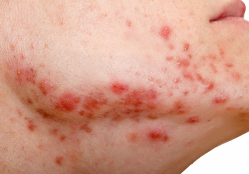
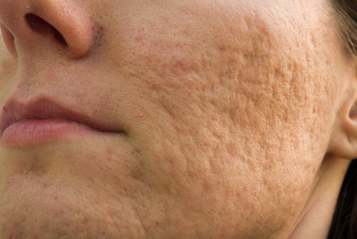
shutterstock_124292974shutterstock_153263516During the first week of my third year medical school dermatology rotation I thought I’d definitely decided that I wanted to become a dermatologist.  I’d been present in the room with my dermatology preceptor and patients who asked about what to do for their acne at least a hundred times, and I knew I could give the same speech as my dermatology teacher.  I later changed my mind about becoming a dermatologist but I still have a great interest in dermatology.  Most people who come into the medical clinic due to problems with acne have tried various over the counter treatments to help with common black heads, white heads or deep cysts from acne.  It’s a common problem for adolescents (85% are effected) and teenagers but also effects many adults as well.  Untreated acne can lead to embarrassment, loneliness and even permanent facial scarring.

So what is acne?  Acne is a skin problem where the openings in the skin where hairs come out (pores) become blocked by skin cells and oil and this creates a plug.  During adolescence the glands that produce oil in the skin enlarge and produce increased amounts of the oily substance we call sebum.  These oil glands are most often found on the face (most commonly an area called the T-Zone of the face – around the eyebrows and nose), neck, chest, upper back and arms.  Even normally, there are bacteria which are present on the surface of the skin and normally they don’t cause any problems in small numbers.  With an increased production of oil on the surface of the skin that occurs during adolescence however, these bacteria reproduce and become present on the surface of the skin in much higher numbers than normal.  The bacteria then combine with the oil and skin cells and become trapped in the pores (hair follicles) and lead to the formation of tender “pimples” that we call acne.

A special type of acne that affects newborn babies is called neonatal acne and usually goes away within a few weeks after birth.  It is related to the hormones from pregnancy and causes small pimples often noticeable on a newborn babies face

There are multiple types or ways we describe acne and grade the severity

  1. Non-inflammatory acne is acne without redness or skin swelling and usually is described as just being whiteheads or blackheads.
  2. Inflammatory acne has redness and swelling and is more severe.  There can be papules, pustules, nodules or cysts present.

Causes of acne:

  1. Sebaceous (oil) glands become more sensitive to hormones during adolescence and become enlarged and produce more oil than normal which increased the likelihood of acne.
  2. Cosmetics that contain oil may clog pores in a similar way to the oil secreted by the sebaceous glands in the skin and can lead to acne.
  3. Frequent or aggressive rubbing with soaps or cleansers can actually increase the production of oil from the skin.  Wash your skin gently.
  4. Some people believe that diet can affect acne.  If you find that you develop worsening oily skin or acne after eating certain foods, avoid these foods if possible.
  5. Psychological stress can increase or worsen acne.

Acne Treatment:  Since there are multiple causes of acne, there is not once simple treatment that works for everyone.  Skin doctors (dermatologists) usually recommend a combination of treatments to reduce acne formation in multiple ways.  Some simple suggestions are outlined below but keep in mind that they may not work for everyone.

  1. Wash your face with a gentle non-soap skin cleanser.  Most of the dermatologists that I’ve trained with have recommended a fragrance free cleanser such as Cetaphil or Dove.  They usually recommend using warm water and washing gently with your hands rather than using a washcloth, skin brush or skin scrubber.  Some of the prescription skin cleansers contain an antibiotic or sulfa base that helps soak up the oil and decrease the bacterial count on the surface of the skin.  In general we don’t recommend washing your face more than twice a day unless you have been sweating excessively or you need to wash off sunscreen.
  2. Most dermatologists don’t recommend picking or squeezing pimples yourself because you might risk causing scars or worsening the infection.  Hearing this however always makes me laugh (usually not out loud in front of the dermatologist though) because they often help patients to pick and squeeze their pimples in a fashion similar to what I think most people would probably do themselves at home.  The difference however is that in the office, they have access to special instruments and tools to help reduce the risk of scarring.
  3. Cautious use of skin moisturizers is recommended.  Remember, your skin is producing oil in abundance and that is a natural skin moisturizer.  If you use too much moisturizer you could worsen the acne by causing plugging of the pores/hair follicles.  If you use a skin moisturizer to help minimize dryness or peeling in certain areas, be sure it’s hypoallergenic or “non-comedogenic” formula to decrease the chance of blocking the skin pores.
  4. Sun screen:  Many of the antibiotics and even topical medications prescribed by medical professionals can increase your chances of getting a sun burn.  Use a sunscreen with at least an SPF of 15 and make sure the sunscreen blocks both UVA and UVB.
  5. Over the counter products:  There is a huge market for acne medications.  Television ads, various articles on the internet and in magazines tout the latest non-prescription skin formula.  Most of these “formulas” that I’ve encountered contain benzoyl peroxide, salicylic acid or a combination of these two ingredients and put their own brand names on them.  I recommend reading the ingredients to determine if the formula you are about to purchase is similar to one you might have already tried.  Benzyl peroxide is an agent that dries the skin and helps decrease the amount of oil.  It is usually applied twice a day.  You should be aware that it can irritate the skin and possibly cause skin redness or flaking.  It can also bleach your pillow case or clothes so be careful not to get it on your favorite items.   If you find that the over the counter product that you try is not working, contact a healthcare provider.  Untreated or inadequately treated acne can lead to permanent scarring of your skin.

Prescription treatments:  Prescriptions for acne can be divided into topical (applied directly to the skin) and non-topical treatments.

A)     Topicals: 

  1. Prescription skin cleansers are topical washes that often contain an antibiotic or drying agent.  They are made to be hypoallergenic and usually do not have a fragrant odor to them.  Examples of a topic skin cleaner include a sulfacetamide/sulfur topical wash.  Multiple companies produce their own brands such as Klaron,  Plexion, Rosanil or Rosula.  These are usually expensive and insurance companies often do not cover them.
  2. Benzoyl peroxide mixed with topical antibiotics: Various pharmaceutical companies make combined products that include benzoyl peroxide with various topical antibiotics such as clindamycin, erythromycin or metronidazole.  Examples are Acanya, BenzaClin, Benzamycin, or Duac.  These topical can be solutions, gels or creams.
  3. Topical antibiotic only:  Many topical antibiotics are produced to help reduce the bacterial count on the skin.  Most of them contain Erythromycin, Clindamycin, Sulfacetamide,  Metronidazole or Dapsone.  Examples are Aczone, Clenia, Cleocin T, Clindagel, Klaron, Metrocream, and Sulfacet-R.
  4. Retinoids:  These medications often produce skin drying and can cause irritation.  Examples are Retin-A, Differin and Tazorac.  Differin tends be less potent and Tazorac is the strongest of the three.  The first time I tried using Retin-A, I woke up with red, irritated, scaly skin that looked worse than the acne.   I recommend starting with a small amount and trying on a small patch of skin first to see how your skin reacts to it.

B)     Oral antibiotics:  The reasons for using an oral antibiotic to treat acne are actually two-fold.  The first reason is that some oral antibiotics have been found to have anti-inflammatory properties in the skin and decrease the inflammatory response leading to the pimples.  The other reason is more straight-forward – to reduce the bacterial count on the skin and thereby decrease the acne.  The most commonly prescribed oral antibiotics for acne at this time are Minocycline or Doxycycline but they should not be used during pregnancy or in young children.

C)     Oral Isotretinoin (previously called Accutane):  It’s also called Amnesteem, Claravis and Sotret.  This medication is most often limited to be prescribed only by dermatologists are physicians who have had special training about this medication.  This is one of the most successful medications that we have for the treatment of severe acne and is usually taken as a pill once or twice a day for 20 weeks.   The reason it is not the first line of treatment for acne and mostly prescribed by dermatologists is because there are some possible serious side effects that can occur when using this medication.  Isotretinoin can cause miscarriage or life-threatening malformations to babies so it cannot be taken by women who are pregnant or at risk of becoming pregnant.  Most dermatologists require women of child bearing age to use  birth control before they will prescribe this medication.  There are strict rules for using the medication regulated by the iPLEDGE program:

  1. Women have to have two documented negative pregnancy tests before starting the medication and continue to take monthly pregnancy tests throughout their treatment.
  2. Any women who might become sexually active (or is already sexually active) with a male partner must use two forms of birth control for at least one month before starting therapy and continue until one month after stopping the medication.
  3. Women of child bearing age who could become pregnant must get their prescription filled at the pharmacy within 7 days of receiving it from their doctor.  Each month a new prescription must be written by their doctor.
  4. Women who cannot become pregnant or men must participate in iPLEGE but do not have to do the pregnancy testing or use birth control.

Side effects of oral Isotretinoin:  I’ve seen many patients who are treated with this medication and I’ve noticed that it works very well for their acne in most cases.  Some of the common side effects that I’ve observed are:

  1. Cracking or sore lips and dry or peeling skin.  Patients may get nosebleeds very easily and have a tough time wearing contact lenses because their eyes dry out.  They may have itchy skin (because of the dryness).  Skin sensitivity to the sun is increased and risk of sunburn is severe.
  2. There can be an increased level of triglycerides (fat) in the blood, it can cause liver damage and cause changes in blood counts when taking this medication so monitoring the blood cholesterol and triglyceride level as well as liver function and blood count is important.
  3. There has been some talk about an increased risk for depression or suicidal behavior when taking this medication but there is not enough evidence to conclude that this is a risk.

Hormone treatment options:  Women with acne are sometimes prescribed a birth control pill to help control acne.  Some hormones and IUDs and injectable hormones however can actually worsen acne, so make sure to talk to your doctor about which birth control might be better for acne if you are considering this form of treatment.

A blood pressure medication called spironolactone has also been used to help with acne because it can effect  hormone levels in women (and thereby decrease acne) but it can also cause an increase in the blood potassium and birth defects so it is usually cautiously.

Where to get more information:

  1. American Academy of Dermatology:  http://www.aad.org/skin-conditions/dermatology-a-to-z/acne
  2. National Library of Medicine:  www.nlm.nih.gov/medlineplus/acne.html


I hope that you have found this information useful.  Wishing you the best of health,

Scott Rennie, D.O.
Board Certified in Obesity Medicine and Family Medicine

This blog is for educational purposes only and does not constitute individual medical advice. Always consult your own physician before making changes to your health, medications, or treatment plan.
Blog: https://doctorrennie.wordpress.com

References:

  1. Haider A, Shaw JC. Treatment of acne vulgaris. JAMA 2004; 292:726
  2. Ozolins M, Eady EA, Avery AJ, et al. Comparison of five antimicrobial regimens for treatment of mild to moderate inflammatory facial acne vulgaris in the community: randomized controlled trial. Lancet 2004; 364:2188
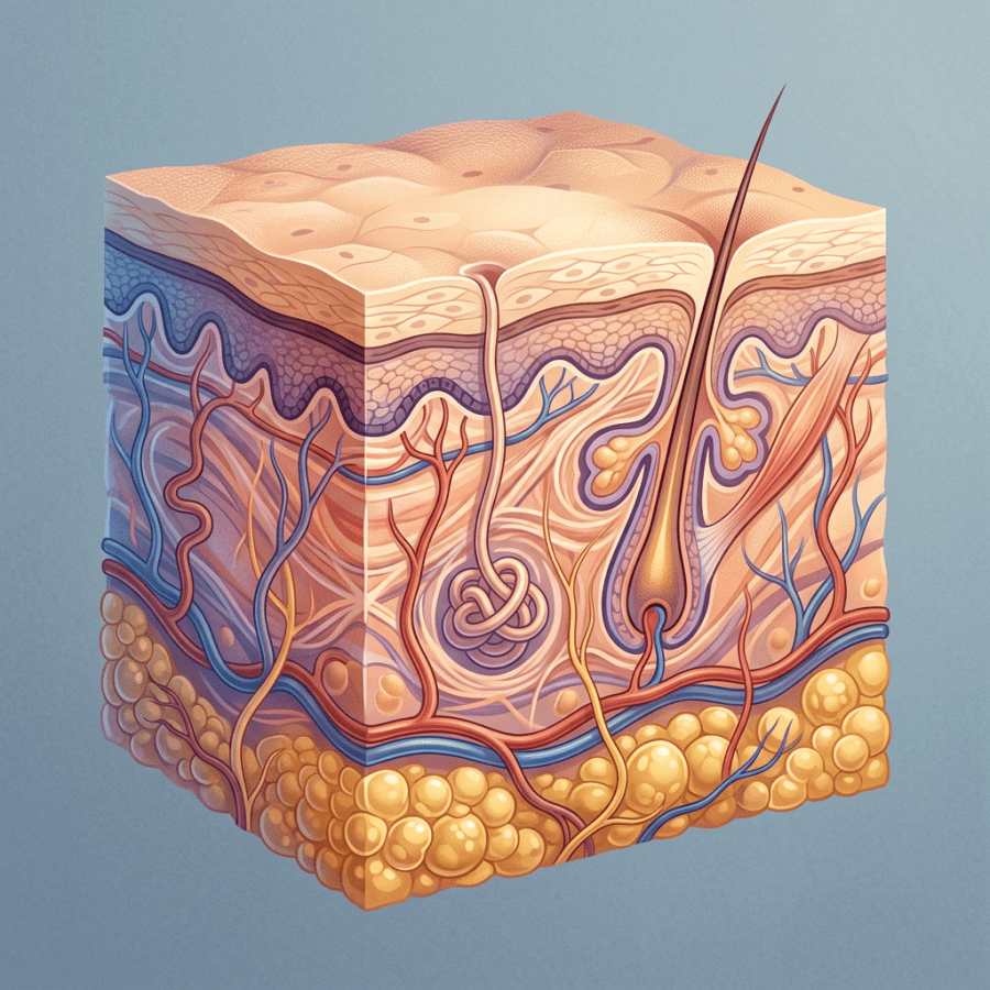
Cross-section illustration of human skin showing epidermis, dermis, hair follicle, sweat gland, blood vessels, and fat cells

What Is Psoriasis? An Overview of This Skin Condition

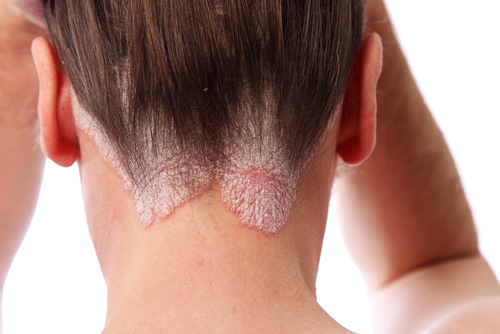
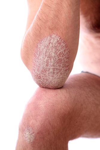
shutterstock_45868720shutterstock_45922807A few times each week a patient will come in to see me due to a rash of unknown cause.  The rash is usually an inflamed, red skin and sometimes it’s a bit thickened and/or covered with a silvery scale.  Since psoriasis often develops in adults, many people are puzzled when they come in and learn they have psoriasis and have lots of questions about the condition.  I’d try to answer some of the most common questions that patients ask me about psoriasis.

What causes psoriasis?  We’re not exactly sure about the cause, but we do know that it seems to have a genetic link (it often runs in families), it involves the immune system and environmental conditions seem to trigger it.  The skin actually is made up of several layers and the top layer of your skin is in a state of shedding old skin cells as new skins cells are produced.  This process keeps the top layer of skin relatively smooth.  Psoriasis is a process in which the outter layer of skin cells grow too quickly and they stop shedding properly.  This leads to a scaly build-up of skin cells that we see on the skin in affected patients.  We believe that the immune system causes certain cells to enter the skin and cause the disorder.

About 40% of patients with psoriasis have family members who also have the condition.

We know that smoking appears to increase the risk of psoriasis.  Certain medications can worsen psoriasis symptoms including beta blockers (used for blood pressure), lithium, and medications that we commonly use to treat malaria.  Stress and anxiety may trigger psoriasis to become noticeable in people who have the genetic risk.  It has also been linked with obesity and increased risk for heart disease.

What are the symptoms of psoriasis?  How do I know if I have it?  A medical provider may determine if you have psoriasis usually by examining your skin.  There is not a blood test that can tell you if you have psoriasis but a doctor may due a biopsy of your skin if it is not clear what is causing your rash.  There are several symptoms of psoriasis including:

  1. Patches of skin more common on certain areas of the body such as the elbows, knees, scalp, genitals, and belly button that may be dry or red and have a white or silver scale.  If you peel the scale off, the skin will likely bleed.
  2. Sometimes people with psoriasis develop a form of arthritis that causes joint pain or aching
  3. At times patients with psoriasis can develop finger or toenail pitting, or crumbling

What can I do about my psoriasis?  Is there a cure?  Unfortunately there is not a complete cure for psoriasis but there are treatment options that can substantially improve the symptoms.  Patients who have severe psoriasis that is highly noticeable to other people may feel embarrassed or feel low self-esteem or anxiety due to the disorder.  The treatment offered to a patient will depend on the severity of the symptoms, the area of the body affected as well as the cost and convenience of treatment and other medical conditions that the patient may have.  Severe psoriasis is usually always treated by a dermatologist (skin specialist) and patients with psoriatic arthritis may be treated by a rheumatologist (joint specialist).  Treatments are organized by whether they are topically applied to the skin or are taken orally or given in an injectable form.

Topical medications:

  1. Moisturizers:  It is very important to keep the skin moist so that the itching and irritation caused by the psoriasis is minimized.  Decreasing the itching and irritation helps to decrease the risk of scarring.  Patients who are constantly scratching their skin may cause increased inflammation and risk damaging their skin and cause thickening and increase the risk for infection.  Greasy ointments or thick creams work better than lotions.
  2. Steroid creams or ointments:  work to decrease the inflammation and redness of the skin.  The most potent creams or ointments work the best for psoriasis but require a prescription.  Sometimes solutions are easier to apply when patients has psoriasis of the scalp.
  3. Tar:  Comes from coal and has been used to treat psoriasis for years.  It is commonly found in shampoos such as Neutrogena T-Gel.  It seems to help decrease the amount of cells produced in the epidermis that actually causes the psoriasis.  Preparations containing tar are non-prescription and over the counter and may be in the form of lotions, creams, oils or shampoos.  They can stain the skin, hair and clothing but are not thought to have any serious side effects.
  4. Ultraviolet Light:  Patients often find that their psoriasis is better in the summer time with exposure to the sun’s ultraviolet light.  Other people actually treat their psoriasis in sun tanning beds if their condition is severe.  The risk of causing skin cancer must be weighed against the effects of psoriasis.
  5. Calcipotriene (Dovonex):  This is a cream that is applied twice a day and slows the growth of the epidermal skin cells.  Another medication called calcitriol (Vectical) is similar to Dovonex and Taclonex is a medication that has calcipotriene and betamethasone (a steroid cream) combined together.
  6. Tazarotene (Tazorac):  This is a vitamin A derivative that is a cream or gel and is applied once a day.  It can cause skin irritation so sometimes it is washed off after 20 minutes.  This medication is similar to Retin-A and Differin.
  7. Calcineurin inhibitors (Protopic and Elidel): are creams that are often used on the face or in skin fold areas where scarring or disfigurement may occur if high potency steroids are used.

Medications that suppress the immune system:  Several medications target the immune system such as Enbrel, Amevive, Remicade, Humira and Stelara.  These are usually reserved for severe forms of psoriasis because they are very expensive and are injections that are either given into the skin or muscle or into a vein over hours in the doctor’s office.  Methotrexate and cyclosporine also suppress the immune system and can increase the risk of the patient developing an infection.

Soriatane is an oral medication that is derived from vitamin A and is called a retinoid.  It may help reduce the symptoms of psoriasis in 3-6 months but should generally not be used in women of child-bearing age as it can cause severe birth defects.

For additional information, feel free to check out the following sites:

  1. Psoriaisis.net:  http://www.skincarephysicians.com/psoriasisnet/whatis.html
  2. National Psoriasis Foundation:  www.psoriasis.org/home/
  3. American Academy of Dermatology:  www.aad.org/skin-conditions/dermatology-a-to-z/psoriasis


I hope that you have found this information useful.  Wishing you the best of health,

Scott Rennie, D.O.
Board Certified in Obesity Medicine and Family Medicine

This blog is for educational purposes only and does not constitute individual medical advice. Always consult your own physician before making changes to your health, medications, or treatment plan.
Blog: https://doctorrennie.wordpress.com

References:

  1. Schon, M, Boehncke, W-H Psoriasis. New England Journal of Medicine 2005; 352:1899
  2. Lebwohl M. Psoriasis. Lancet 2003; 361:1197
  3. Strober BE, Siu K, Menon K. Conventional systemic agents for psoriasis. A systematic review. J Rheumatol 2006; 33:1442.
Cryosurgery device, magnifying glass, tools, and liquid nitrogen vial on blue cloth

What to do about the common skin wart

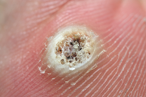
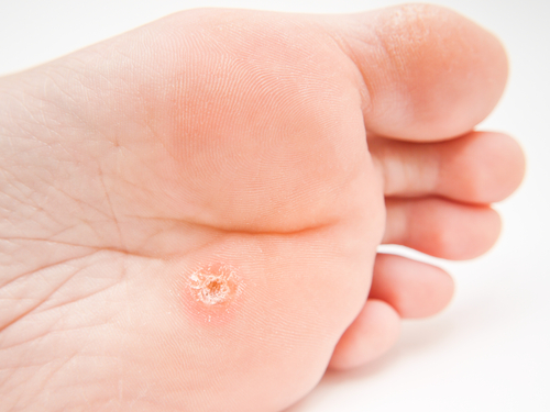
  shutterstock_115521190shutterstock_148362410I often have people come into the clinic and ask me to treat warts, mostly on their hands or their feet.  Some people aren’t aware of what these are so I thought a brief discussion might help people identify warts and also mention some common treatments. Common skin warts are generally non-dangerous raised and round or oval shaped skin growths that often stick up compared to the surrounding skin.  If they’ve been present for months or years, they can sometimes become rather large or form patches that appear like a cauliflower shaped lesion.  Sometimes they are identified by tiny black spots or dots that are small, clotted blood vessels but some people call them “seeds.” What causes a wart?  Warts are actually the result of a virus (human papillomavirus) and is spread by touching someone else’s skin who has a wart.  They’re also often spread by picking at existing warts and touching other areas on your own body.  The virus lives in skin surrounding the wart and can be spread easily by scratching are removing some of the virus under your finger nails.  Warts can also be spread by coming into contact with skin cells that have fallen off an infected persons foot.  It can actually take up to six months after exposure to the virus for a wart to appear. What are the most common areas where warts occur on the body?  The most common areas of the body for warts are:
  1. Fingers
  2. Hands
  3. Knees
  4. Elbows
  5. Around the fingernails (periungual warts)
  6. Feet (plantar warts)
  7. Face
  8. Lower legs
How do I know if I have a wart or if the skin lesion is due to something else?  Usually a medical provider can diagnose a wart based on how it looks.  A biopsy is not usually required. Once I know that I have a wart, how do I get rid of it?  Warts can be very difficult to treat and there are many different options for treating warts.  The treatment of choice often depends on where there wart is located and how sensitive the skin is.  Some possible treatment options are:
  1. Leave it alone – about 67% of all warts will go away within two years even if not treated.  Most people treat then however because they can spread or become larger over time.
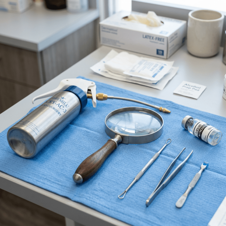
  2. Liquid nitrogen:  In the doctor’s office, we usually use this very cold liquid to freeze the skin around the wart.  It can be painful so it can be a difficult treatment for young children to tolerate.  We often need to treat a wart several times using liquid nitrogen and if the wart is large, we may need to trim the top part of the wart off to make the treatment more effective.
  3. Salicylic acid:  Over the counter patches employ this kind of treatment.  Usually a liquid or patch is applied to wart and left in place for several days.  It is often helpful to soak the skin in warm water for 10-20 minutes before applying the acid to soften the skin.  Treatment with salicylic acid can be painful and cause redness to the skin and even bleeding.  Many people find that using a nail file or pumice stone is helpful to gently remove the dead skin from the surface of the wart every few days during the treatment.  You should be cautious when doing this however because there is a high risk of spreading the virus/warts to other areas on the body when using a file or stone.  I usually recommend using a new file or stone each time to help prevent spreading the wart virus.  Most people don’t realize that you need to keep applying the acid each day for 1-2 weeks even after the wart is gone because the virus can be present on the skin even if no wart is visible.  This helps ensure that the wart does not return weeks or months later.
  4. Duct tape:  The sticky tape easily found in most home improvement stores has been helpful to some people with warts.  They apply it directly to the skin over the wart and leave it in place for about a week.  It’s not entirely clear how the treatment works, but my thought is that the tap sticks to the surface of the skin where the wart is present and the tape on the skin causes moisture to build up and this makes it easier to remove the dead skin cells (containing the wart virus) when the tape is removed.  Many people use an emery board or pumice stone to remove the excess skin after removing the tape and then reapply the tape for another week.  It may take up to 4 weeks for the wart to go away using this treatment.  We usually don’t recommend using duct tape if you have diabetes because if you cover your skin and a bacterial infection begins, you might not be able to see it starting and an infection may get very large before it is noticed.
  5. Cantharidin:  This is a liquid that is prescribed by healthcare providers such as a dermatologist and applied directly to the wart on the skin.  It may cause a blister to appear over the wart after 2-24 hours of treatment.  It is usually just placed on the skin once and often dermatologists will recommend using salicylic acid for a week after the skin heals to decrease the chances of the wart coming back.
  6. Imiquimod:  Aldera is the other name for this prescription cream that is applied at bed time several times per week.  It works by stimulating the immune system to fight off the wart virus.  It is rather expensive and is usually prescribed for genital warts or another type of virus called condyloma acuminate.  It can also be used to treat small skin basal cell skin cancers or pre-cancers.
  7. 5-Fluorouracil:  This cream which also goes by the name Carac, Efudex or Fluroplex is applied to flat warts twice a day for 3-5 weeks.  We also use this cream to treat small skin pre-cancers and superficial basal cell cancers.  It can cause skin irritation especially for those people who get lots of sun exposure.
  8. Shave excision:  This is a procedure where the skin is cut away or removed where the wart is present on the body.  This procedure is not very common for treating warts because it can cause permanent skin scarring or keloid formation and may also require stiches after the procedure.
  9. Immunotherapy:  A dermatologist (skin doctor) may inject a medication directly into the wart that triggers the body’s natural immune system to attack the virus.  These medications called contact sensitizers are not widely used because they are highly potent, expensive and require careful handling to avoid causing unintentional allergic reactions.
Should I see a doctor to treat my wart?  I’d recommend seeking the help of a medical provider if you are not sure that the skin growth is a wart, if it’s not improving with home treatment, if you have questions about what treatment is best for you or if you have been treated for warts before and have developed a complication such as a skin infection or scar. Where can I get more information?  The following sources may be helpful:
  1. American Academy of Dermatology:  www.aad.org/skin-conditions/dermatology-a-to-z/warts
  2. Medline Plus:  www.nlm.nih.gov/medlineplus/ency/article/000885.htm
References:
  1. Gibbs S, Harvey I. Topical treatments for cutaneous warts.  Cochrane Database Syst Rev 2006
  2. Moed L, Shwayder TA, Chang MW. Cantharidin revisted: a blistering defense of an ancient medicine. Arch Dermatol 2001; 137:1357
  3. Muzio G, Massone C, Rebora A. Treatment of non-genital warts with topical imiquimod 5% cream. Eur J Dermatol 2002; 12:347
  I hope that you have found this information useful.  Wishing you the best of health,

Warts are stubborn and mostly harmless. The conditions I spend my time on now are the quiet ones that do damage without symptoms, starting with why high cholesterol matters even when you feel fine.

Scott Rennie, D.O.
Board Certified in Obesity Medicine and Family Medicine

This blog is for educational purposes only and does not constitute individual medical advice. Always consult your own physician before making changes to your health, medications, or treatment plan.
Three blank white squeeze tubes on a laboratory countertop

A comparison of topical steroid medications

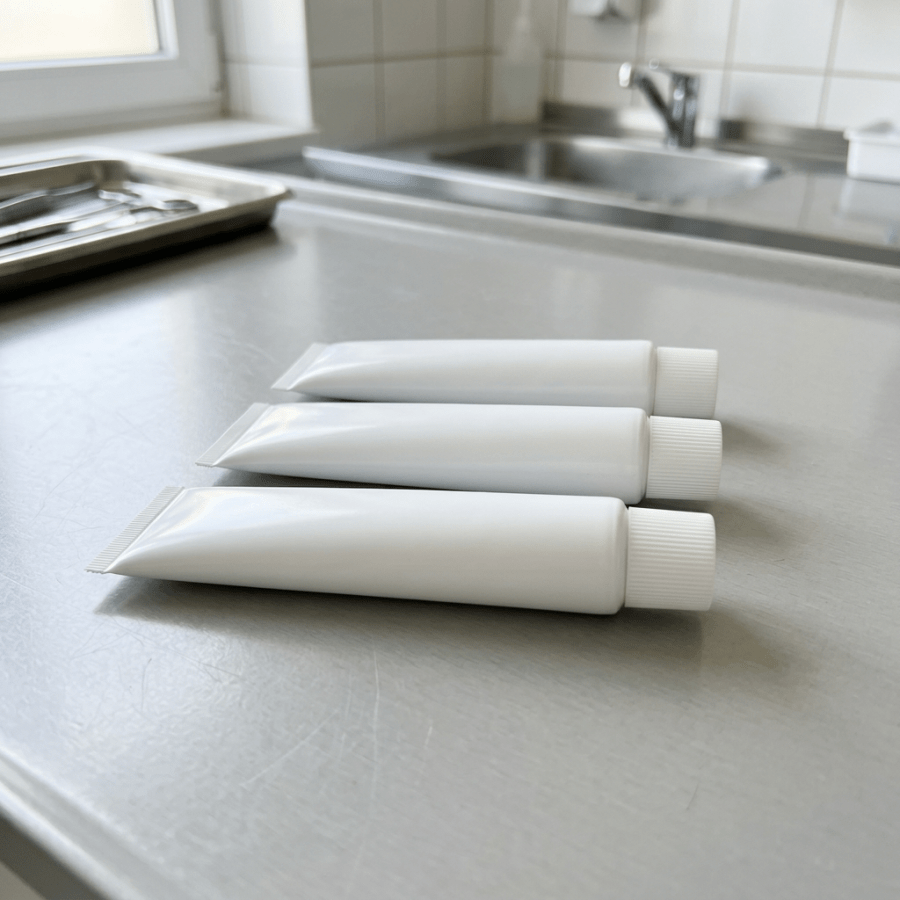
shutterstock_47533036Many patients come into the clinic and are confused about steroid creams, ointments, lotions, sprays, foams, and gels.  They come in many different forms and vary in their degree of potency.  They are generally grouped into classes according to their strength.  The medical provider’s choice in prescribing  a particular topical steroid medication depends on many factors including the medical condition (ie. eczema, psoriasis, bee sting, allergic reaction, etc.) the location of the body that is being treated and of course the patient’s ability to afford the prescription. There are some potentially harmful consequences that can occur if a steroid cream is used inappropriately.  For example, applying a high potency steroid cream to an area of the body such as the face or groin can cause permanent thinning of the skin. Here is a list of the different classes or categories that topical steroids are grouped into: Class 1:  Superpotent Class 2:  Potent Class 3:  Upper Mid-Strength Class 4:  Mid-Strength Class 5:  Lower Mid-Strength Class 6:  Mild Class 7:  Least Potent Over the counter topical steroids are generally in the class 7 or least potent category. Some examples of topical steroids and their respective classes include:
CLASS 1—Superpotent
Clobex Lotion/Spray/Shampoo, 0.05% Clobetasol propionate
Cormax Cream/Solution, 0.05% Clobetasol propionate
Diprolene Ointment, 0.05% Betamethasone dipropionate
Olux E Foam, 0.05% Clobetasol propionate
Olux Foam, 0.05% Clobetasol propionate
Temovate Cream/Ointment/Solution, 0.05% Clobetasol propionate
Ultravate Cream/Ointment, 0.05% Halobetasol propionate
Vanos Cream, 0.1% Fluocinonide
Psorcon Ointment, 0.05% Diflorasone diacetate
Psorcon E Ointment, 0.05% Diflorasone diacetate
CLASS 2—Potent
Diprolene Cream AF, 0.05% Betamethasone dipropionate
Elocon Ointment, 0.1% Mometasone furoate
Florone Ointment, 0.05% Diflorasone diacetate
Halog Ointment/Cream, 0.1% Halcinonide
Lidex Cream/Gel/Ointment, 0.05% Fluocinonide
Psorcon Cream, 0.05% Diflorasone diacetate
Topicort Cream/Ointment, 0.25% Desoximetasone
Topicort Gel, 0.05% Desoximetasone
CLASS 3—Upper Mid-Strength
Cutivate Ointment, 0.005% Fluticasone propionate
Lidex-E Cream, 0.05% Fluocinonide
Luxiq Foam, 0.12% Betamethasone valerate
Topicort LP Cream, 0.05% Desoximetasone
CLASS 4—Mid-Strength
Cordran Ointment, 0.05% Flurandrenolide
Elocon Cream, 0.1% Mometasone furoate
Kenalog Cream/Spray, 0.1% Triamcinolone acetonide
Synalar Ointment, 0.03% Fluocinolone acetonide
Westcort Ointment, 0.2% Hydrocortisone valerate
CLASS 5—Lower Mid-Strength
Capex Shampoo, 0.01% Fluocinolone acetonide
Cordran Cream/Lotion/Tape, 0.05% Flurandrenolide
Cutivate Cream/Lotion, 0.05% Fluticasone propionate
DermAtop Cream, 0.1% Prednicarbate
DesOwen Lotion, 0.05% Desonide
Locoid Cream/Lotion/Ointment/Solution, 0.1% Hydrocortisone
Pandel Cream, 0.1% Hydrocortisone
Synalar Cream, 0.03%/0.01% Fluocinolone acetonide
Westcort Cream, 0.2% Hydrocortisone valerate
CLASS 6—Mild
Aclovate Cream/Ointment, 0.05% Alclometasone dipropionate
Derma-Smoothe/FS Oil, 0.01% Fluocinolone acetonide
Desonate Gel, 0.05% Desonide
Synalar Cream/Solution, 0.01% Fluocinolone acetonide
Verdeso Foam, 0.05% Desonide
CLASS 7—Least Potent
Cetacort Lotion, 0.5%/1% Hydrocortisone
Cortaid Cream/Spray/Ointment Hydrocortisone
Hytone Cream/Lotion, 1%/2.5% Hydrocortisone
Micort-HC Cream, 2%/2.5% Hydrocortisone
Nutracort Lotion, 1%/2.5% Hydrocortisone
Synacort Cream, 1%/2.5% Hydrocortisone
  I hope that you have found this information useful.  Wishing you the best of health,

Scott Rennie, D.O.
Board Certified in Obesity Medicine and Family Medicine

This blog is for educational purposes only and does not constitute individual medical advice. Always consult your own physician before making changes to your health, medications, or treatment plan.

Gloved medical professional arranging surgical instruments on a sterile operating table

Pilonidal Cysts – A pain in the rear

Photo credit:  http://www.pilonidalcystcure.com

A pilonidal cyst tends to announce itself in a particular way.  They usually have complaints of difficulty sitting down or laying on their back because of pain.

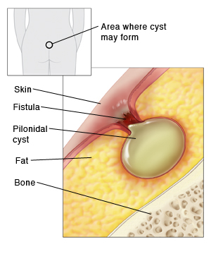
A pilonidal cyst is a fluid-filled sac that forms above the crease of the buttocks.  The cysts are usually red, inflamed and most often infected when I see them.  In many cases the cysts are hard, but sometimes they have a small hole in them where some fluid or blood has been draining.  Pus (milky yellow/green fluid) is sometimes draining from them as well.  The drainage often has a foul smell.

Cause:  The cause of these cysts is not clear. They may form when a hair is forced under the skin or when a hair follicle ruptures. Or, a person may be born with the cyst. These cysts are often diagnosed in people between the ages of 16 and 26. But people of any age can have a pilonidal cyst. And they affect both men and women.

Treatment:  We usually refer patients to a surgeon who can drain the cyst or remove it with surgery.  They are often very painful, so performing an incision and drainage in the urgent care or clinic is often not possible because the patient requires a higher level of pain control and possibly conscious sedation to tolerate the procedure.  To drain a cyst, the medical provider will first numb the area and then cut open the cyst with a scalpel and push the fluid out.  They will then wash the area to clean it thoroughly and sometimes they might pack the empty cyst with gauze or leave a drain in place so that it doesn’t fill up with blood or pus again immediately.  The drain or gauze can then be removed in a few days.  We usually try to provide the patient with medication for pain, and often recommend sitting on a doughnut-like pillow with a hole in the center to help with pain.

Removing the cyst entirely often requires a more extensive surgical procedure because pilonidal cysts often have a sinus (small channel) that goes deep under the skin and can lead to another cyst or be deep enough to almost touch the sacrum or tailbone area.  Patients with multiple cysts often need an even larger surgical procedure and consequently the recovery time can be much longer for multiple or larger cysts.


I hope that you have found this information useful.  Wishing you the best of health,

Carrying extra weight is one of the recognized risk factors for pilonidal disease and for it coming back after surgery. Weight is the other half of my practice now, so if that is on your list, here is what Wegovy and Zepbound cost without insurance.

Related Reading

Abscesses and MRSA: What To Do About a Skin Infection

When to Use Telemedicine vs. Urgent Care: How I Spot the Sick One

Scott Rennie, D.O.
Board Certified in Obesity Medicine and Family Medicine

This blog is for educational purposes only and does not constitute individual medical advice. Always consult your own physician before making changes to your health, medications, or treatment plan.
Tweezers removing debris from beneath a person’s fingernail

Splinter (Sliver) Removal


Photo credits: http://www.lemamme.it/sos-cosa-fare-quando-il-bambino-si-fa-male/

http://www.aafp.org/afp/2003/0615/p2557.html

Splinters look like the simplest possible problem, right up until one of them is not.  I thought I’d discuss some different types of splinters and some techniques that you can use at home to remove them.  In addition, it’s important to know when to have it taken out in person for help with that process.

Splinters are actually very common, and children and adults need it taken out in person usually with a small foreign body embedded into the superficial or subcutaneous soft tissues of the hands or feet.  Wood, glass and metallic splinters are the most common types.  Many splinters can be removed at home by our patients, which leaves the physicians only the deeper and larger splinters or retained splinters that have been broken down during an attempt at removal.  If splinters are not removed completely, they may cause complications such as infection and inflammation.

When to see a medical provider:  I recommend that you see a medical provider instead of trying to remove the splinter yourself if there is lots of redness around the splinter, increased swelling or pus draining from the wound.

The severity of the reaction and type of reaction that the body has to a particular splinter.  Some examples of reactions to retained foreign materials include:

1)   Glass – Mild reaction and the body attempts encapsulation

2)   Blackthorns – Severe reaction with inflammation from the plant alkaloids

3)   Wood – Severe reaction with inflammation from the oils and resins

4)   Cactus Spines – Moderate to severe reaction from the fungal coating on the plant and possibly an allergic reaction

5)   Rose thorns – Moderate to severe reaction with inflammation from the fungal coating on the plant

6)   Sea urchins – Moderate to severe reaction with inflammation and possible infection

7)   Metal – Mild reaction and the body attempts encapsulation

8)   Plant spines – Mild to severe reaction from the toxins in the plant

9)   Animal spines – Mild to severe reaction from the toxins in the animal spines

10)  Plastic – Mild reaction and the body attempts encapsulation

Sometimes splinters can be difficult to detect, especially if they are deep or very small.  Clues that a splinter is present might be swelling, tenderness, a mass, or soft tissue infection such as cellulitis, or an abscess.  Sometimes we might use a diagnostic test such as a x-ray, ultrasound, CT scan or MRI to detect a splinter if it’s deep and difficult to see.

Removal:  We try to remove splinters quickly before inflammation or infection occurs.  There are several ways to remove a splinter, some of those might include:

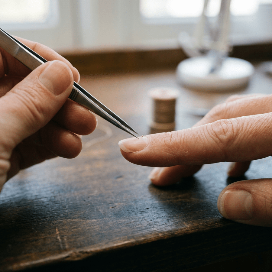
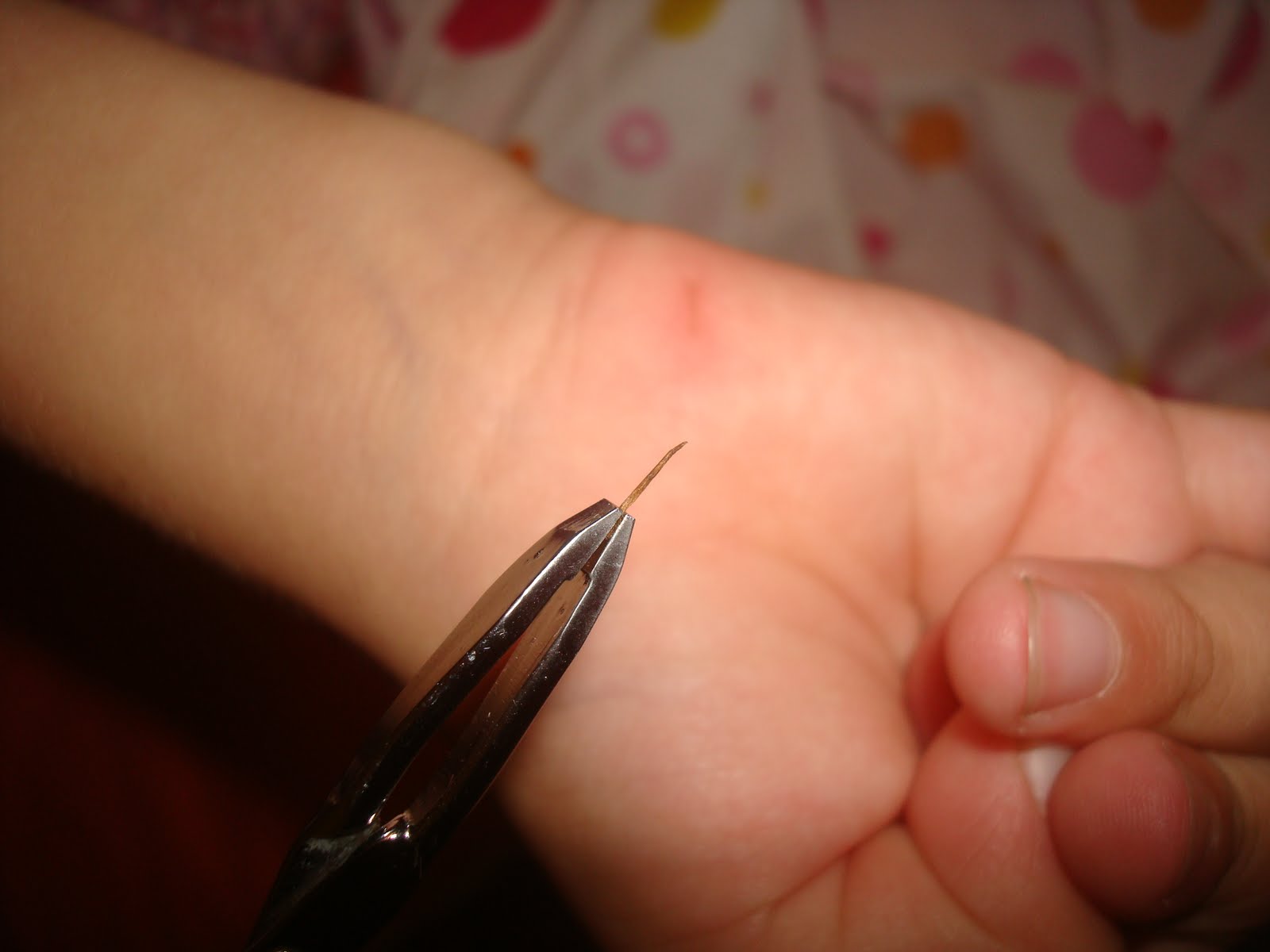
1)   Using tweezers: Make sure you have the right kind of tweezers.  The sharp tipped splinter tweezers work best most of the time.  If the sliver is sticking out of the skin, clean the pair of tweezers with alcohol and carefully pinch the sliver as close to the skin as possible and gently slide it out.

2)   Use a small needle:  If the sliver isn’t sticking out of the skin, often a needle can help push the sliver out.  First clean the needle with alcohol and then push the sliver from the bottom (the pointed end that entered the skin first) and angle upward toward the puncture hold in the skin’s surface.  Try to start at the bottom of the sliver so you don’t break it and leave sliver shreds in the skin.

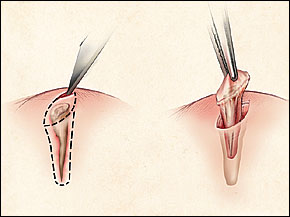
3)   Use a razor blade to make a small slice in the skin over the sliver:  If the sliver is deep and using tweezers or a needle isn’t working well, you can clean a razor blade in alcohol and make a shallow incision parallel to the sliver, just above it.  I know this sounds scary, but the top layer of skin is made of dead skin cells that do not have nerves, so this is not likely to hurt unless you cut too deeply.  Once the incision is made, gently part the skin and pick out the sliver.

4)   Clean the wound:   Whatever method you use to remove the sliver, make sure you clean the wound with soap and water after the sliver is removed.  Puncture wounds are often laced with bacteria and are very likely to become infected.

5)   Antibiotics:  Apply a small amount of antibiotic ointment such as bacitracin over the puncture wound.

6)   Examine the area at least once a day for the next several days to look for increasing redness, swelling or pain.  If any of these occur, I recommend that you see a medical provider because this might be the start of an infection.


I hope that you have found this information useful.  Wishing you the best of health,

A splinter you cannot reach is one of the few things a video visit genuinely cannot fix. I wrote about the conditions that still need an in person visit.

Related Reading

Abscesses and MRSA: What To Do About a Skin Infection

When to Use Telemedicine vs. Urgent Care: How I Spot the Sick One

Scott Rennie, D.O.
Board Certified in Obesity Medicine and Family Medicine

This blog is for educational purposes only and does not constitute individual medical advice. Always consult your own physician before making changes to your health, medications, or treatment plan.
Sole of foot with dry, peeling skin and calluses

Calluses and Corns: Causes, Prevention, and Treatment

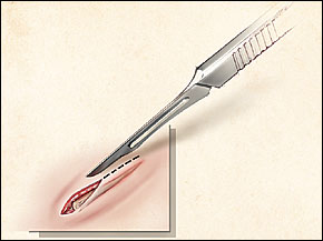
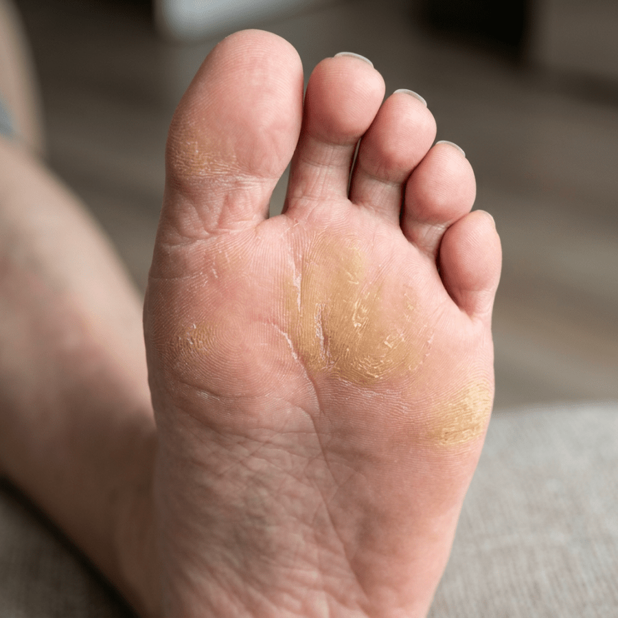
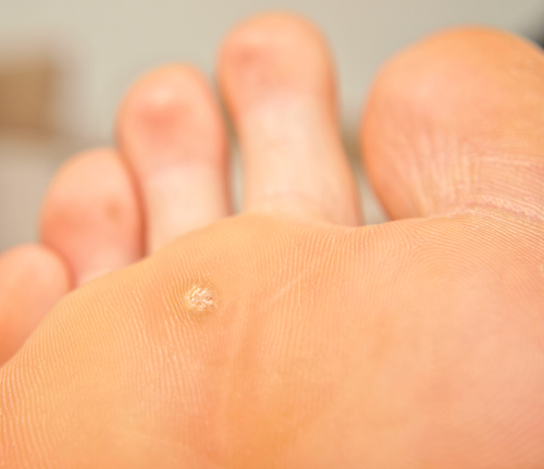
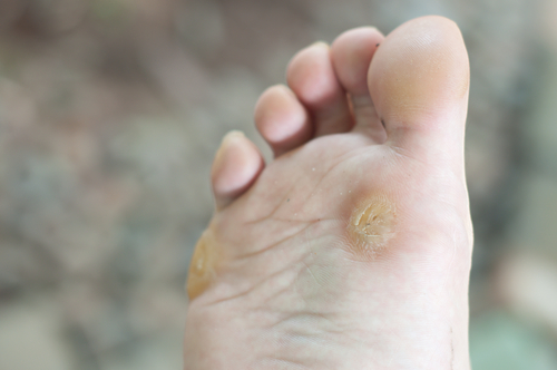
shutterstock_113610118shutterstock_93910513I can’t tell you how many patients come in to the clinic because of a callus or corn that’s bothering them – too many to count!  Corns and calluses can cause significant pain, especially when they’re on the feet because we often get them in areas over pressure areas.  When they get thickened they can re-distribute your weight onto other areas of your feet and that may lead to foot instability or worsening pain. Calluses are usually on the hands and feet and basically just thickened areas of skin that form when something rubs or presses on these areas over prolonged periods of time. Corns are thickened areas of skin that are often on the soles of the feet or sides of the toes and look like a small nodule that has a hard center.  Corns are usually more painful because they are often over a smaller area and press harder on a more specific area of the foot. Causes:  Possible reasons that calluses or corns develop include: 1)   Wearing shoes that don’t fit properly for your feet – either too loose or too tight 2)   Walking barefoot 3)   Wearing shoes without socks 4)   Calluses on the hands can be caused by repetitive sports such as rowing, golfing, tennis or biking without gloves Prevention: 1)   Get shoes that fit properly!  It’s important to be proactive and to prevent calluses or corns if they bother you.  There are special shoe stores that work with foot doctors to help you select shoes that fit your feet properly.  Some people are born with narrow or wide feet and that makes it more difficult to find shoes that fit properly.  If you are getting painful corns or calluses, take time and visit a store where someone can help you to pick out shoes that fit you well 2)   Avoid going barefoot or wearing shoes without socks 3)   If you have spots on your feet that rub inside your shoes, you can get special pads that prevent rubbing Treatment:  I often work with patients who come into the clinic to trim the corn or callus down so that it reduces the pressure in the affected area.  I use a scalpel to carefully trim away the thickened skin.  A foot doctor (podiatrist or orthopedic surgeon who specializes in treating conditions of the foot/ankle) can make special orthotic devices that can help reduce the pain and help prevent recurrence of corns/calluses.  Some people treat calluses themselves by purchasing special pads that contain medications to burn or dissolve the thick skin.  I highly recommend that patients with diabetes be seen and treated by a medical provider to reduce the risk of infection.  Diabetic patients are at increased risk of foot infections and should be seen for yearly foot examinations even if they don’t have calluses or corns.   I hope that you have found this information useful.  Wishing you the best of health,

Scott Rennie, D.O.
Board Certified in Obesity Medicine and Family Medicine

This blog is for educational purposes only and does not constitute individual medical advice. Always consult your own physician before making changes to your health, medications, or treatment plan.