
Doctor and patient discussing health across a medical office desk

What is Hidradenitis suppurativa?

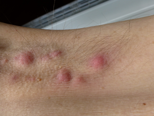
shutterstock_148330382A patient came into the clinic today and said she had a couple lumps in her left armpit (axilla) that have been very painful and draining some clear liquid.  She told me that she had one before in her right arm pit and she had it treated by taking antibiotics and a doctor also performed an incision and drainage. Hidrandenitis suppurativa (HS) is a condition in which there is red, swollen painful bumps in places where the skin rubs together.  The nodules can make it difficult to move because they are so painful.  They can also smell foul and drain pus or blood.  These bumps may go away on their own, but often stay for weeks to months and often come back. Causes:  This is not caused by being unclean – it’s not an infectious disease. Often the area may become irritated by shaving in that area or a certain deodorant or antiperspirant.   You cannot spread this to anyone else as it’s not contagious.  It is generally a genetic condition that is more common in first-degree relatives that have the condition. The bumps or nodules are usually located in the: 1)   Armpits (axilla) 2)   In the groin 3)   Under the breasts (in women) 4)   On the inner thighs 5)   Buttocks 6)   Around or near the anus Often the skin hardens and scars around the painful nodules and some can form tunnels under the skin. Treatment:  Possible treatment options include: 1)   Antibiotic liquids or gels that you put on the affected areas – these actually work to reduce inflammation rather than treat infection 2)   Antibiotic pills – to reduce inflammation 3)   Injections of steroid medications into the areas to bring down the inflammation 4)   Some women take hormone treatments to improve their condition 5)   Surgery Things you can do to reduce your symptoms: 1)   If you are overweight, lose weight because this condition is more common or severe in people who are overweight 2)   Try to avoid activities that cause your skin to rub against itself 3)   Do not wear tight-fitting clothes 4)   For people with recurrent infections, the use of an antibiotic lotion such as clindamycin 1% applied to the area twice a day can help prevent recurrence. 5)   Show and wash the tender areas everyday gently.  Do not scrub with a washcloth, brush or loofah 6)   Avoid smoking 7)   Use antiperspirants rather than deodorants 8)   Avoid exposure to hot, humid environments as much as possible   I hope that you have found this information useful.  Wishing you the best of health,

Updated for 2026: There Are Real Drugs For This Now

When I wrote this in 2012, hidradenitis suppurativa was a condition I could describe far better than I could treat. There were no approved medications for it. Antibiotics, drainage, and surgery, and a lot of people cycling through all three for years. That has changed, and if you were told years ago that nothing much could be done, you were told something that was true then and is not true now. Three biologics now carry FDA approval for moderate to severe disease. Adalimumab, a TNF inhibitor, was first, approved in 2015. Secukinumab, which blocks interleukin 17A, was approved on October 31, 2023, dosed at 300 milligrams every four weeks with the option to move to every two weeks if the response is not enough (1). Bimekizumab, which blocks both IL-17A and IL-17F, followed on November 20, 2024, with phase 3 responses holding out to forty eight weeks (2). Two approvals in about a year, after a decade with one. That is the most movement this condition has seen in my career.

Staging Still Drives the Decision

Hurley staging has not changed and it is still how severity gets sorted. Stage one is abscesses without tunnels or scarring. Stage two adds recurrent abscesses with sinus tracts and scarring. Stage three is diffuse involvement with multiple interconnected tunnels (3). The stage is what decides whether this is a topical and antibiotic conversation or a biologic conversation. It is worth knowing yours and asking for it by name.

What This Looks Like Over Video, and Where It Does Not Work

I can do a fair amount here. History, how long, what triggers a flare, what has already been tried and how far each was pushed. The effect on your life, which in this condition is enormous and routinely under-asked. Photographs of known lesions over time are genuinely useful for tracking whether something is working. Where video runs out is staging and starting a biologic. Counting nodules and tunnels, and mapping how far the disease actually extends, needs hands and an in-person look. Starting a biologic also needs screening bloodwork first, tuberculosis testing and hepatitis serologies among them. That is a referral, not a video prescription, and I would be doing you a disservice to pretend otherwise. The most useful thing I can do from here is name it correctly and get you to a dermatologist, because the average person with this condition spends years being told they have recurrent boils.

When To Be Seen Promptly

Fever, feeling systemically unwell, or redness spreading fast, which suggests infection beyond the usual flare. A lesion tense and painful enough to need draining. And any long standing wound in advanced disease that will not heal, changes character, or starts behaving differently, because squamous cell carcinoma arising in chronic hidradenitis is rare but real and is the reason nobody should be managing stage three alone.

The Bottom Line

If your information about this condition is more than a few years old, it is out of date in a way that matters. Find out your Hurley stage, and if it is moderate or severe, ask specifically about biologics. This is no longer a condition where the honest answer is to keep draining them.

Sources

1. Novartis. FDA approves Cosentyx (secukinumab) for hidradenitis suppurativa. October 31, 2023. https://www.novartis.com/news/media-releases/fda-approves-novartis-cosentyx-first-new-biologic-treatment-option-hidradenitis-suppurativa-patients-nearly-decade 2. UCB. FDA approval of Bimzelx (bimekizumab) for moderate to severe hidradenitis suppurativa. November 20, 2024. https://www.ucb.com/newsroom/press-releases/article/ucb-receives-us-fda-approval-for-bimzelxr-bimekizumab-bkzx-as-the-first-il-17a-and-il-17f-inhibitor-for-adults-with-moderate-to-severe-hidradenitis-suppurativa 3. American Academy of Dermatology. Hidradenitis suppurativa: diagnosis and treatment. https://www.aad.org/public/diseases/a-z/hidradenitis-suppurativa-treatment

Related Reading

Abscesses and MRSA: What To Do About a Skin Infection When to Use Telemedicine vs. Urgent Care: How I Spot the Sick One Scott Rennie, D.O.

Board Certified in Obesity Medicine and Family Medicine

This blog is for educational purposes only and does not constitute individual medical advice. Always consult your own physician before making changes to your health, medications, or treatment plan.

Sunlight breaking through heavy grey cloud over a city waterfront

It’s Cloudy in Seattle: Can You Still Get a Sunburn?

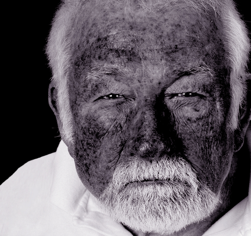
shutterstock_5810650Recently I’ve been noticing patients are coming in with sunburns even though the weather has been cloudy.  They are often unaware that the sun is causing damage to their skin at the time, and come in later with severe sunburns.  Even on cloudy days, it is important to protect your skin because UV radiation can pass through the clouds and cause sunburns.  In addition, UV rays can be reflected off surfaces like sand, snow, cement and water. Prevention:  Here are several ways to help prevent sunburn: 1)   Avoid sun exposure:  If you plan to be out in the sun during the day, keep in mind that the suns rays are strongest between 10:00am and 4:00pm in the continental U.S. and avoiding exposure during these hours decrease your risk of burning.  Wearing a topical sunscreen as well as clothing that blocks the suns rays are helpful in reducing your chance of sunburns. 2)   Find Shade:  Areas that don’t have direct sunlight reduce your chances of sunburn.  These areas include under trees, an umbrella or structures such as porches, or tents.  A topical sunscreen is still recommended while sitting in the shade because your skin is still exposed to some UV radiation. 3)   UV Index:  One way to predict your risk of sunburn on a given day is a numerical value called the UV index.  It is a number between 0 and 11+, in which 0 indicates a lower risk of sun exposure and 10 indicates high risk with 11+ an extreme risk.  You can find the UV Index online. 4)   Sunscreen:  There are many sunscreen products on the market, and most of them work by protecting the skin via absorbing the radiation or reflecting it.  SPF or Sun Protection Factor is a numerical indicator that gives us some idea of how much protection the sunscreen offers against UVB (Ultraviolet B) burns.  It’s important to look for a sunscreen that protects against both UVA (Ultraviolet A) and UVB rays – sometimes they label these products broad-spectrum.  Here are some additional tips. A)  I often get asked what SPF rating is best.  My first response is usually “the maximum you can get,” because I realize that even low levels of ultraviolet radiation increase your risk for sun damaged skin, skin cancers and a poor cosmetic outcome with increasing age.  The American Academy of Dermatology recommends an SPF of 30 or greater on sun exposed skin with protection of UVA and UVB and recommends a higher SPF in your are fair-skinned or will be out in the sun for longer periods of time or anticipate intense exposure (such as on a beach or skiing trip). B)  Use enough:  I find that most people don’t use enough sunscreen, and they don’t put it on soon enough.  Your really need about 2 tablespoons of lotion to cover an adults arms, legs, neck and face.  If you want to cover your back and chest, you will need more than that.  You should also apply it at least 15-30 minutes before going out in the sun for it to become active. C)  Reapply:  Even if the sunscreen bottle says that it’s sweat-proof, or water-proof, I recommend reapplying every 2-3 hours or after drying off with a towel or swimming.  There is some evidence that suggests that after being out in the sunlight for 20 minutes, you should reapply the sunscreen even if you’re not in the water or haven’t been sweating profusely. D)  Protect your lips:  Make sure your remember to protect your lips with lip balm that has an SPF of 30 or higher and reapply frequently. E)  Buy new sunscreen each year:  Chemical sunscreens become less effective with time.  Leaving them in the sun or where it is hot, such as in the car may speed this degradation process.  Expired sunscreen is likely less effective and reduces the SPF rating. Definition:  Sunburns occur when the skin is burned by UV radiation.  Often sunburns are not severe, but it’s the exposure over years that increase your risk of skin cancer, wrinkles and other cosmetic concerns.  In todays society, we often think about the immediate gratification which might include a suntan, however often the harmful consequences come years later often after we are no longer spending as much time in the sun.  I show my younger patients who have sunburned skin several photos of older patients who’ve spent years in the sun or had sunburns over years.  Hopefully that helps them understand the consequences that come with repeated exposure so they can make more informed decisions about protecting their skin from harmful radiation. Symptoms:  Sunburns are often not immediately apparent because the redness and pain develop 3-5 hours after being out in the sunshine.  Redness of the skin that is hot and painful to touch is common.  There may also be blistering and swelling over the affected areas.  The redness is usually at it’s worst by 12-24 hours after sun exposure and this fades over 72 hours. Causes:  Melanin is a pigment in the skin that causes your skin to appear dark or light colored.  Your skin can temporarily increase the amount of melanin to help protect from burns (suntan).  The amount of ultraviolet radiation that is needed to burn your skin depends on several factors: 1)   Melanin:  The amount of melanin in your skin affects how quickly you can get burned.  People with light colored skin and light hair generally have a higher risk of sunburn compared with patients with dark colored skin.  Some individuals with a low amount of melanin can burn in less than 15 minutes. 2)   Location:  There is increased UV radiation due to more direct sunlight near the equator so individuals who are in these locations are at more risk of sunburn (Hawaii for example). 3)   Medications:  Certain medications can increase the risk of sunburn including ibuprofen, some blood pressure medications such as hydrochlorothiazide (HCTZ), and some antibiotics such as tetracycline. Complications:  Premature skin aging, permanent discoloration of the skin, wrinkles, skin cancers such as malignant melanoma, basal cell and squamous cell carcinomas, cataracts (the lens of the eye becomes cloudy). Treatment: 1)   Stay out of the sun until the redness and pain go away.  Repeated sun damage after a recent burn is even more harmful. 2)   After noticing a sunburn, I often recommend immediately taking ibuprofen or Aleve to help with the pain 3)   Cool compresses, and aloe-based lotions and sprays 4)   Sprays with a local anesthetic that numbs the skin such as Solarcaine may help decrease the pain but they do not decrease the long-term risks of skin cancer and sun damaged skin. If you have had repeated sunburns, a history of skin cancer, or strong family history of skin cancers, I recommend that you see a dermatologist at least every year for a head to toe skin examination. To find a Dermatologist in your area, the American Academy of Dermatology’s Website has a very useful locator:  http://www.aad.org/find-a-derm   I hope that you have found this information useful.  Wishing you the best of health,

Scott Rennie, D.O.
Board Certified in Obesity Medicine and Family Medicine

This blog is for educational purposes only and does not constitute individual medical advice. Always consult your own physician before making changes to your health, medications, or treatment plan.
Several small raised skin growths on a person’s neck

Skin Tags (Acrochordons): What They Are and Look Like

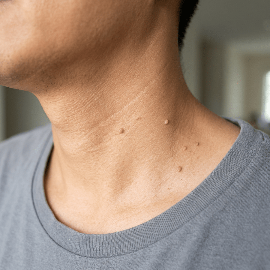
Photo credit:  http://www.your-doctor.net/dermatology_atlas/english/?id=38 Patients often come into the urgent care and ask about lesions on their skin.  It’s a very good practice to have skin lesions examined by a medical provider because without actually seeing the lesion, it can be difficult to make the proper diagnosis.  As you probably know, some skin lesions can be cancerous and so proper diagnosis is critical to ensure you get the right treatment and prevent complications. Skin tags are non-cancerous lesions that are an outgrowth of normal skin.  They occur in about 25% of adults and are more common as we age.  They commonly occur at areas of skin friction such as in the armpit (axilla), on the neck, under the breast tissue, or in the groin.  They can become painful when jewelry or clothing rub on them.  If they get twisted, the blood supply to the skin tag can tear or become compromised and they sometimes change to a red or black color. Diagnosis:  I recommend that you have any skin lesion that you’re unsure about examined by a medical provider.  If you have a history of skin cancer or family history of certain types of skin cancer, I recommend being examined by a dermatologist at least once a year and perhaps even more frequently.  The diagnosis of skin tags is usually fairly easy based on the appearance, but they must be differentiated from other types of skin lesions that may look similar. Treatment:  If you have healthcare insurance, it may not provide coverage for removal of skin tags if they are being removed only for cosmetic reasons.  Usually if they are painful or bleeding however, health insurance will cover the treatment for removal.    Some possible treatment options for removal include: 1)   Using forceps and fine grade scissors – these lesions often bleed vigorously so larger lesions may need suturing or cauterization 2)   Cryosurgery or liquid nitrogen treatment.  This freezing treatment is often done by super-cooling fine tipped forceps in liquid nitrogen and then gently squeezing the “stalk” of the skin tag to freeze it.  The procedure of freezing and un-thawing is similar to treating warts. 3)   Electrodessication Recurrence:  Unfortunately, skin tags can come back soon after they are treated. To find a Dermatologist in your area, the American Academy of Dermatology’s Website has a very useful locator:  http://www.aad.org/find-a-derm   I hope that you have found this information useful.  Wishing you the best of health,

Scott Rennie, D.O.
Board Certified in Obesity Medicine and Family Medicine

This blog is for educational purposes only and does not constitute individual medical advice. Always consult your own physician before making changes to your health, medications, or treatment plan.

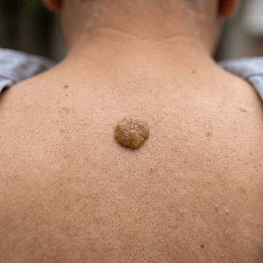
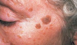
Raised, textured skin growth on a person’s upper back

Seborrheic Keratosis: The Skin Growth That Looks Stuck On

Photo credit:  http://medicine.academic.ru/7367/Seborrheic_keratosis

Patients often come in to see their doctor because of skin lesions that they are unsure about.  I encourage anyone with a skin lesion they are unsure about be evaluated by a medical provider because some lesions can be cancerous or pre-cancerous.

Seborrheic keratosis (SK’s) are very common, often developing after age 50 and occasionally as a young adult.  They are non-cancerous lesions and are more common in some families (genetic).  There is no way to prevent the development of new lesions.

They are non-cancerous but have been reported in association with a number of other skin malignancies, most commonly basal cell carcinomas and infrequently, melanoma.  The sign of “Leser-Trelat,” is the sudden onset of multiple seborrheic keratoses in association with skin tags and acanthosis nigrans.  This has been associated with a variety of cancers including gastrointestinal and lung cancers.

Diagnosis:  The appearance of the lesions is usually characteristic “stuck-on” or “warty” looking and they may be tan, light brown or dark-brown to black.  They are most commonly on the trunk of the body, face and arms/hands.  They are often scaly.  Microscopic examination is sometimes needed if they are small or atypical.

Treatment:  These lesions do not need to be treated, but due to cosmetic reasons they can be removed.  Insurance will usually not pay to have them removed unless they become painful, or bleed.  They can be treated by:

1)   Excisional biopsy – we send the specimen for examination under the microscope to rule out cancer if the lesion is suspicious

2)   Shave excision

3)   Cryotherapy with liquid nitrogen – sometimes a lighter skin pigment may occur after treatment and healing

Again, I encourage anyone with a skin lesion that they are unsure about be evaluated by a medical provider because some lesions can be cancerous or pre-cancerous.

To find a Dermatologist in your area, the American Academy of Dermatology’s Website has a very useful locator:  http://www.aad.org/find-a-derm


I hope that you have found this information useful.  Wishing you the best of health,

Scott Rennie, D.O.
Board Certified in Obesity Medicine and Family Medicine

This blog is for educational purposes only and does not constitute individual medical advice. Always consult your own physician before making changes to your health, medications, or treatment plan.
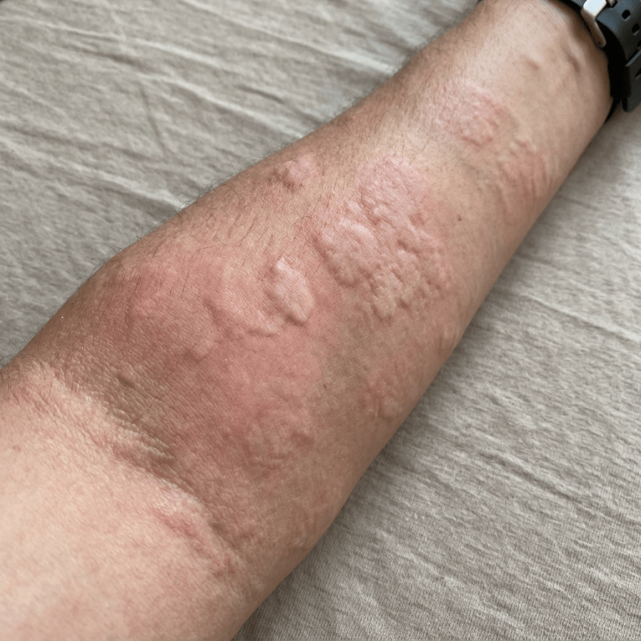
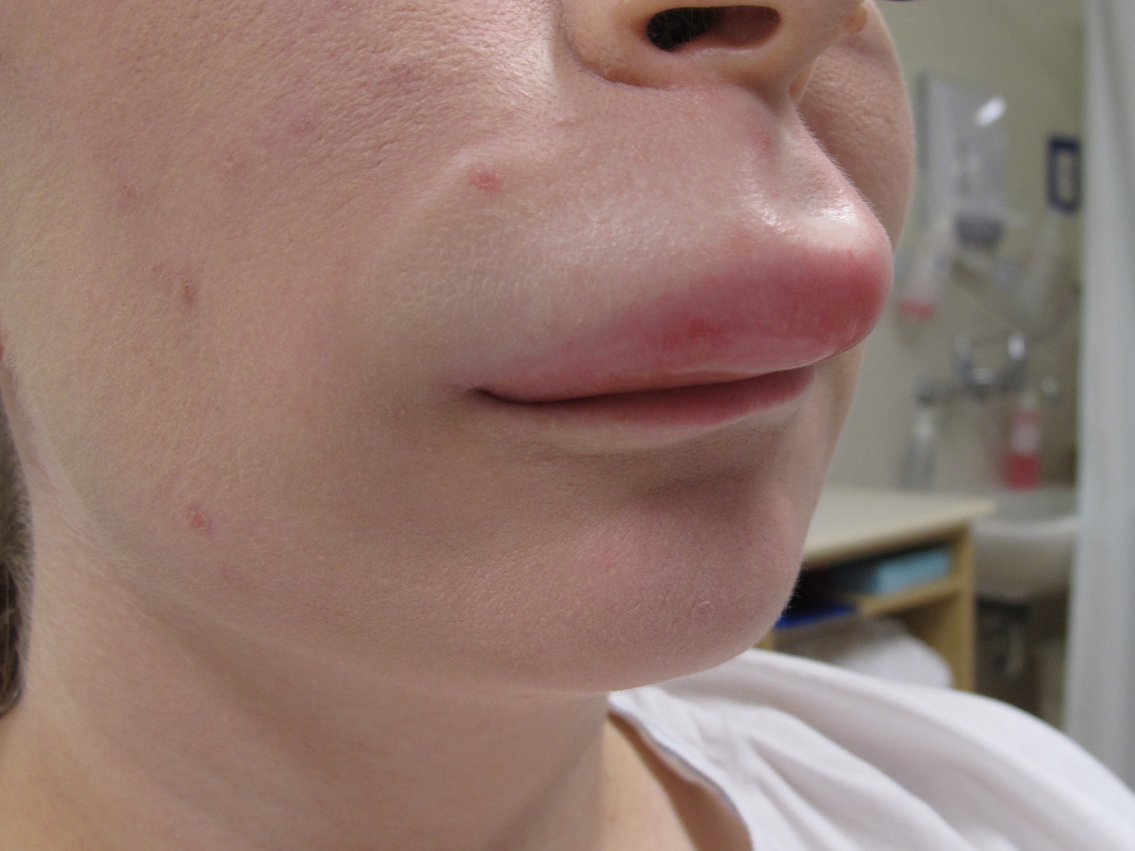
Raised pink welts covering a person’s forearm

Hives and Angioedema: Signs of an Allergic Reaction

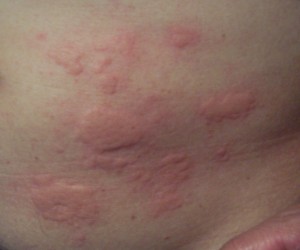
urticaria_hives-300x250 Photo credit:  http://drhuiallergist.com/allergy/urticaria-and-angioedema/ Some patients come to the urgent care with swelling or puffiness under the skin, usually in the face, eyelids, ears, mouth, tongue, hands, feet or genital area.  The most common place that we see this is on the upper lip but it can occur in many areas on the body.  We call this condition angioedema. Hives are another common problem that sometimes occur with angioedema.  They are red, raised patches of skin that are generally very itchy.  Urticaria is the medical term for hives.  They are raised areas of skin that itch intensively and are red with a pale center.  About 20% of people get hives at some time during their lives.  When someone comes into contact with a substance that they are allergic to, the body releases histamine from mast cells.  In most cases, hives appear and the disappear within several areas, and so the rash may look like it migrates or spreads around the body.  Sometimes the red raised areas get bigger or merge together and form larger hives.  Hives are generally not painful and do not have blood-blister type areas in them.  Hives usually do not appear with fever and joint pain. Causes of Hives:  There are several possible and may include: 1)      Infections – viral infections that cause the common cold are the cause of hives in 80% of children 2)     Medications 3)     Painkillers such as codeine or morphine 4)     IV contrast dye 5)     Insect stings 6)     Food allergies – typically occur with 30 minutes of eating the food.  Typical foods that cause hives include milk, eggs, peanuts or other nuts, soy, wheat, fish or shellfish. 7)     Physical contact – After touching a certain substance such as animals, certain plants, raw fruits/vegetables or latex (found in balloons, latex gloves and condoms) Causes of Angioedema:  In people who get angioedema  for the first time, it might be because of a new allergy.  Allergies are common to several of the following and can cause angioedema or hives: 1)      Medicines such as antibiotics or aspirin 2)     Foods such as eggs, fish, nuts or shellfish 3)     Insect stings 4)     Exercise can cause angioedema or hives 5)     Environmental substance such as a plant, animal, laundry detergent, soap, perfume latex, etc. 6)     Many times we are unsure of what caused the angioedema or hives to occur 7)     Certain blood pressure medications called “ACE Inhibitors” such as lisinopril, enalapril, captopril (and many others) can cause angioedema 8)     Ibuprofen/Advil or Aleve Treatment:  Minimizing exposure to the allergic agent is the first step.  Depending on the severity of symptoms, your medical provider may provide treatment including antihistamines such as Benadryl, Prednisone or other steroid medications  (either pills or shots) . Prevention:  Avoiding foods, medicines or exposure to insects that you have allergies to can decrease your chances of developing angioedema or hives.  Some people take antihistamines every day to help prevent getting angioedema if they get it frequently. When to seek medical care:   Make sure you are evaluated by a medical provider right away if you have any of the following: 1)      Trouble breathing 2)     Tightness in the throat 3)     Nausea/vomiting 4)     Abdominal pain/cramping 5)     Passing out or fainting   I hope that you have found this information useful.  Wishing you the best of health,

Scott Rennie, D.O.
Board Certified in Obesity Medicine and Family Medicine

This blog is for educational purposes only and does not constitute individual medical advice. Always consult your own physician before making changes to your health, medications, or treatment plan.
Golden retriever wearing a harness and leash in a sunny park

Animal Bites: What to Do if Your Neighbor’s Dog Bites You

shutterstock_54807220Quite a few patients come to the urgent care and see me for an animal bite.  Usually it’s a dog or cat bite, but sometimes I’ve seen patients who’ve been bit by a squirrel or raccoon and then there’s the rare person who is worried that a bat might have bit them.  In the US, two to five million people get bit each year and children are bitten much more often than adults.

Most people worry about the possibility of rabies, although an infection in the skin is much more common.  Bites on the hands can be serious because the skin surface is close to underlying bones and joints.

Dog bites:  Most dog bites are to boys between the ages of 5-9 years old, and the most common areas to be bit are on the head and neck for the younger children.  The right hand is the most frequent site of injury for older children and adults.  The complications of the bite range in severity from tearing away a body part to deep open cuts, crush injuries, to minor scratches.   In Washington State, we have not had a rabies case from a dog bite in many years and do not routinely give rabies treatments for individuals bitten by a dog unless it is known to have rabies infection.

Cat bites:  Claws or teeth can cause wounds from cats.  67% of cat bites are on the arms or hands and scratches are most often on the face.  Puncture wounds that are deep from a cat bite is concerning because bacteria from the cat’s mouth can go deep into the wound and cause infection to the underlying bones or joints.  Infections from cat bites generally cause increased redness to the skin, swelling and pain as early as 12-24 hours after the bite and progress quickly.

Rodent bites:  Rats are the most common rodents that cause bites, but squirrels, hamsters, rabbits and guinea pigs are generally all treated the same way as cat bites.

Other bites:  Bites from raccoons, skunks, fox, coyote or bats should be evaluated right away even if the bite is small and does not look like it’s infected.  These animals can carry rabies and medications are generally given to prevent rabies infection in individuals who are bit by these types of animals.

Treatment:  The first step is to clean the wound with soap and lots of water.  In the event of bleeding, gauze pads can be applied and pressure used to stop the bleeding once the wounds have been washed.

When to seek medical care:

1)   The bleeding has not stopped after applying pressure for 15 minutes

2)   There is a broken bone or serious injury such as large or deep laceration

3)   The bite victim has medical problems such as diabetes, cancer, HIV or takes medication that weaken the immune system

4)   The animal is from one of the animals listed above which have a high risk of rabies

Antibiotics:  Skin infections are the most common complication of an animal bite.  High-risk wounds such as on the face, or involving the bone or a joint are usually treated with an antibiotic.  Many experts also recommend antibiotics to treat people bitten by cats because of the high risk of infection from cat bites.  Most of the time oral antibiotics such as Augmentin are given to treat animal bites however in severe bites with infections that are already present, we will use IV antibiotics.

Tetanus immunization:  Tetanus is a serious, and potentially life threatening infection that can be transmitted by animal or human bite.  If the last vaccine was greater than 5 years ago, the patient should receive a tetanus vaccine.

Sutures (aka stiches):  Wounds on the face are usually closed to avoid developing a scar however due to the risk of infection, some bites may not be sutured immediately.  They may be flushed with a sterile solution and left open for 72 hours after injury and watched closely for signs of infection.

Infected bite wound:  If bite wounds are not treated appropriately right after the injury, an infection may develop.  Surgical treatment and antibiotics may be required at that point.

For more information on animal associated hazards, a useful resource is the centers for disease control article: http://wwwnc.cdc.gov/travel/yellowbook/2012/chapter-2-the-pre-travel-consultation/animal-associated-hazards.htm


I hope that you have found this information useful.  Wishing you the best of health,

Scott Rennie, D.O.
Board Certified in Obesity Medicine and Family Medicine

This blog is for educational purposes only and does not constitute individual medical advice. Always consult your own physician before making changes to your health, medications, or treatment plan.
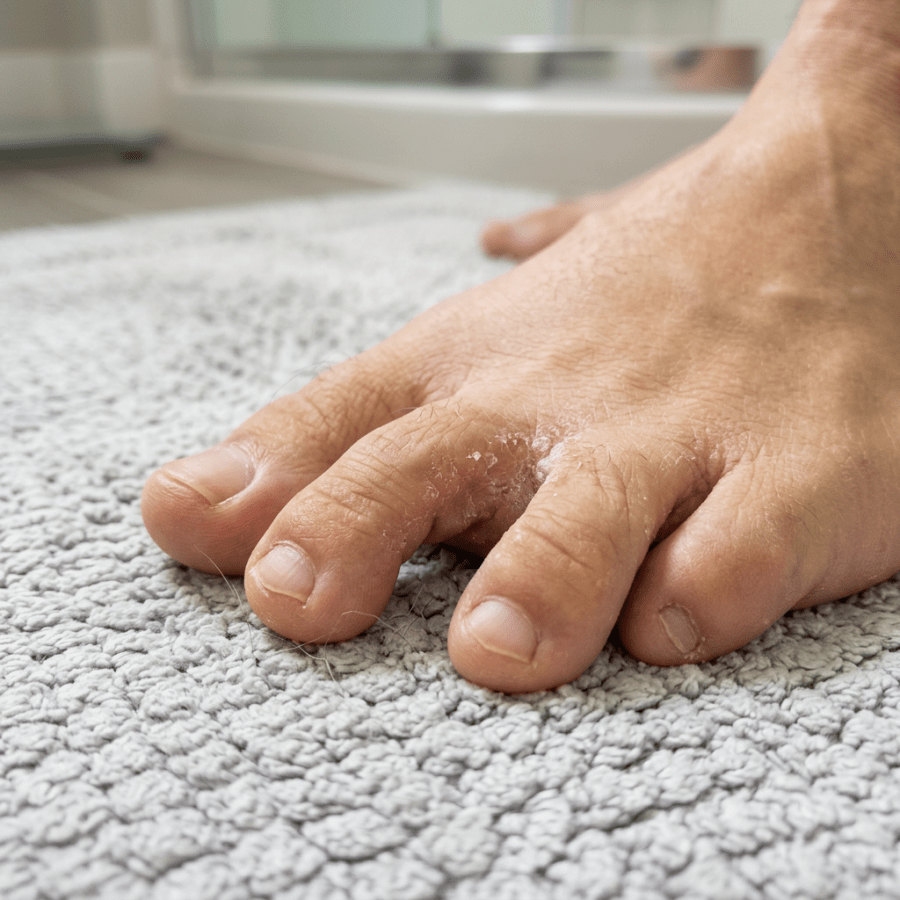
Close-up of toes with dry, peeling skin on a textured towel

Ringworm, Athlete’s Foot and Jock Itch and Fungal Nail Infections – “There Are Fungus Among Us”

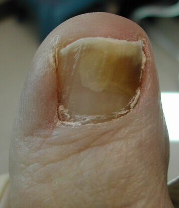
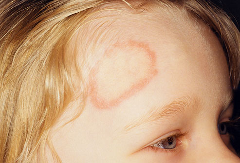
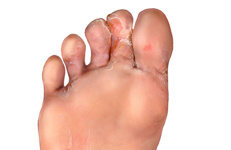
Photo credit:  http://fluconazole.biz/fluconazole-resistant-tinea/ People come to their doctor quite frequently due to fungal infections of the skin or nails.   One might think that because of the name, “Ringworm,” it would be caused by a worm, but it is actually caused by a fungus.  The shape of the rash looks like a ring and thus it got its name.  It’s a red, itchy rash on the skin and is also caused tinea. Four different type of ringworm infections are named for the body-part that is affected: 1)   Tinea capitis affects the head or scalp and is mostly seen in children 2)   Tinea pedis affects the feet and is called athlete’s food because athlete’s common acquire it in the locker room 3)   Tinea cruris affects the groin and is also called jock itch because it also commonly affects athletes or “jocks” presumably because of the moisture on the skin that the fungus prefers 4)   Tinea corporis is a term for fungal infections that affect other body surfaces Ringworm is a contagious infection and can be spread by someone else who is infected or even by an infected dog or cat.  It may be spread in a shower stall, locker room floor or pool area.  Often small skin shavings from the infected person are left behind in these areas and an unknowing person touches the infected area with their feet or other body part. Tinea capitis:  a fungal infection of the scalp may be related to cradle cap in children.  It causes a red scaly rash and can lead to bald patches.  It rarely affects adults.  We treat scalp infections with either oral or topical medications or a combination of both.  Children often improve with the use of gentle massage of the scalp during a bath to remove the scaly areas and then use of an antifungal medical on the scalp to kill the bacteria.  Adults and children are usually treated with oral medications and treatment can take 2-12 weeks depending on the severity of the infection. Tinea pedis (athlete’s foot):  the skin of the feet (often between the toes of the 3-5th toes) become itchy, red, tender, cracked and scaly.  We also see it on the soles of feet and in this area it is usually scaly and simply looks like dry skin.  Sometimes blisters form on the feet, especially between the toes.  It’s very important to treat athlete’s foot in diabetic patients because the fungal infection predisposes the patient even worse – a bacterial infection in the feet.  Diabetic patients can have a decreased sensation on their feet, so they can develop worsening infections without realizing it.  We usually start by removing the dead, scaling skin from the feet, and then treat athlete’s foot with a topical antifungal cream such as Lamisil AT twice a day for up to 12 weeks. The fungus can live on the skin even after the scaling skin is gone, so it’s important to treat the infection even for 1 week after the infection is totally gone.  This infection can be very difficult to treat because if you miss a day of applying the cream, the fungal infection can grow rapidly.  It’s important to keep the feet as dry as possible because fungal infections grow better in warm, damp areas. Tinea corporis (body infection):  often occurs when the fungal infection is transferred from one part of the body to another.  It is commonly seen in high school wrestlers.  It usually appears as a circular or oval scaly area with an outer red edge that is slightly raised while the center is usually flat and skin colored.  We treat this infection with a topical antifungal medication twice a day for 1-2 weeks.  Wrestlers may not be allowed to compete (by their school policy) until their infection clears. Onychomycosis (fungal nail infection):  a fugal infection of the nail that causes the nail to thicken, and change color such as white, yellow or brown.  This type of infection happens most commonly in the toenail but can also happen in fingernails. Fungal nail infections are common and don’t usually lead to serious long-term problems.  It is usually very easy to diagnose a fungal infection of the nails just by examining them.  Most over the counter medications don’t work for fungal nail infections.  In fact even prescription medications don’t work very well, and most fungal nail infections return after being treated with oral antifungal medications. In addition, the oral medications used to treat fungal infections of the nails can cause severe liver disease, so I usually don’t recommend treating this type of infection and instead recommend keeping the nails trimmed to prevent nail fracture or trauma. Preventing ringworm: 1)   Do not share clothing or sports equipment/towels with others 2)   Wear slippers or sandals at the gym, local pool or public shower 3)   Wash with soap and shampoo after skin-to-skin contact with others 4)   Avoid tight-fitting clothing to allow the skin to dry 5)   With athlete’s foot, put socks on before underwear to prevent spread of the fungal infection to other parts of the body including the groin 6)   Treat pet fungal infections to prevent spread to humans 7)   If a family member has a fungal infection, make sure they get treatment right away To find a Dermatologist in your area, the American Academy of Dermatology’s Website has a very useful locator:  http://www.aad.org/find-a-derm   I hope that you have found this information useful.  Wishing you the best of health,

Athlete’s foot and thickened fungal nails matter more than they look, because the cracks they leave between the toes are a common entry point for a deeper foot infection. That risk is highest in diabetes, which is why I wrote a separate post on how to check and protect your feet when you have diabetes.

Updated for 2026: There Is a Resistant Ringworm Now

When I wrote this in 2012, ringworm was a nuisance with a reliable answer. Put an antifungal cream on it, wait a couple of weeks, done. For most people that is still true. But a genuinely new problem has arrived and it is worth your knowing about, because it changes what to do when the cream does not work. Trichophyton indotineae emerged on the Indian subcontinent and has spread internationally, including to multiple US states. It is frequently resistant to terbinafine through mutations in the squalene epoxidase gene. One documented US case had a terbinafine minimum inhibitory concentration of 16 micrograms per milliliter, which is to say the drug was doing essentially nothing (1). It tends to look different. Extensive rather than a tidy ring. Often on the face, trunk, and groin at once. Sometimes a ring inside a ring. Intensely itchy, widespread, and stubborn in a way ordinary ringworm is not. It has also been reported as possibly sexually transmitted (2). The frustrating part is that an ordinary fungal culture cannot tell it apart from garden variety Trichophyton. Identifying it takes specialized gene sequencing available at only a handful of labs. Itraconazole usually works, but often needs more than three months. Terbinafine resistant Trichophyton rubrum, the far more common species, is also increasing in the United States (3).

What That Means Practically

Mostly it means stop assuming that treatment failure is your fault for not applying the cream long enough. If you have had an adequate course of a topical antifungal, or a proper course of oral terbinafine, and the rash is still spreading, that is not a reason to run the same drug again. That is a reason to have the diagnosis reconsidered and, if it still looks fungal, to change class rather than repeat. For ordinary tinea nothing has changed much. Body and groin ringworm respond to a topical antifungal over about two weeks, and the terbinafine class tends to clear it slightly faster than the azoles. Athlete’s foot takes longer, roughly four weeks with an azole. For toenails, oral terbinafine 250 milligrams daily remains first line, and current guidance is to confirm the diagnosis with testing before committing someone to months of an oral antifungal (3).

The Video Visit Version

Most of what I need for this one I can get from a photograph and a few questions, which makes it a reasonable fit for a virtual visit. What helps me is a well lit close up, plus a wider shot showing how far it extends. Tell me how long it has been there, what you have already tried and for how long, whether anyone else at home or any pet has it, and whether you have been treating it with a steroid cream, because a steroid on ringworm changes its appearance and makes it worse. Where I will send you elsewhere is toenails. If we are talking about months of an oral antifungal, I want the diagnosis confirmed rather than assumed, and roughly half of thickened toenails turn out not to be fungal at all.

When To Be Seen In Person

Extensive or rapidly spreading tinea that has already failed proper treatment, which raises the question of the resistant species and needs specialized testing. Scalp involvement, which needs oral treatment and cannot be handled with cream. Pain, warmth, or spreading redness suggesting a bacterial infection on top, particularly if you are diabetic. And any widespread fungal infection if your immune system is suppressed.

The Bottom Line

Ordinary ringworm is still ordinary and still responds to cream. What is new is that a failed course now means something. If two weeks of the right treatment has not touched it, do not just buy another tube. Get it looked at again.

Sources

1. Caplan AS, et al. Notes from the Field: First Reported US Cases of Tinea Caused by Trichophyton indotineae. https://www.ncbi.nlm.nih.gov/pmc/articles/PMC10208369/ 2. Potential Sexual Transmission of Antifungal-Resistant Trichophyton indotineae. Emerging Infectious Diseases. 2024;30(4). https://wwwnc.cdc.gov/eid/article/30/4/24-0115_article 3. Diagnosis and Management of Tinea Infections. American Family Physician. October 2025. https://www.aafp.org/afp/2025/1000/tinea-infections

Related Reading

Scabies Infection: The Mite Bite When to Use Telemedicine vs. Urgent Care: How I Spot the Sick One Scott Rennie, D.O.

Board Certified in Obesity Medicine and Family Medicine

This blog is for educational purposes only and does not constitute individual medical advice. Always consult your own physician before making changes to your health, medications, or treatment plan.
Honeybee collecting nectar from a purple wildflower

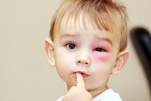
Bee and Insect Stings

shutterstock_116816458Patients often come into the urgent care clinic due to bee or insect stings as the weather improves.  Being stung is often painful and can also be anxiety provoking because some people have serious or life-threatening allergic reactions to stings and require quick treatment. Sting Reaction:  Immediately after being stung, most people have a sharp or burning pain as well as redness or swelling at the site of the sting.  The swelling and pain usually improve within a few hours.  About 10% of people will develop severe redness and swelling after a sting, and this is called a large localized reaction.  These large red areas may reach up to 4 inches in diameter over 1-2 days, and then slowly resolve over 5-10 days. If you have a large area of inflammation after a sting, this does not mean that you will have an anaphylactic reaction if stung again. Rarely, some individuals will develop a severe allergic reaction, called anaphylaxis.  Only 5-10% of people with a large localized reaction will have an anaphylactic reaction. Symptoms of severe allergic reaction (usually develop quickly): 1)   Hives, redness or swelling of skin away from the area that was stung – ie. face or lips if being stung on the hand 2)   Shortness of breath, hoarse voice or difficulty breathing 3)   Abdominal pain, nausea, vomiting or diarrhea 4)   Feeling lightheaded, dizzy or passing out Treatment for local skin reaction: 1)    If the insect has left a stinger, remove it as soon as possible after being stung.  Flicking or scraping the stinger out is sufficient. 2)    Apply a cold or damp washcloth wrapped around ice to the area 3)    Take an antihistamine such as Benadryl or Zyrtec if you develop itching 4)    A pain medicine such as Ibuprofen or Aleve may be helpful Treatment for severe allergic reactions:  These are a medical emergency that can lead to death if not treated quickly.  Do not drive yourself to the hospital – call 911.  A shot of epinephrine (adrenaline) may be helpful to prevent more severe allergic reactions in some patients.  Epinephrine is prescription only and should only be used if there is concern about a possible life threatening reaction.  Training on how and when to use epinephrine should be given at your medical provider or pharmacy. Prevention:  Usually bees or wasps are not aggressive when they are away from their nests.  The usually only sting after being hit, stepped on or swatted.  Wearing a white or light-colored clothing may help reduce the chance of being attacked if you are near a nest.  If you are eating outside, keep food and drinks covered and clean up spills quickly.  Watch for yellow jackets inside of drink containers.  If you find a nest near your home, do not try to get rid of it yourself.  Call a pest control professional. If you are being swarmed or stung, cover your mouth and nose with your hand and retreat to inside a building or an enclosed vehicle.   I hope that you have found this information useful.  Wishing you the best of health,

Scott Rennie, D.O.
Board Certified in Obesity Medicine and Family Medicine

This blog is for educational purposes only and does not constitute individual medical advice. Always consult your own physician before making changes to your health, medications, or treatment plan.
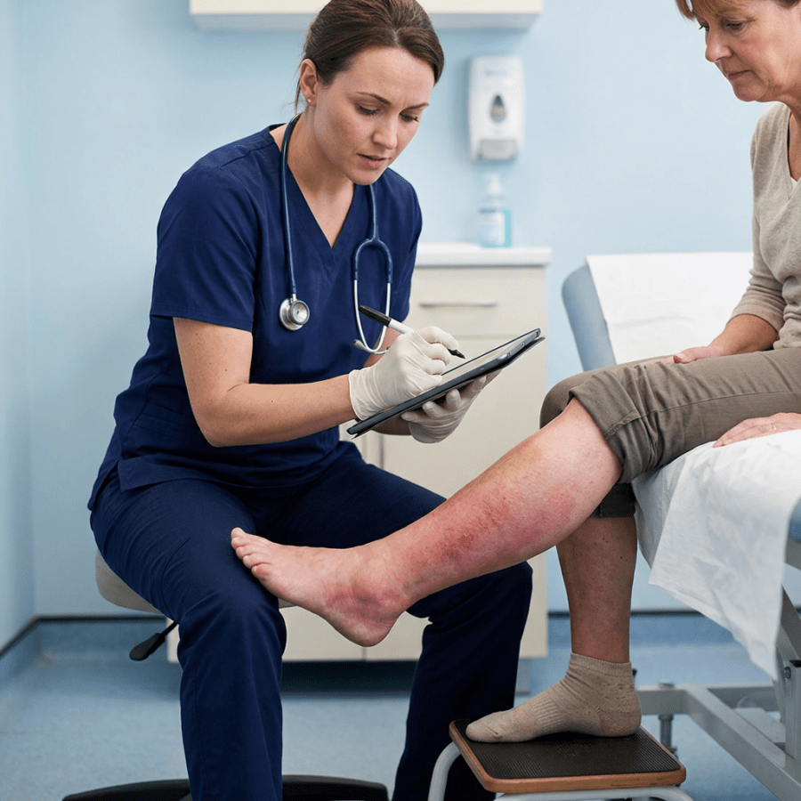
Nurse documenting examination of patient's reddened lower leg

Cellulitis – a soft tissue/skin infection – is it MRSA?

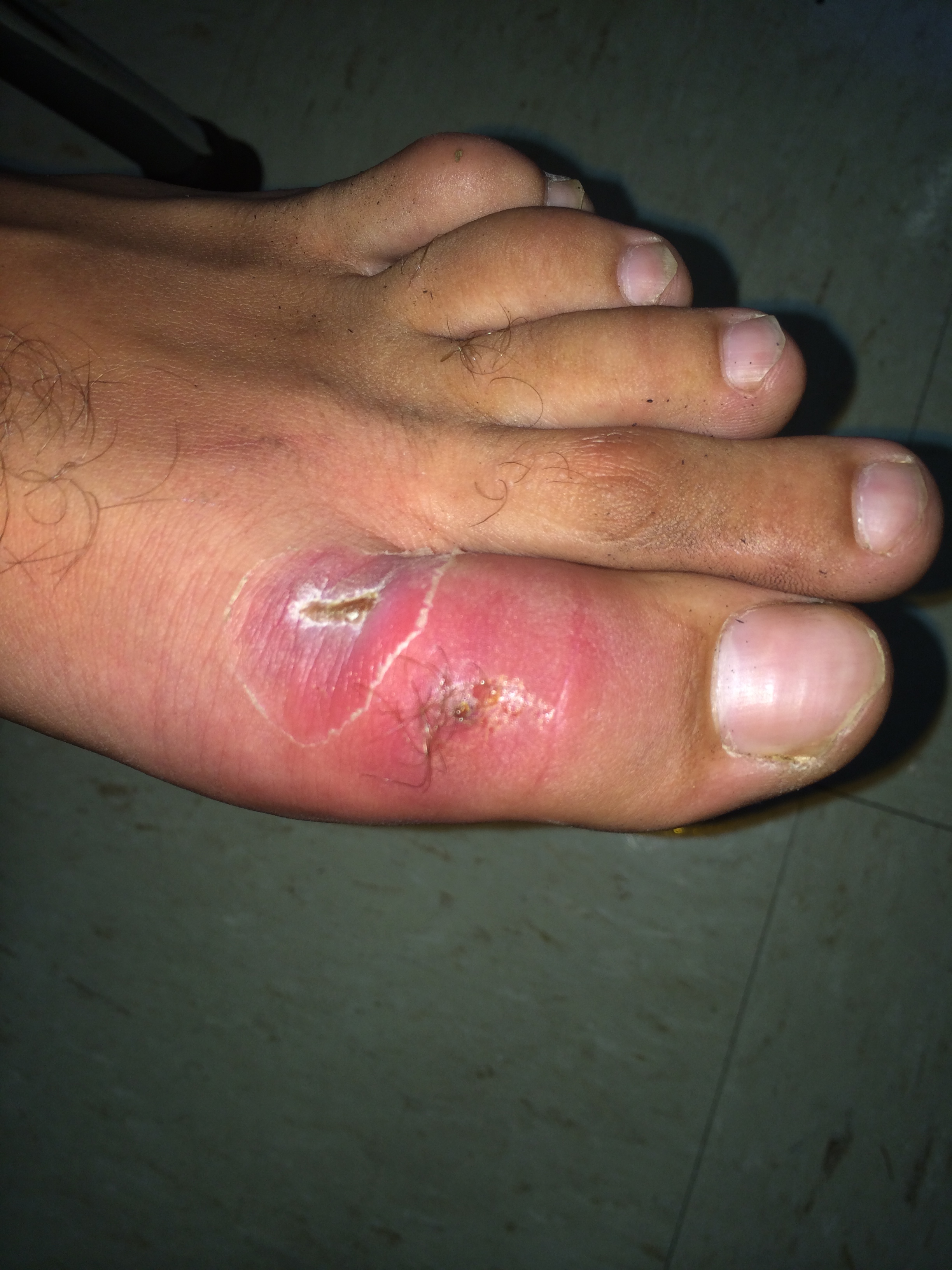
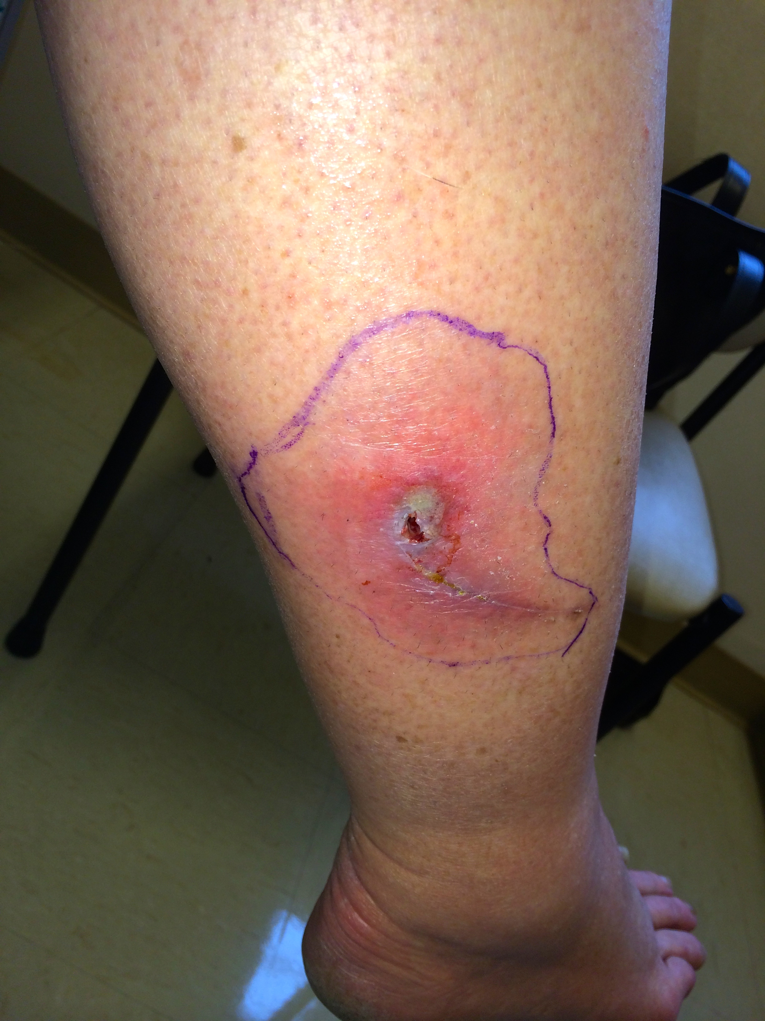
IMG_5864Cellulitis is an infection of the skin and soft tissues.  It is common and most often it’s caused by bacteria that normally live on the skin.  These bacteria don’t normally cause any problems unless the skin is scratched, torn or punctured.  The most common bacteria that cause cellulitis are streptococci “Strep” or staphylococci “Staph.” Most of the time cellulitis is mild and heals with the use of antibiotics, however it can become severe and cause an infection that spreads throughout the body if left untreated.  It can also lead to deep infections called abscesses. Risk factors: 1)   Chronic skin condition such as eczema or psoriasis 2)   Accumulation of fluid (edema) due to poor circulation possibly from heart failure, liver disease, or removal of lymph nodes 3)   Recent injury, wound, cut or laceration to the skin 4)   Current skin infection such as athlete’s foot or impetigo 5)   Being overweight Symptoms:  Pain, tenderness, increased warmth, redness and swelling in a distinct area of skin.  The skin can be smooth and shiny in this area.  Fever and chills are not usually present. Other skin infections:  different kinds of infections that can be confused with cellulitis include an abscess or boil.  These are different because they are usually bumpy, raised and filled with pus.  Abscesses are usually caused by a staph bacteria which may be MRSA.  Cellulitis is more commonly caused by “strep” bacteria. Treatment:  We commonly use antibiotics and also treat the underlying problem that caused the skin infection (such as athlete’s food or increased edema, eczema or psoriasis) if present. Keeping the area elevated to reduce inflammation is also helpful.  It is important to keep the infected area clean and dry.  Showering or bathing can be done normally, but pat the area dry with a clean towel afterwards to dry the area completely.  You can also use a bandage or gauze to protect the skin if needed. Antibiotics are often used and will be tailored to the individual patient.  The patient’s specific area of infection, medical history, history of allergies and examination will be important in determining the proper antibiotics to use.  Sometimes the antibiotics will need to be given by IV in more serious cases of cellulitis. Treatment time:  In most cases, the swelling, warmth and redness should improve within 1-3 days after starting the antibiotics.  We often use a Sharpie marker and draw a circle around the area of redness so that we can examine the red area over a few days to see if it’s increasing, decreasing or staying the same.  Symptoms of redness and swelling can persist for up to 2 weeks but it should be gradually decreasing.  If it’s not improving, you should call or return to your health care provider for re-evaluation.   I hope that you have found this information useful.  Wishing you the best of health,

Cellulitis of the lower leg often starts at a crack or a small cut on the foot, and with diabetic neuropathy that break can go unnoticed until the whole leg is red. It is the reason I keep after people about the diabetic foot check and what to look for.

Updated for 2026: Pus or No Pus Decides the Antibiotic

The most useful thing I can add to this post is a distinction that was fuzzy in 2012 and is now the thing the whole treatment decision turns on. Is there pus, or is there not. If there is an abscess, a pocket of pus you can feel giving under your finger, that is usually staph, MRSA is on the table, and it needs drainage plus an antibiotic that covers MRSA. I have written about that separately. If it is plain cellulitis, meaning spreading red, warm, tender skin with no drainable pocket, that is usually strep, and a cephalosporin like cephalexin is the right drug. Adding MRSA coverage on top does not help, and this has been tested directly. Moran and colleagues randomized patients with uncomplicated cellulitis to cephalexin plus trimethoprim-sulfamethoxazole or cephalexin alone. Cure was 83.5 percent against 85.5 percent (1). No benefit, two drugs, more side effects. So if you have been handed two antibiotics for a red leg with no abscess, that is worth a conversation. The 2014 infectious disease guidelines remain the operative document here and have not been superseded (2).

Draw a Line Around It

This is the single most useful thing you can do, and it is more valuable to me on a video visit than almost anything else. Take a pen and trace the edge of the redness. Write the time next to it. Then photograph it. When we talk tomorrow, or if you end up in an urgent care instead, that line answers the only question that really matters, which is whether this is advancing, holding, or retreating. Redness that has crossed well past the line in a few hours is a different problem from redness that has not moved. Without the line, everyone is guessing from memory, and memory is bad at this.

What I Can and Cannot Tell From a Screen

I can see color, extent, and whether there is an obvious pocket. I can ask about fever, chills, how fast it came on, whether there was a break in the skin, and how your blood sugars have been. What I cannot do is feel it. Warmth and firmness are things I am taking your word for, and the difference between cellulitis and something deeper and far more dangerous is partly a matter of how the tissue feels and how much pain there is relative to how it looks. That last one matters. Pain out of proportion to the appearance is a red flag I take seriously, and it is a reason for me to send you in rather than treat from here. Good photographs help more than people expect. Natural light, the same angle each time, and something for scale.

When To Be Seen, Today

Fever or chills. Redness advancing visibly past your marked line. Pain that seems far worse than the skin looks, or skin that is dusky, blistering, or numb. Cellulitis on the face. Any of this in someone diabetic, immunosuppressed, or with poor circulation. And no improvement after forty eight hours on the right antibiotic. The rapidly advancing, severely painful ones are surgical emergencies, not prescriptions, and they are the reason I would rather over-refer than under-refer on this particular condition.

The Bottom Line

No pus means a cephalosporin, and adding a second antibiotic for MRSA does not improve anything. Pus means drainage. Draw a line around the redness and note the time, because that line is the best data anyone will have.

Sources

1. Moran GJ, et al. Effect of Cephalexin Plus Trimethoprim-Sulfamethoxazole vs Cephalexin Alone on Clinical Cure of Uncomplicated Cellulitis. JAMA. 2017;317(20):2088-2096. https://jamanetwork.com/journals/jama/fullarticle/2627970 2. Stevens DL, et al. Practice Guidelines for the Diagnosis and Management of Skin and Soft Tissue Infections: 2014 Update. Clin Infect Dis. 2014;59(2):e10-e52. https://www.idsociety.org/practice-guideline/skin-and-soft-tissue-infections/

Related Reading

Abscesses and MRSA: What To Do About a Skin Infection Am I Truly Allergic to Penicillin? When to Use Telemedicine vs. Urgent Care: How I Spot the Sick One Scott Rennie, D.O.

Board Certified in Obesity Medicine and Family Medicine

This blog is for educational purposes only and does not constitute individual medical advice. Always consult your own physician before making changes to your health, medications, or treatment plan.

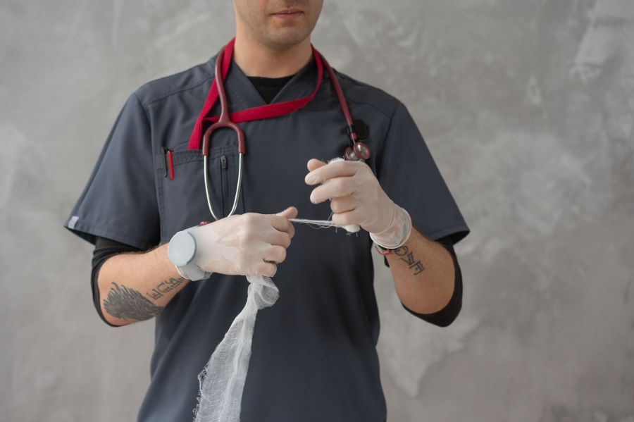
A clinician in scrubs and gloves preparing a length of sterile gauze for a dressing.

Abscesses – What to do about MRSA (Methicillin-resistant Staphylococcus aureus)

IMG_5872“Staph” infections are most often caused by the organism Staphylococcus aureus, a bacterium that is carried on the skin of about 30% of healthy individuals.  Most of the time, these bacteria do not cause any symptoms. They just hang out on the skin because the skin is a natural layer of defense against infections.  If the skin is damaged, even from a small scratch however, Staph can cause anything from a mild skin infection to a severe, life-threatening illness, especially in young children or older adults. The difference between normal Staph infections and MRSA Infections:  Staph infections in the past were treated with antibiotics such as penicillin.  Some strains of Staph have unfortunately become resistant to penicillin and related antibiotics.  These strains of MRSA cannot be cured with traditional penicillin-related medications.  MRSA is now found in up to 70% of people in the community with a diagnosed staph infection. The spread of MRSA:  Many people become “colonized” with MRSA and that means that they carry the bacteria on their skin or in their nose.  It doesn’t cause any problems unless there is an opening in the skin from a scratch or cut.  You can become colonized with MRSA bacteria in several ways: 1)   Touching the skin of someone else who has been colonized with MRSA 2)   By touching or inhaling small droplets from breathing, coughing or sneezing 3)   By touching a contaminated surface such as a phone headset, countertop or door handle Community MRSA:  You can more easily pick up MRSA by: 1)   Being overweight or obese 2)   Shaving body hair 3)   Sharing personal items or equipment that is not cleaned 4)   Skin trauma such as turf burns, cuts or sores 5)   Physical contact with someone with a draining cut or sore that has MRSA Symptoms:  A skin infection is the most common symptom and it may be mistaken for a spider bite.  A raised, red lump that is tender or cluster of  “pimples,” or large tender lump that drains pus may also be present.  If the bacteria enter the bloodstream, it is possible to develop an infection in areas other than the skin.  A staph infection can occur in a heart valve, inside a bone, a joint or in an implanted device such as an IV line, pacemaker or replacement joint.  Symptoms of these types may include fever, fatigue as well as swelling in the infected area. Diagnosis:  To actually diagnose a true MRSA infection, a culture of the infected area must be performed.  A small sample of bacteria or pus collected from the skin or bloodstream is collected and allowed to grow in an environment that the bacteria thrive in, and then we test these bacteria for resistance to certain antibiotics, including penicillin. Treatment:  in patients with community acquired MRSA we use antibiotics that may be different than patients who develop MRSA in the hospital.  This is because community acquired MRSA is usually sensitive to antibiotics such as Bactrim, clindamycin, or doxycycline.  MRSA that is acquired in the hospital often has to be treated with antibiotics given through an IV such as Vancomycin. A surgical procedure called an I&D (incision and drainage) can be done to drain the pus and heal the infection. If you’ve had an incision and drainage done at your doctor’s office:  After having a surgical procedure done at your doctors office, the wound is often packed with sterile gauze to allow the wound to heal from the bottom upwards.  This way, the wound doesn’t close up at the top and leave a space underneath the top of the skin where bacteria can start growing and cause an infection.  This type of wound needs special attention and observation each day. Basics of wound care after an I&D (incision and drainage): 1)   Keep the bandage/wound clean and dry. 2)   Only remove the bandage to clean around the wound and follow the advice of your medical provider on how to do this. 3)   The packing gauze should be removed little by little until the wound heals completely.  The decision on how much to remove (if any) should be made by a medical provider but this can be explained to the patient on an individual basis (ie. Wounds heal differently depending on the situation). 4)   You may need to wear a splint to decrease movement to the area and allow wound healing. 5)   Antibiotics and pain medication may be prescribed. Prevention:  The CDC has made several recommendations about how to prevent and control MRSA in our communities: 1)   Wash hands with soap and water paying special attention to the fingernails, wrists and between the fingers. 2)   If a sink is not available, alcohol-based hand sanitizers are a good alternative. 3)   Cover all cuts and scrapes and keep them clean until healed. 4)   Don’t touch wounds or bandages from other people 5)   Avoid sharing towels, razors, clothing, uniforms, towels, brushes, combs, make-up and towels. 6)   Athletes should shower after every sports activity and use soap and clean towels. 7)   Exercise equipment at sports clubs and schools should be wiped down with an alcohol-based solution after using it. Care for family members:  Our guidelines do not recommend that family members with MRSA be treated with antibiotics.  Proper preventative measures as described above, should be used. Should I be tested?:  Experts do not recommend widespread testing for MRSA because of the small risk of infection.  Currently only 4 out of 10,000 people in our communities develop a MRSA infection per year.   I hope that you have found this information useful.  Wishing you the best of health,

Recurrent boils and abscesses are one of the reasons I end up asking about someone’s blood sugar, since diabetes both invites skin infections and slows the healing after they are drained. If that is where you are, start with what a new diagnosis of type 2 diabetes actually means. Most of my writing now is obesity and metabolic medicine, including what Wegovy and Zepbound cost without insurance.

Updated for 2026: What Has Changed Since I Wrote This

I first published this in 2012. Most of it held up. One part did not, and the change matters enough that I would rather correct it here than leave it sitting. Back then the teaching was that a small abscess, once drained, did not need antibiotics. Drainage was the treatment. Antibiotics were for people who looked sick, ran a fever, or had redness spreading past the lump. That is what I wrote. It is what most of us were taught. Two randomized trials changed it. In 2016, Talan and colleagues published a placebo-controlled trial in the New England Journal of Medicine. Everyone got drainage. Half then got trimethoprim-sulfamethoxazole and half got placebo. Cure was 92.9 percent with the antibiotic against 85.7 percent with placebo (1). The following year Daum and colleagues ran a similar trial in smaller abscesses, adults and children together, comparing clindamycin, TMP-SMX, and placebo after drainage. Cure came in at 83.1 percent, 81.7 percent, and 68.9 percent. Clindamycin had fewer recurrences, 6.8 percent against 13.5 percent, and more diarrhea (2). Drainage still does the heavy lifting. But adding an antibiotic afterward buys a real, measurable improvement, and I now recommend it for most drained abscesses instead of holding it back for the ones who look ill. One limit worth knowing, because it cuts the other way. The benefit is specific to pus. In 2017 Moran and colleagues tested cephalexin plus TMP-SMX against cephalexin alone in uncomplicated cellulitis with no abscess. No difference, 83.5 percent against 85.5 percent (3). No pocket of pus, no reason to add MRSA coverage.

What This Actually Means on a Video Visit

I practice entirely by video, so let me be straight about what I can and cannot do with an abscess on a screen. I can look at it. I can ask you to press on it and tell me whether it feels like a firm knot or a soft pocket that gives under your finger. I can ask how fast it came up, whether you have had fever, how your blood sugars have been running. What I cannot do is drain it. If there is pus under there, somebody has to open it, and that somebody has to be in the room with you. So here is the honest version. Send me a photo of a red, painful, fluctuant lump and I am going to tell you it needs to be drained, and help you work out where to go. Once it has been drained, the antibiotic afterward is something I can handle from here without any trouble.

When You Need To Be Seen Today

Fever or chills. Redness spreading well past the edge of the lump. An abscess on your face, particularly near your eyes or nose. Poorly controlled diabetes, or a suppressed immune system. No improvement forty eight hours after drainage and antibiotics. Any of those and you belong in front of someone in person, not on a screen (4).

The Bottom Line

Cutting it open is still what fixes an abscess. The 2012 version of this post told you that was usually enough by itself. The evidence since then says an antibiotic afterward makes a real difference, and that is how I practice now.

Sources

1. Talan DA, et al. Trimethoprim-Sulfamethoxazole versus Placebo for Uncomplicated Skin Abscess. N Engl J Med. 2016;374(9):823-832. https://pmc.ncbi.nlm.nih.gov/articles/PMC4851110/ 2. Daum RS, et al. A Placebo-Controlled Trial of Antibiotics for Smaller Skin Abscesses. N Engl J Med. 2017. https://pmc.ncbi.nlm.nih.gov/articles/PMC6886470/ 3. Moran GJ, et al. Effect of Cephalexin Plus Trimethoprim-Sulfamethoxazole vs Cephalexin Alone on Clinical Cure of Uncomplicated Cellulitis. JAMA. 2017;317(20):2088-2096. https://jamanetwork.com/journals/jama/fullarticle/2627970 4. Stevens DL, et al. Practice Guidelines for the Diagnosis and Management of Skin and Soft Tissue Infections: 2014 Update. Clin Infect Dis. 2014;59(2):e10-e52. https://www.idsociety.org/practice-guideline/skin-and-soft-tissue-infections/

Related Reading

When to Use Telemedicine vs. Urgent Care: How I Spot the Sick One Allergic Antibiotic Drug Reactions: Am I Truly Allergic to Penicillin? Cellulitis – a soft tissue/skin infection – is it MRSA? What is Hidradenitis suppurativa? Pilonidal Cysts – A pain in the rear Epidermal Inclusion Cysts Explained: What Are They? Scott Rennie, D.O.

Board Certified in Obesity Medicine and Family Medicine

This blog is for educational purposes only and does not constitute individual medical advice. Always consult your own physician before making changes to your health, medications, or treatment plan.