Woman washing her hands at a kitchen sink with irritated forearm

Skin burns – 1st degree, 2nd degree, 3rd degree and what to do about them

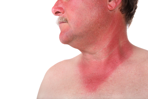
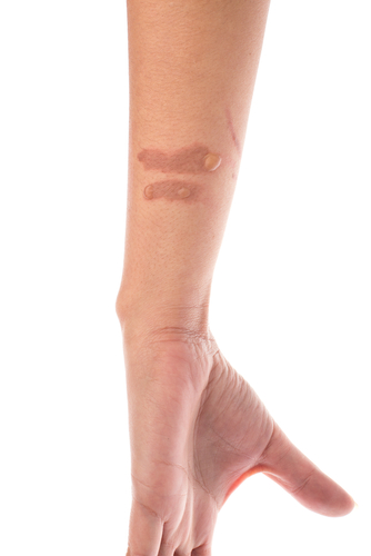
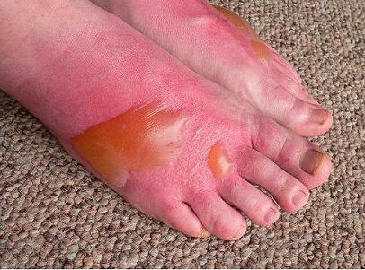
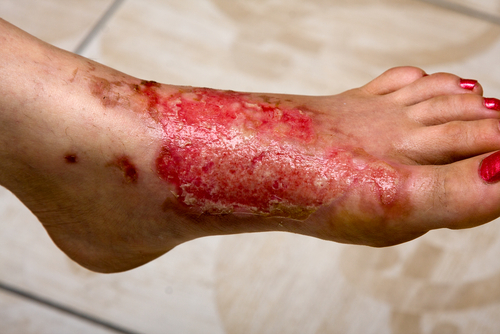
Patients frequently come into the medical clinic with burns.  Several possible sources of exposure can lead to skin burns including hot water or steam, hot objects or flames, chemical, electricity or exposure to the sun. Since moderate to severe burns can cause serious complications, it is important to recognize the signs of a serious skin burn so you can be evaluated by a healthcare provider. When to seek help: 1)    If the burn is on or near a joint such as the fingers, feet or hands 2)    If the burn is on the genital area 3)    If the burn goes all the way around (encircles) an arm, leg, foot, finger or your chest 4)    If the burn is larger than 3 inches or deep 5)    If the patient is younger than 5 years old or older than 70 years old 6)    If there is increasing pain, swelling, redness or pus-like discharge or fever > 100.4 degrees F. Burn classification:  The classification system that we used in the past was based on the thickness of the burn.  The new classification is as follows: 1)  Superficial burn or 1st degree involves only the top layer of skin.  They heal within 3-6 days.shutterstock_56132251 2)  Partial-thickness  or second degree burns involve the two top layers of skin.  These burns are painful with air movement or temperature changes and form blisters.  Partial thickness burns turn white when pressed.  These burns usually health within 7-21 days.  The skin that was burned may become darker or lighter color permanently in some patients. shutterstock_127655633 3)  Deep partial-thickness burns were previously called 3rd degree burns and are painful with deep pressure.  They form blisters and do not turn white with pressure.  These burns take longer than 21 days to heal and usually develop a scar that may be severe.  If the skin blisters immediately, it is at least a deep partial-thickness burn.  bb2_2 4)  Full Thickness Burns:  Extend through all layers of the skin and destroy it completely.  The burned area usually does not hurt and is waxy white to leathery gray in color and the skin is usually dry.  Full-thickness burns do not heal without surgery.  These burns used to be called 4th degree burns.  shutterstock_101309467 Treatment:  Most of the time small superficial or partial-thickness burns can be treated at home, but deeper wounds should be evaluated by a medical provider.  Treatment in the home: 1)   Clean the wound:  If clothing is stuck to the wound, seek care by a medical provider.  Gently wash the burned skin with soap and water.  Do not use alcohol, iodine or other cleansers. 2)   Cool the wound:  After cleaning, you may apply a cold compress or soak the skin in cool water for comfort and reduce pain.  Do not put ice directly on the wound itself however. Infection prevention:  Apply a small amount of antibiotic ointment such as bacitracin or silvadine cream may be applied.  A minor burn may be covered with a bandage or dressing.  If there are blisters, these should be covered with a clean bandage or dressing.  Do not pop the blisters with your fingernail as this may increase the risk of infection.  Leave the blisters alone or gentle pop them on the side of the blister sterile needle if they are large.  Use a non-stick dressing such as Vaseline impregnated gauze or Telfa dressing to cover the wound. If your tetanus has not been updated within 5 years, you need a tetanus booster if the burn is superficial partial-thickness or deeper. Burn Prevention: 1)   Keep matches, lit candles and lighters out of the reach of children 2)   Do not smoke when sleepy 3)   Use a cool-mist humidifier instead of a warm mist humidifier 4)   Apply sunscreen liberally to prevent sunburn and stay out of the sun when possible 5)   Be careful when placing a child in a car seat or stroller because the metal or vinyl can become hot 6)   Set your hot water thermostat to lower than 120 degrees F 7)   Keep children away from fireplaces, hot stoves and ovens 8)   Children’s sleepwear should be non-flammable.   I hope that you have found this information useful.  Wishing you the best of health,
A burn is one of the problems where a camera only tells you so much, and I have written about which problems a video visit can handle and which ones need to be seen in person. The rest of my work these days is obesity medicine, where the argument right now is whether Ozempic and Zepbound cause muscle loss.

Updated for 2026: The Cooling Advice Has Gotten More Specific

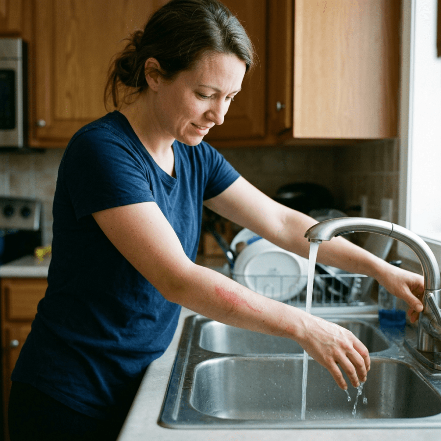
When I wrote this in 2012 the standard advice was to run cool water over a burn. That was right, but it was vague, and vague advice gets applied badly. Here is the current version. Cool running tap water, somewhere between 46 and 77 degrees Fahrenheit, for at least twenty minutes, started within the first half hour. There is evidence the window stays useful out to about three hours, so if an hour has already passed, still do it (1). Not ice. Ice constricts the blood supply to skin that is already injured and makes the wound deeper. I still hear ice recommended and it is worth saying plainly: skip it. Plain tap water is fine. It does not raise the infection risk compared with sterile solutions, and the twenty minutes of cooling matters far more than what the water came out of (1).

Silver Sulfadiazine Has Fallen Out of Favor

This is the part of the old post I would write differently today. For decades silver sulfadiazine cream was the default. It is no longer where the evidence points. There is reasonable concern that it impairs the cells doing the healing, slowing things down and possibly worsening scarring, and the trial support for it was never as strong as its popularity suggested (1). What has replaced it is less glamorous and works better. Either a topical antimicrobial under simple gauze changed every twelve to twenty four hours, or one of the modern occlusive dressings, silver foam, hydrocolloid, or silicone, left alone for as long as two weeks. The second approach hurts less and gets opened less often, which is why I favor it, particularly in children (1). Antibiotic pills are not part of routine burn care. An uncomplicated burn does not need them, and giving them anyway does not prevent infection. One thing that has not changed: tetanus. A partial or full thickness burn in someone whose shots are incomplete or unknown needs Td or Tdap, and immune globulin if the primary series was never finished (1).

What I Can and Cannot Do With a Burn Over Video

Burns are one of the harder things to assess on a screen, and I would rather tell you that than pretend otherwise. Depth is the problem. On camera I can usually tell superficial from something deeper, but the line between a deep partial thickness burn and a full thickness one is a judgment I would rather make with the wound in front of me. Good lighting and a close, steady photo help more than people expect. So does telling me exactly what caused it, how hot, and how long the contact lasted. What I can do well from here is talk you through the first twenty minutes while it is happening, sort out dressings and pain control, check your tetanus status, and tell you honestly when this is beyond what a camera can settle.

Burns That Need a Burn Center, Not a Video Visit

The American Burn Association keeps a referral list and it is worth knowing (2). Any full thickness burn. Partial thickness burns covering ten percent or more of the body. Any deep burn on the face, hands, feet, genitals, perineum, or across a major joint. Anything suggesting smoke inhalation, including facial flash burns or singed nasal hair. Chemical burns. All electrical burns, and high voltage or lightning injuries immediately. Pain you cannot get on top of. And essentially any significant burn in a child.

The Bottom Line

Twenty minutes of cool running water, measured, not guessed. No ice. Skip the silver sulfadiazine your medicine cabinet may still be holding. And know which burns are past the point where any of this applies.

Sources

1. Rice PL, Orgill DP. Outpatient Burn Care: Prevention and Treatment. American Family Physician. April 15, 2020. https://www.aafp.org/afp/2020/0415/p463 2. American Burn Association. Guidelines for Burn Patient Referral. https://ameriburn.org/burn-care-team/resources/guidelines-for-burn-patient-referral

Related Reading

When to Use Telemedicine vs. Urgent Care: How I Spot the Sick One Abscesses and MRSA: What To Do About a Skin Infection Scott Rennie, D.O.

Board Certified in Obesity Medicine and Family Medicine

This blog is for educational purposes only and does not constitute individual medical advice. Always consult your own physician before making changes to your health, medications, or treatment plan.
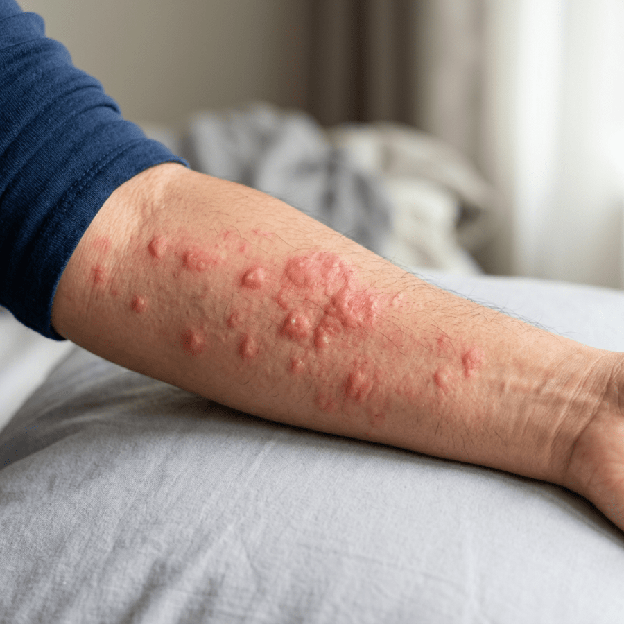
Forearm with multiple raised red bumps and irritated skin

Hives – What am I allergic to?

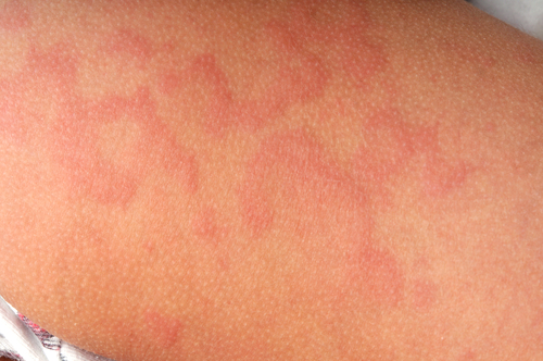
shutterstock_46021174The medical term for hives is urticaria and they are described as raised areas of skin that itch intensely and they are red and often have a pale color to the center.  They are common and occur in at least 20% of people at some point in their lives. Why hives develop:  This skin rash occurs as the body’s immune system is exposed to an allergen (such as a food or chemical) that it inappropriately believes could be harmful.    The body then responds by activating it’s army of immune cells in the skin to fight off the allergen.  When these immune cells (called mast cells) are activated, they release histamine that is a natural chemical that causes itching, redness and swelling of the skin.   In most cases, hives appear and can reappear suddenly – within several hours. Skin appearance:  Hives can occur anywhere on the body and actually appear and disappear and give the appearance that the rash is actually moving around the body.  They are raised areas of skin that itch and these red areas may enlarge and merge together.  The itching is usually the most bothersome symptom and it can be severe enough to interfere with work or sleep. Angioedema:  Some people who get hives also have puffiness of the face/lips, eyelids, mouth, hands, feet and genitalia.  Swelling usually affects one side of the body more than the other and may give the sensation of fullness or discomfort in the area of swelling. Anaphylaxis:  Hives can occur as part of a serious allergic reaction called Anaphylactic shock.  You should see a medical provider right away if you have symptoms along with hives such as: 1)   Trouble breathing 2)   Tightness in the throat 3)   Nausea/vomiting 4)   Cramping or abdominal pain 5)   Passing out 6)   Dizziness or light-headedness Types of hives:  Hives are usually classified based on how long they’ve been present.  Acute (brief) hives, vs. chronic (long-standing) or physical (triggered by a physical stimulation such as sun exposure or cold). Acute hives:  These are the type that we see most often in the medical clinic.  They usually don’t last beyond a few days to a week or two.  Triggers that can cause acute hives include: 1)   Infections:  Viral infections actually have been found to cause more than 80% of the cases of hives in children.  The hives usually last a week or two. 2)   Medications:  Many different kinds of drugs can cause hives including nonsterioidal anti-inflammatory drugs (NSAIDs) such as aspirin, ibuprofen or Aleve.  Antibiotics can also cause hives, as well as narcotic pain medication such as morphine or codeine. 3)   Insect stings:  Stings from bees, wasps, hornets or fire ants can cause hives around the sting.  If you get hives all over your body after a sting, this could be sign of anaphylaxis, and you should be seen by a medical provider as soon as possible. 4)   Food allergies:  food-associated hives usually occur within 30 minutes to an hour after eating the food.  Common foods which can cause hives include milk, eggs, peanuts or other nuts, soy, wheat, fish or shellfish. 5)   Physical contact:  After you touch a certain substance that you are allergic to, you may get hives.  Common substances that can cause hives are plants, raw fruits and vegetables, latex (found in balloons, certain gloves, condoms, etc.) Chronic hives:  Many people have hives that occur daily or almost every day.  We call hives that last longer than 6 weeks chronic hives.  Sometimes they can even last for years.  Hives are frustrating and can effect how you look but it is important to remember that hives are not contagious, they are rarely permanent, they are not life-threatening and symptoms are treatable in most people. For most people who get chronic hives, the cause is unknown.  Chronic hives can be a sign of other medical problems including thyroid or liver problems, chronic infection or lupus.  People with these problems usually have symptoms other than just hives however. Physical hives:  Hives can be triggered by exposure to cold (they often appear as the skin warms up), changes in body temperature or sweating, vibration, pressure, exercise, sunlight or water.  Some people can develop reddened, raised lines if the skin is stroked firmly or scratched.  This is called dermatographism. Diagnosis:  Most people do not need testing. The diagnosis can be made by the medical provider by taking a history and performing a physical examination.  Sometimes testing is done if symptoms are not resolving within six weeks.  Skin testing may be ordered by an allergist to test for food and medication allergies.  Blood tests can also be performed to look for other underlying medical problems.  A skin biopsy is a small sample of skin that is removed and examined under the microscope.  The biopsy can sometimes be helpful for patients who have chronic hives and it is usually performed by a dermatologist (skin doctor). Treatment: 1)  Avoid the cause/trigger.  Many times the cause in not found and the hives usually disappear over days or weeks anyway. 2)  Antihistamines – there are two types of antihistamines.  The older type such as Benadryl often work well, but can cause drowsiness, and are usually taken multiple times per day.  The newer types of antihistamines such as Zyrtec, Claritin or Allegra have fewer side effects are usually taken only once or twice a day. 3)  Oral steroids such as prednisone at a high dose are often used in combination with antihistamines.  We are careful with the use of steroid medications because long-term use can cause serious side effects if taken of long periods of time (months to years). 4)  Topical steroids or creams:  Sometimes topical creams may help with the itching, however we usually don’t use topical steroid creams on the hives because the rash may move from one area of the body to another rapidly.   I hope that you have found this information useful.  Wishing you the best of health,

Hives were everyday family medicine for me. These days I write more about metabolic and menopause care, including the FDA pulling the black box warning off menopause hormone therapy.

Scott Rennie, D.O.
Board Certified in Obesity Medicine and Family Medicine

This blog is for educational purposes only and does not constitute individual medical advice. Always consult your own physician before making changes to your health, medications, or treatment plan.
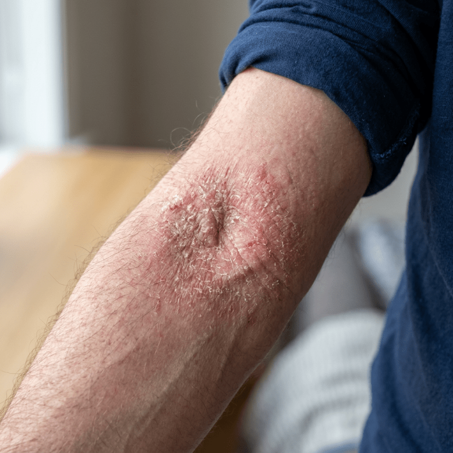
Dry, red, cracked skin surrounding a person's elbow

Dry, Itchy Skin: Could It Be Eczema or Dermatitis?

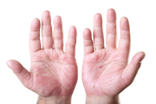
shutterstock_106764923Eczema (atopic dermatitis) is a problem that is characterized by dry, itchy, scaly and often red skin.  There seems to be a genetic predisposition for eczema in some people.  People who have eczema seem to have more allergies than those who do not. Causes:  The epidermis or outside layer of skin seems to get irritated by environmental irritants.  Patients who have this can be extremely sensitive to water – and if they repeatedly put their hands in water, it may be more common.  Other times, there are certain skin products that seem to bring it on such as makeup, and petroleum products are a common cause in those who work in the automotive industry.  For healthcare workers, latex gloves can cause severe irritation to the skin.  In children, it has been linked to food allergies. Symptoms:  Most individuals have their first symptom of eczema before the age of 5.  The symptoms are often characterized by itching, small bumps, skin flaking and patches of redness.  Scratching the skin can cause worsening symptoms.  It is more commonly found in certain areas of the body, which help in the diagnosis. Most common areas of the body affected: 1)   Hands/fingers of healthcare workers 2)   In infants – front of arms, legs, cheeks or scalp may be red/scaly. 3)   Back of the neck, elbow creases and backs of the knees as well as the face, wrists and forearms may be affected. Diagnosis:  There is no specific tests to diagnose eczema. The diagnosis is made by taking a medical history and performing a physical examination. Cure:  There is no cure for eczema – it is controlled/treated. Treatment:  It typically improves and then flares (worsens) periodically.  Eczema is not curable, but it is controllable with proper self-care and medications such as: 1)   Keeping skin hydrated with skin emollients (creams and ointments that moisturize the skin and prevent it from drying out).  The best emollients for people with eczema tend to be thick creams or ointments such as (Eucerin or Vaseline petroleum jelly).  They are most effective when applied immediately after bathing.  Lotions contain more water than creams and are less effective. 2)   Avoid hot baths (greater than 10-15 minutes) because they actually can dry the skin out 3)   Topical steroids – prescription strength steroid creams or ointments may be recommended and are usually applied twice a day. 4)   Protopic and Elidel are often effective for eczema but don’t work as quickly as topical steroids.  The are useful in sensitive areas such as the face or groin and can be used in kids under age 2. 5)   Oral Steroids are very rarely used to treat a severe eczema flare.  There are potential side effects, therefore it is not the most common treatment. 6)   Ultraviolet light therapy (phototherapy) can control eczema, but is expensive and may increase the patient’s risk for skin cancer so is usually only used if other therapies are not successful. 7)   Oral antihistamines can be useful for itching, but do not usually help with the eczema. 8)   Wet dressings may also help soothe the skin, and reduce itching and redness but do not treat the eczema, but rather help with symptoms. Factors that make eczema worse:  Reducing or eliminating some of these factors may help: 1)   Heat, perspiration or chronic exposure to water (such as handwashing) 2)   Emotional stress/anxiety 3)   Rapid temperature changes 4)   Exposure to certain irritating chemicals/soaps/detergents/perfumes.   I hope that you have found this information useful.  Wishing you the best of health,

Scott Rennie, D.O.
Board Certified in Obesity Medicine and Family Medicine

This blog is for educational purposes only and does not constitute individual medical advice. Always consult your own physician before making changes to your health, medications, or treatment plan.
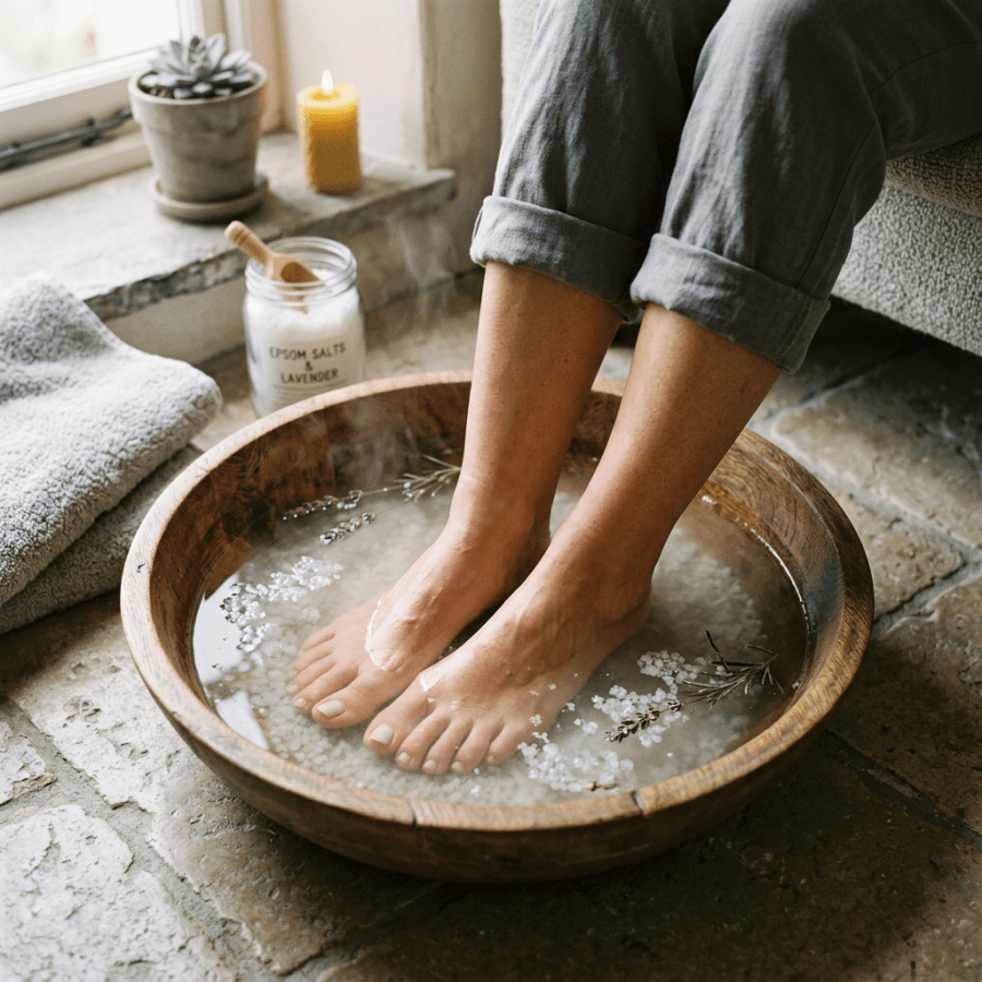
Feet soaking in wooden bowl with steaming water, lavender, and Epsom salts

Ingrown toenails and paronychia

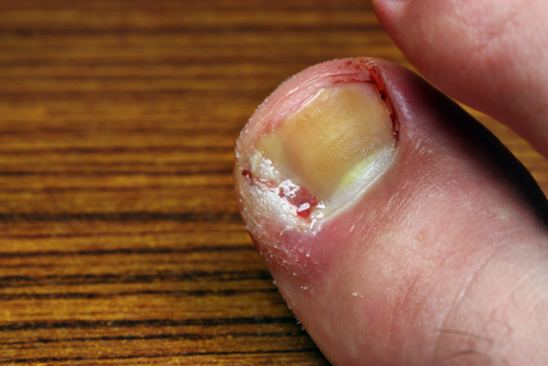
shutterstock_149255009I see patients who come into the clinic for ingrown nails, most often toenails but even ingrown fingernails can bring people to the doctor. Paronychia: an inflammation involving the lateral and proximal fingernail folds.  It can be acute (rapid onset) or chronic (long-standing). Causes:  occupations where the hands are in the water frequently, nail biting, thumb sucking, overzealous manicuring and even diabetes can lead to fingernail infections. Ingrown toenail:  The big toe is most commonly affected.  Signs and symptoms can include pain, swelling, drainage and granulation tissue.  It’s more common to get ingrown toenails from poor fitting shoes, toe trauma, excessive trimming of the lateral nail plate, or in patients who have a over-curvature of the nail which can be hereditary or acquired. Acute nail infections are usually accompanied by redness, and pain at the sides of the nail, sometimes with a blister that can be filled with purulent material If the patient hasn’t been biting their nails, the infection is most often caused by Staphylococcus aureus or Steptococcus pyogenes or a combination of both.  Different bacteria are often present if the patient is a nail biter. Chronic nail infections are often associated with eczema, however it can also be complicated by a Candida or yeast infection of the nails. Diagnosis:  In the clinic we can usually tell that there is a nail infection by the appearance of the nail folds on examination.  If a purulent fluid collection is present, the diagnosis is even easier to make. Treatment:  If the infection has come on rapidly (acute onset), the treatment usually involves warm compresses or soaks to the affected finger or toe for 20 minutes three times/day.  Antibiotics that are taken orally may be helpful in severe causes.  Topical antibiotics such as triple antibiotic ointment might be helpful after soaking the digit, but there isn’t a lot of research to support it. If there is an abscess present, incision and drainage is usually helpful in addition to the above treatments.  We usually perform a digital block to numb the finger or toe for comfort before the surgery is performed.  Treatment with antibiotics is common and we usually use them for 5 days after a surgical drainage and for 7-10 days if there is no drainage.  The choice of the antibiotic depend on whether the patient has been biting the nails, and the patient’s history of drug allergies.  Possible treatment options include dicloxacillin, Keflex (no no MRSA in suspected) or Bactrim.  We may also add metronidazole or clindamycin to the regimen if the patient has been biting their nails. For ingrown toenails that are mild, we may place a cotton wedge or dental floss underneath the lateral nail plate to relieve the pressure and doing the soaks in warm soapy water for 10-20 minutes 3x/day.  If the ingrown nail is severe however, we usually perform a partial nail removal after doing a digital block to relieve the discomfort.  Antibiotics may be prescribed after partial nail removal may be helpful, however evidence has not shown that antibiotics decrease the healing time. Follow-up after 3-4 days to assess treatment. Recurrent ingrown toenails:  May require treatment with permanent nail ablation surgery using both surgical excision plus phenol ablation (chemical matricectomy).  The keeps the toenail from returning. Post-op care after toenail removal:  You should be able to put weight on both feet immediately after surgery, but walking will be uncomfortable.  Some mild bruising and bleeding is normal after foot surgery. We usually apply some antibiotic ointment and 4×4” gauze, tube gauze and paper tape.  After 24-48 hours, soak the affected toe in warm, soapy water and reapply antibiotic ointment and a clean bandage.  This should be done 3-4x/day for 1-2 weeks after the procedure.   Keep your foot and leg elevated while sitting or lying down and make sure your bandages are clean and dry at all times.  We ask patients not to wear shoes for 3 days and recommend antibiotics and anti-inflammatory medications for 10 days.   I hope that you have found this information useful.  Wishing you the best of health,

For most people an ingrown toenail is a nuisance. With diabetes or neuropathy it is a different problem, because an infected nail edge on a foot you cannot feel well is how some ulcers start, which is why I wrote about protecting your feet if you have diabetes.

Updated for 2026: Mostly Confirmed, With One Number Worth Having

I went looking for what had changed here since 2012 and the honest answer is: not much. No major society has rewritten this in the last few years. The approach I described holds up. So rather than manufacture a revolution, here is what has been sharpened. For an ingrown toenail bad enough to need a procedure, the thing that matters is whether the nail matrix gets treated, not just the nail edge removed. Partial nail avulsion combined with chemical destruction of the matrix using phenol gets cure rates above 95 percent. Avulsion alone does not. A 2025 trial of 140 patients put recurrence at 1.43 percent with phenol against 10 percent without (1). So if you are offered a procedure for a recurring ingrown nail, the question worth asking out loud is whether the matrix is being treated. That single detail is the difference between fixing it and doing it again next year.

Chronic Paronychia Is Not an Infection

This is the part I most want to correct, because it is treated wrongly all the time. Acute paronychia, the hot painful swelling beside a nail that comes on over a day or two, is an infection. Warm soaks, and if there is a pocket of pus it needs draining. Once it is drained, oral antibiotics usually are not necessary. They get reserved for spreading cellulitis, a suppressed immune system, or someone who is genuinely unwell (2). Chronic paronychia, meaning six weeks or longer, is a different animal. It is an irritant dermatitis, driven by repeated wet work and exposure, and treating it with antifungals or antibiotics does not work because it is not an infection. The treatment is getting the hands out of the water, barrier protection, and a topical steroid or calcineurin inhibitor. It takes weeks to months to settle, and people abandon treatment long before that (2). If you have had a swollen nail fold for two months and have been through three courses of antibiotics, that is the pattern.

What This Looks Like Over Video

A photograph does most of the work here, and this is one where I want more than one angle. From directly above and from the side, so I can see whether the nail fold is simply inflamed or whether there is a pocket of pus lifting it. What I need from you beyond that is duration, which is the single most useful question, along with what your hands or feet are exposed to during the day, whether you are diabetic, and what has already been tried. I can settle the chronic cases from here, and those are the ones most often mismanaged. What I cannot do is drain anything or take a nail off, so if there is pus, or if the nail needs a procedure, my job becomes telling you that clearly and getting you to someone who can do it.

When To Be Seen, Sometimes Urgently

Spreading redness, red streaking, or fever. Any toe infection at all if you have diabetes or poor circulation, where the threshold to be seen should be much lower than you think. Severe pain in the finger pulp, which can mean a felon. And pain on straightening the finger with a sausage shaped swelling, which suggests flexor tenosynovitis and is a same day surgical problem, not something to sleep on.

The Bottom Line

For a nail that keeps growing in, ask whether the matrix is being treated. For a nail fold that has been swollen for months, stop taking antibiotics for it and start treating the skin.

Sources

1. Comparison of Partial Nail Avulsion With or Without Phenolization in the Management of Ingrown Toenails. Cureus. May 2025. https://pmc.ncbi.nlm.nih.gov/articles/PMC12147675/ 2. Acute and Chronic Paronychia. American Family Physician. 2017;96(1):44-51. https://www.aafp.org/pubs/afp/issues/2017/0701/p44.html 3. Ingrown Toenail Management. American Family Physician. 2019;100(3):158-164. https://www.aafp.org/pubs/afp/issues/2019/0801/p158.html

Related Reading

Abscesses and MRSA: What To Do About a Skin Infection When to Use Telemedicine vs. Urgent Care: How I Spot the Sick One Scott Rennie, D.O.

Board Certified in Obesity Medicine and Family Medicine

This blog is for educational purposes only and does not constitute individual medical advice. Always consult your own physician before making changes to your health, medications, or treatment plan.
Red first aid kit with assorted bandages, scissors, gloves, thermometer, antiseptic, and medication arranged on white surface

Basic Wound Care Tips for Non-Medical Professionals

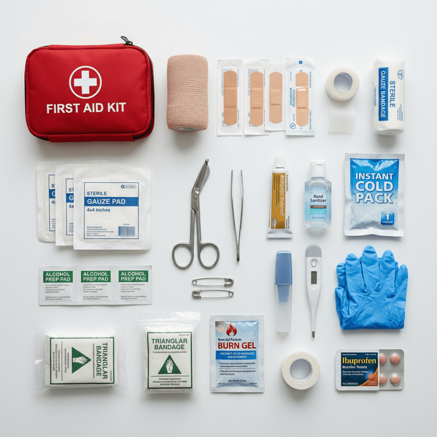
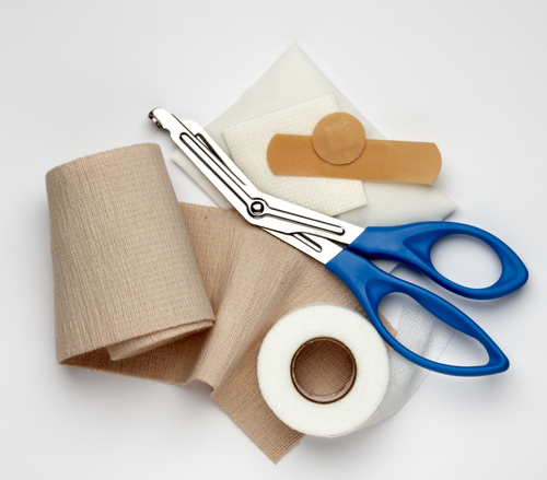
shutterstock_43127152I find that many patients who come into the clinic with a wound have questions about how to take care of it.  The wound might be a post-surgical incision or even a minor cut.  Hopefully this will help with some of those questions. Do I need stitches?:  The best way to know for sure is to be examined by a medical provider, but some basic things we look at: 1)   Depth of the skin wound – if it does not go all the way through the skin, it usually doesn’t need stiches 2)   The size of the wound, whether it’s wide, or jagged.  If it’s large, wide or jagged – there is a higher likelihood of needing stiches 3)   Where on the body the wound is – in areas of the body with lots of stress on the wound, the stitches help hold the skin together better than tape, or a butterfly bandage. 4)   How long ago the injury occurred:  If you got cut 5 days ago, there is less chance sutures would be appropriate because wound healing has already begun. 5)   If it was an animal bite or dirty wound:  We will sometimes put stitches in a wound that was created by an animal bite, but these are generally dirty wounds and we are very careful not to bring the skin together too tight so that the wound can drain. What to do at home – basic guidelines: 1)   Clean the cut or scrape with soap and water.  If there is a piece of glass, or foreign object in the wound that will not come out, you will need to see a medical provider 2)   Stop the bleeding with direct pressure on the wound.  Press a clean cloth or bandage on the wound for 20 minutes minimum without releasing.  If the bleeding is not stopping, contact a medical provider. 3)   Put a thin layer of antibiotic ointment on the wound edges – not in the wound.  I recommend bacitracin (mupirocin) and not Neosporin. 4)   Cover the wound with bandage/gauze to keep the wound clean and dry.  Change the bandage 1-2 times every day until the wound heals 5)   Examine the wound for signs of infection – see below.  Healing time for most wounds is 7-10 days but this can vary. When to see a medical provider: 1)   The wound is deep and/or you can see fatty tissue, muscle, bone or dirty/debris within the wound 2)   You are having severe pain 3)   There are signs of infection (usually begin on about 2-4 days after the wound injury) such as:  Fever, redness, swelling, increased warmth around the wound, pus draining from the wound or red streaks on the skin around the wound. 4)   The wound is from a bite 5)   The wound is a puncture wound caused when a sharp object goes through the skin into the tissue underneath.  There is a higher chance of infection with these kinds of wounds. Do I need a tetanus shot?:  You may need a tetanus booster depending on when your last one was.  If you have deep wound, it is best to contact a medical provider if you’re unsure of whether you need a tetanus vaccine. Abscess:  An abscess is an infection under the skin where bacteria (pus) collect.  A surgical procedure called an I&D (incision and drainage) can be done to drain the pus and heal the infection. If you’ve had an incision and drainage done at your doctor’s office:  After having a surgical procedure done at your doctors office, the wound is often packed with sterile gauze to allow the wound to heal from the bottom upwards.  This way, the wound doesn’t close up at the top and leave a space underneath the top of the skin where bacteria can start growing and cause an infection.  This type of wound needs special attention and observation each day. Basics of wound care after an I&D (incision and drainage): 1)   Keep the bandage/wound clean and dry. 2)   Only remove the bandage to clean around the wound and follow the advice of your medical provider on how to do this. 3)   The packing gauze should be removed little by little until the wound heals completely.  The decision on how much to remove (if any) should be made by a medical provider but this can be explained to the patient on an individual basis (ie. Wounds heal differently depending on the situation). 4)   You may need to wear a splint to decrease movement to the area and allow wound healing. 5)   Antibiotics and pain medication may be prescribed.   I hope that you have found this information useful.  Wishing you the best of health,

Scott Rennie, D.O.
Board Certified in Obesity Medicine and Family Medicine

This blog is for educational purposes only and does not constitute individual medical advice. Always consult your own physician before making changes to your health, medications, or treatment plan.