
Woman in a beanie holding a steaming mug by a fireplace

Hypothermia: Staying Safe When It’s Cold Out There

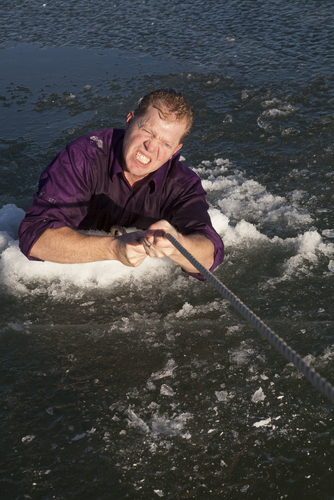
shutterstock_171146414I credit the information that I’ve learned and posted here to Eric Weiss, MD who is an international expert in Wilderness Medicine and Hypothermia. Patients with accidental hypothermia come to medical providers year-round and in all climates.  Hypothermia is defined as a decrease in core body temperature below 95 degrees F (35 degrees C). For purposes of emergency management and resuscitation, hypothermia can best be characterized as either mild or severe.  In mild hypothermia (core temperature 87.8-95 degrees F), the victim is conscious, still shivering and generally not prone to developing abnormal heart rhythms.  In severe hypothermia (core temperature below 87.8 degrees F), the patient has altered level of consciousness, diminished or absent shivering and is prone to abnormal heart rhythms. Mild hypothermic victims can still generate heat through shivering so they generally do well without intensive rewarming.  In severe hypothermic patients, active rewarming techniques such as extracorporeal blood rewarming, inhalation therapy, peritoneal lavage, thoracic cavity lavage or thoracotomy with mediastinal irrigation may be needed. Pre-hospital Treatment of Mild Hypothermia:  Preventing further heat loss and facilitate rewarming are the goals.  The rescuer should remove all wet clothing and replace it with dry clothing, insulate the patient with sleeping bags, blankets, extra clothing or other suitable material.  Use insulation underneath the patient as well as on top.  Encourage drinking of warm fluids and sugary drinks if they can swallow without aspirating (inhaling the drink into their lungs).  It is not uncommon to observe a continued decline in core temperature after a hypothermic patient is removed from the cold environment and external warming is initiated.  This phenomenon is called core temperature afterdrop. Pre-hospital Treatment of Severe Hypothermia:  Careful handling is necessary because these patients are prone to develop abnormal heart rhythms through rough handling.  If in the backcountry, consider helicopter transport to prevent jostling that might occur with an overland evacuation.  Keep them horizontal when possible to minimize orthostatic hypotension.  Provide oxygen if you have it.  Administer a minimum of 500ml of heated (98.6-105.8 degrees F) IV normal saline or D5NS.  Lactated ringers should be avoided because when the liver is cold, it poorly metabolizes lactate.  Consider intraossious (I/O) infusion for alternative pathway for fluid replacement for a dehydrated patient who you cannot get IV access. Hot water bottles or heat packs can be placed in the axillae and groin area and along the neck where large blood vessels course near the surface.  Hot water bottles should be wrapped with insulation to prevent thermal burns. In a severely hypothermic patient, they may feel/look clinically dead. Breathing may be difficult to detect if the breathing rate is significantly depressed. The rescuer should listen to the chest and palpate over carotid or femoral arteries for at least 1 minute to detect a pulse.   If the patient has any sign of life, chest compressions should not be initiated as they may precipitate ventricular fibrillation (abnormal heart rhythm). At a core temperature of 20 degrees C, cardiac arrest is tolerated for up to 30 minutes without clinically significant neurologic or neuropsychological deficits.  This knowledge and the fact that a dead victim may be clinically indistinguishable from one that is severely hypothermic and alive has lead to the adage that “No one should be pronounced dead, until they are warm and dead.” A serum potassium greater than 10mmol/L in a non-hemolyzed specimen however has been proposed as a reasonable ceiling for viability. Negative Pressure Rewarming (Thermarescue):  A non-invasive way of creating a direct thermal pipeline between the skin and body core.  The patients forearm is fitted through an acrylic sleeve with an air tight seal around the arm.  Vacuum pressure of -40 mm Hg is established and the thermal load is applied via a chemical heating pad. If you are interested in learning more about wilderness medicine, a great resource for information is the wilderness medicine society:  http://www.wms.org/   I hope that you have found this information useful.  Wishing you the best of health,

Scott Rennie, D.O.
Board Certified in Obesity Medicine and Family Medicine

This blog is for educational purposes only and does not constitute individual medical advice. Always consult your own physician before making changes to your health, medications, or treatment plan.
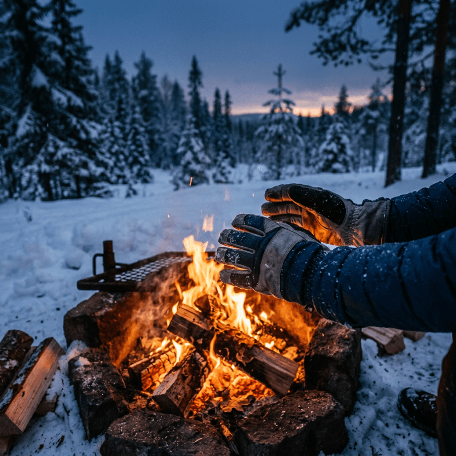
Gloved hands warming over a campfire in a snowy forest

Frostbite and cold induced injuries

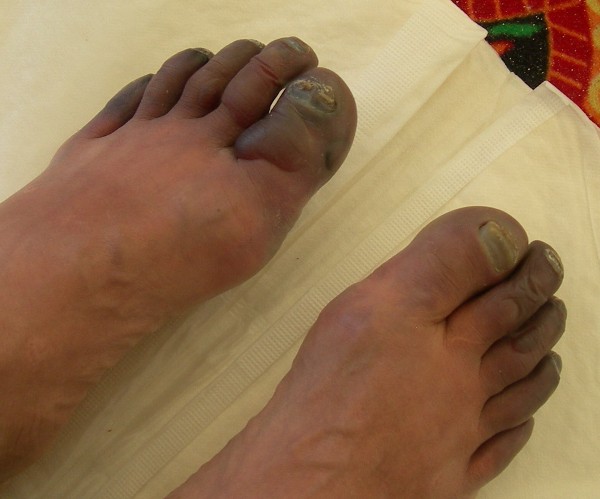
Photo credit:  http://www.everester.org/BlogViewer.aspx?Id=DCD57FDDA64B2206   Much of the information presented here comes from Peter Hackett, MD who is an expert in Wilderness Medicine and especially mountain and high altitude medical illness and care. Frostbite:  A severe, localized cold-induced injury.  Tissue destruction of frostbite is due to both immediate cold-induced cell death and more gradual development of localized inflammatory process and tissue ischemia.  Following exposure to subfreezing temperatures, ice crystals form outside the tissue cells.  If freezing is rapid, ice crystals may also form inside cells.  The initial cellular damage and subsequent inflammatory process are made worse in the setting of thawing followed by refreezing of the area. Areas most often affected:  Ears, nose, cheeks, chin, fingers and toes Causes:  Anything that increases localized heat loss or decreases heat production.  Exposure to wind, or conductive heat loss due to contact with metal or water 1)   Inadequate insulation 2)   Circulatory compromise 3)   Dehydration 4)   Moisture Behavioral risk factors: 1)   Mental illness 2)   Alcohol and drugs 3)   Fear, apathy, panic Vascular Stage- Post thaw:  Recovery of circulation and then thrombosis, ischemia, necrosis and even gangrene can result.  Intracellular ice and tissue death occurs if there is refreezing. Classification of frostbite injuries: 1)   Mild or Superficial (no tissue loss) 2)   Severe or Deep (with tissue loss) 3)   Historical classification 1st to 4th degree had no clinical usefulness Treatment (Pre-Hospital): 1)   Supportive care for trauma, and dehydration (splint/pad affected area) 2)   Avoid additional heat loss – remove wet clothing 3)   If frozen and rescue is near keep frozen unless you can do a warm water thaw and there is no danger of refreezing. 4)   Do not rub frost bitten areas as this can cause further tissue damage 5)   If already thawed, avoid refreezing 6)   Avoid using stoves or fires to rewarm frostbitten tissue Treatment (Hospital): 1)   Hydration 2)   Wound care – early debridement or amputation of dead or dying areas while preserving viable tissue 3)   Pharmacology – Dextran, NSAIDs, Nifedipine 4)   Imaging studies (X-rays may show coincidental trauma related fractures or cold-induced soft tissue swelling), Technetium (Tc)-99 scintigraphy is commonly used to predict long-term tissue viability. 5)   Sympathetic block or surgery if needed Methods of Thawing: 1)   Rapid rewarming in warm water (37-41 degrees C) is optimum 2)   Gradual thawing – often unavoidable 3)   Harmful methods – delayed thawing with ice or snow or excessive heat Non-freezing Cold Injury:  Frostnip, Chilblain/Pernio, Trenchfoot, Raynaud’s Frostnip:  Cold-induced, localized parasthesias that resolve with warming and there is no permanent tissue damage. Pernio/chilblain:  Localized inflammatory lesions that can result from acute or repetitive exposure to damp coldness above the freezing point.  Lesions are swollen, often reddish or purple and may be painful or itchy.  Pernio is most common in young women but both sexes and all age ranges may be affected. Trenchfoot (immersion foot):  Injury to the sympathetic nerves and small blood vessels of the feet.  First described in 1914 during WWI during trench warfare.  It results from prolonged exposure of the feet to a combination of dampness and cold.  Tight-fitting boots exacerbate the condition.  Feet, and sometimes hands are red, swollen and can be extremely painful and often are covered with bloody blisters.  Tissue loss can occur. Prevention: 1)   Pay attention to weather forecasts (predicted high and low temperatures, forecasted precipitation and wind chill index) 2)   Dress appropriately for the weather 3)   Have an emergency plan when going into remote areas 4)   Avoid alcohol consumption and smoking 5)   Avoid exposure to metal surfaces 6)   Maintain adequate calorie intake 7)   There is inadequate evidence to support the use of applying emollients to exposed skin to prevent frostbite and it is not suggested 8)   Carry emergency supplies in the backcountry or in remote areas in case your group becomes stranded 9)   Use a buddy system for monitoring 10)  Perform an equipment and clothing check 11)  Keep hydrated 12)  Carefully wash and dry feet 13)  Do not sleep in wet socks 14)  Avoid tight socks and shoes 15)  Do not add socks for warmth, get a larger shoe 16)  Rewarm gently, do not use a strong heat source 17)  Do not rub the skin, use passive skin-to-skin contact 18)  Elevate the feet above the level of the heart 19)  Consider antiperspirant with aluminum hydroxide for a week before exposure Raynaud’s Vasodilation Training: (Physician Sports Med, March 1990; vol18 no3) 1)   Immerse hands in hot tap water 2)   Stay indoors for 5 minutes, then in the cold for 10 minutes, then back indoors for 5 minutes (one cycle) 3)   Do 3-6 cycles per day every other day 4)   50 cycles established effect, but variable Tips to control moisture and cold: 1)   Wear socks with moisture-wicking capabilities 2)   Change socks and dry them frequently 3)   Use foot powders that control fungus and absorb moisture 4)   Wear shoes with adequate draining capabilities or make holes 5)   Consider waterproof socks, either SealSkinz, Waterproof MVT, Seirus Neo-Sock or Stormsock 6)   When resting or sleeping, take off wet shoes and socks to allow feet to breath and socks to dry against your body in your sleeping bag If you are interested in learning more about wilderness medicine, a great resource for information is the wilderness medicine society:  http://www.wms.org/   I hope that you have found this information useful.  Wishing you the best of health,

Scott Rennie, D.O.
Board Certified in Obesity Medicine and Family Medicine

This blog is for educational purposes only and does not constitute individual medical advice. Always consult your own physician before making changes to your health, medications, or treatment plan.
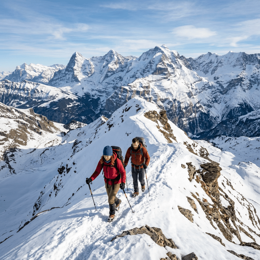
Two hikers climb a snowy mountain ridge with snow-covered peaks behind them

Altitude Illness

shutterstock_163362512Much of this information comes from Peter Hackett, MD, a wilderness medicine expert on mountaineering and altitude related illness and treatment. Altitude illness is usually due to the stress of decreased oxygenation in the setting of an individual who is not acclimatized.  It can happen at any altitude over 8,000 feet and usually occurs during the initial ascent. Two areas of the body that are most affected by altitude illness are the brain and the lungs.  When altitude illness strikes the brain it is divided into two groups called Acute Mountain Sickness (AMS) and High Altitude Cerebral Edema (HACE).  Lung injury is usually due to lung edema called High Altitude Pulmonary Edema (HAPE). Risk factors for altitude illness: 1)   Genetic susceptibility 2)   Live at an altitude of less than 3000 feet 3)   Fast rate of climb/ascent 4)   Past history of high altitude illness (HAI) 5)   Age less than 50 years old (for Acute Mountain Sickness – AMS) 6)   Heavy exertion/exercise 7)   Pre-existing illness (especially for High Altitude Pulmonary Edema – HAPE) Acute Mountain Sickness (AMS): Diagnostic Criteria 1)   Recent gain in altitude 2)   Headache and any of the following
  1. Gastrointestinal upset
  2. Fatigue or weakness
  3. Dizziness or lightheadedness
  4. Difficulty sleeping
3)   Feels like a hangover Treatment of Acute Mountain Sickness:  Usually gets better on it’s own.  Average duration of symptoms is about 16 hours.  It may persist for weeks at higher altitudes however.  There may be progression to High Altitude Cerebral Edema (HACE) with or without High Altitude Pulmonary Edema (HAPE).  It responds well to descent/treatment. 1)   Oxygen therapy 2)   Descent 3)   Hyperbarics 4)   Acetazolamide (Diamox) – 125 to 250mg every 8-12 hours – start taking the day before travel until day 2 or 3 at altitude. 5)   Hyperventilation 6)   Dexamethasone 4mg every 6 hours – careful because this can lead to adrenal failure if used at high doses and if it’s not tapered gradually. 7)   Treat symptoms of headache with ibuprofen/naproxen, codeine, etc. and nausea with Zofran or Phenergan Acetazolamide Prophylaxis:  125-250mg twice a day (5mg/kg/day) starting the day before travel and continued until day 2 or 3 at altitude.  If allergic to sulfonamides (sulfa) be cautious.  Side effects of the medication are dose related.  More commonly a feeling of numbness/tingling, metallic taste in the mouth, generalized fatigue, nausea and blurry vision can occur. Prevention of altitude sickness: 1)   Go up slowly in staging – avoid a sea level to 9,000 foot climb in one day 2)   Sleep at a max of 2000 feet higher elevation each night 3)   Acclimatize to 10-12,000 feet before going any higher High Altitude Pulmonary Edema (HAPE):  Symptoms Early:  Fatigue, weakness, dry cough, shortness of breath with activity.  May progress to increased respiratory rate, increased heart rate. Late:  Pink or blood-tinged sputum from lungs, crackles heard with stethoscope in the right axilla/arm pit. Treatment for HAPE:  Oxygenation is the highest priority.  Descend with minimal exertion.  For mild/moderate cases use bed rest with oxygen.  For severe illness use high flow oxygen with descent and perhaps a hyperbaric bag.  There is some thought about using pulmonary vasodilators such as calcium channel blockers, nitric oxide, Viagra and/or Dexamethasone. Preparation:  Take a medical kit with Diamox and dexamethasone and albuterol inhalers.  If you’re with medical providers, you may have access to nifedipine or Viagra also which may be helpful. For more information: 1)   www.altitudemedicine.org 2)   www.hypoxia.net 3)   Auerbach’s Wilderness Medicine 4)   DuPoint Travel Medicine 5)   Tintinelli Emergency Medicine If you are interested in learning more about wilderness medicine, a great resource for information is the wilderness medicine society:  http://www.wms.org/   I hope that you have found this information useful.  Wishing you the best of health,

Scott Rennie, D.O.
Board Certified in Obesity Medicine and Family Medicine

This blog is for educational purposes only and does not constitute individual medical advice. Always consult your own physician before making changes to your health, medications, or treatment plan.
Open suitcase packed with clothes beside a passport and sunglasses

Essential Foreign Travel Tips: What to Know Before You Go

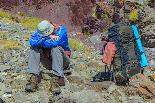
shutterstock_144889000The information listed below is a smattering of information gained from real life experience, Wilderness Medicine Conferences, and is mostly credited to Gene Allred, MD a very experienced physician and world traveler. Two major categories for foreign travelers: A)  The senior traveler (age 65 and above) who have the resources to travel and may have a bucket list.  They have chronic medical conditions and in many foreign countries, access to medical care and medications is more difficult.  Think about access – i.e. there is probably not a wheelchair ramp available in many areas.  Bring your medications.  I don’t know how many times I’ve had patients travel here to Hawaii and not bring enough of their blood pressure medications with them, and I’m sure it happens when the go other places too.  Many seniors may might not get altitude sickness but may have cardiac strain from lower levels of oxygen or increased physical exertion.  Plan accordingly. B)  The adventure/exotic traveler (all ages) who tend to participate in more dangerous activities – mountain climbing, rock climbing, wilderness expeditions like kayaking the Nile river, etc.  These travelers have a higher incidence of trauma from MVA or injury from their activities.  Homicides in some areas may be more of a problem than an MVA.  We don’t think twice about putting on our seatbelt when we get in a car here in the USA, so don’t get in a taxi or bus without a seatbelt (the roads are often worse in other countries and there may be less lighting and more people).  If you do get into an MVA abroad, the medical care is probably not going to be as good as you would get in first world countries. Causes of death in foreign travelers: 1)   Almost 50% are from heart attacks and strokes, the other 50% is from trauma (motor vehicle accidents, drowning, falls and homicide) – also be aware that most counties outside of the US, Canada, Australia, New Zealand, Europe and Japan don’t screen their blood for HIV or Hepatitis C. 2)   Only 1% of death in foreign travel results from Infectious disease Most dangerous aspect for foreign travel:  Motor Vehicle Accidents – account for 25% of fatalities of U.S. travelers.  In Langos, Nigeria buses are called danfos “flying coffins.”  The rate of motor vehicle deaths compared to the USA: 1)   Sri Lanka – 23x more deaths from MVA than USA 2)   Turkey – 44x more deaths from MVA than USA 3)   China has 2% of the worlds driver’s and 15% of the fatalities – WHO 2007 4)   In Ghana 722 drivers were selected at random and 21% had blood alcohol level >80 mg/dl; 4% of the bus drivers had this level.  70% of the trauma patients here are transported by taxi or bus, and 22% and transported by private vehicle.  5% are transported by police and only 3% of trauma victims are transported by ambulance. In the USA, 60% of the motor vehicle crash deaths occur among the drivers.  In the 3rd world countries, 90% occur among passengers, pedestrians and cyclists.  Urban pedestrians account for 50-70% of the deaths due to motor vehicle accident. In contrast, the leading cause of injury/illness in foreign travel is infectious disease.  Up to 75% of travelers become ill from infectious disease, however only 1% of the deaths of international travelers are from infectious disease. Recommendation:  Before you travel, consult your physician, CDC or travel clinic.  Let your medical provider know the type of activities you are planning including whether going to a remote region, participating in a strenuous activity (cycling, mountain climbing, swimming in fresh water, or diving, etc.) Resources: http://www.travelhealthassist.com http://www.cdc.gov/travel http://travel.state.gov/travel/tips/safety/safety_1747.html Everyone needs Hepatitis A immunization when traveling to a high endemic area. Hepatitis A and B vaccinations are a lifetime investment. There are stories about bogus drugs and dirty needles used in certain areas. New Delhi, India (CNN) 2/21/09 – “Authorities were carrying out raids in India’s western Gujarat state for bogus drugs and recycled syringes after a hepatitis B outbreak left 32 people dead, officials said Saturday.”             *  Five medical practitioners were also arrested for violations             *  One arrested for reusing injection syringes             *  111 cases of hepatitis B in the district over two weeks with 32 dead I’m not saying that you should bring your own needles when traveling, but the news report above is frightening. Before your travel, consider the following: 1)  Plan at least three months ahead (buy your tickets, look into what visas you might need, if there a restrictions  and plan what to bring with you). 2)  Look at the CDC website – http://www.cdc.gov/travel and study up on where you are going and what diseases you need to be protected against. 3)  Get excellent travel insurance – there are many companies that offer this – International SOS is one of them https://www.internationalsos.com/en/.   Read the fine print and make sure the company that you go with doesn’t say something like “will evacuate to most appropriate facility” and something more specific like “Will evacuate to USA, Canada, Japan, Australia, New Zealand, or Europe” 4)   Decide what medical resources are available in the foreign country 5)  Plan for diarrheal illness – bring antibiotics (sometimes you might take prophylaxis) and medications to help slow the bowls such as Imodium. 6)   How will I get around while there?  Avoid car crashes – don’t get on a crowded bus, a motorcycle or scooter.  Wear your seatbelt! 7)  Stay sober when out in public – you are more of a helpless target if you’re drunk.  If you drink – do it in the hotel where it’s safer. 8)  Don’t look rich – avoid wearing fancy jewelry/watches or you’ll look like a good target. 9)   Problems of counterfeit drugs – i.e. take own medications 10)   Take a copy of your medical history/prescriptions, driver’s license, passport and visas.  Leave one copy at home with a friend and bring the original and one copy with you. 11)   Consider the season you are traveling in when going to a foreign country i.e. rainy vs. dry.  In malaria endemic regions, this may dramatically influence your risk 12)   Traveling in motor vehicles at night in third world countries substantially increases your risks of injury or death 13)   Acclimatize yourself to the altitude 14)   Bring sunblock 15)   Think about heat exposure 16)  Take drinking water precautions (chlorine dioxide, etc.) 17)  Be wary of swimming in fresh water or going barefooted – consider infections due to Schistosomiasis, Leptosirosis or Cryptosporidium 18)  Think about safety/security – 9% of U.S. travel fatalities are due to homicide.  9000 homicides in Sao Paulo in 1999, compared with 700 in New York City. 19)  Avoid dangerous situations – khakis = N. American, avoid going out alone at night, don’t wear expensive jewelry, carry no more cash than you need for the day and don’t flash roll of bills.  Use a hotel safe when available.  Fanny packs and purses are “one-stop” shopping targets – be aware of “slash and grab.”  Don’t accept food from strangers – “drug and rob scam,” Beware of “spilled food scam” (e.g. mustard), use licensed taxis over unlicensed taxis.  Scan your passport and then email it to yourself so you’ll be able to retrieve a copy. 20)  Protect yourself from insects – Dengue fever, Malaria (1-2 million deaths/year), West Nile Virus, etc.   The higher the concentration of DEET, the longer lasting it is – 99% DEET = 10 hours, 30% DEET is safe in kids down to 2 months of age.  DEET + Permethrin is > 99% effective.  Most malaria is Chloroquine resistant.  Three drugs of choice for malaria are Mefloquine, doxycycline and malarone (primaquine is 2nd line option). 21)  The unexpected usually happens.  Accept the things that are beyond your control.  Resign yourself to the fact that your luggage will be lost, your hotel reservations will be canceled, and your last flight out will leave without you.  They key is to then decide what you are going to do about it.  No matter how bad you think things are now, they can always get worse.  Always have a contingency plan.  Research alternative travel arrangements such as accommodations, research climate, local health risks, etc.  Instead of letting problems accumulate, deal with each challenge “head on.”  Work to “fix” one problem at a time and then move on to the next. If you are interested in learning more about wilderness medicine, a great resource for information is the wilderness medicine society:  http://www.wms.org/   I hope that you have found this information useful.  Wishing you the best of health,

Scott Rennie, D.O.
Board Certified in Obesity Medicine and Family Medicine

This blog is for educational purposes only and does not constitute individual medical advice. Always consult your own physician before making changes to your health, medications, or treatment plan.
Lifeguard seated on an elevated chair overlooking a busy outdoor swimming pool

Drowning and Near Drowning: Water Accidents Explained

shutterstock_106770593I credit the information that I’ve learned and presented here to Karen Van Hoesen, MD who is an expert in Wilderness Medicine and has specialized training in Dive Medicine and water related injuries and illness. Drowning is the 3rd most common cause of accidental deaths in the U.S, and the leading cause of death in children < 5 years old.  Every year drowning accounts for a least 500,000 deaths worldwide and about 4,000 fatalities in the US.  2005 WHO Definition:  “Drowning is the process of experiencing respiratory impairment from submersion/immersion in liquid.” There are several factors that are common to drowning: 1)   Age:  More common in toddlers and teenage boys 2)   Location:  Most frequent in home swimming pools, bathtubs and buckets 3)   Sex:  More common in male than female 4)   Race:  Happens more commonly in ethnic minorities 5)   Drugs:  Being under the influence of alcohol contributes greatly to drowning accidents (more than 50% of adult drowning deaths are believed to be alcohol related). 6)   Trauma:  More common secondary to diving or falls 7)   Drowning is the leading cause of death in Scuba The effects of drowning on the organ systems involve spasm of the larynx, and flooding of the lungs with water causes loss of surfactant (a fluid that helps keep the small air sacs of the lung open).  The decreased oxygenated blood flow leads to decreased cardiac output and abnormal heart rhythms may occur. Decreased oxygenated blood to the brain causes damage to the brain in multiple areas.   Decreased blood flow to the kidneys can lead to kidney failure. Clinical presentation of the drowning victim:  There is a broad spectrum to how they look clinically.  They can be alert or comatose.  They can have signs of decreased blood flow such as blue tone to the skin, or have signs of coughing, increased respiratory rate, increased heart rate and low grade fever.  A chest X-ray may be normal or can show signs of severe damage to the lungs called ARDS (acute respiratory distress syndrome).  Imaging of the brain might show swelling. Early complications (within 4 hours) of Near Drowning: 1)   Spasm of the upper airway 2)   Vomiting with breathing in vomit 3)   Hypothermia 4)   Seizures Late complications (after 4 hours) of Near Drowning: 1)   ARDS – Lung failure 2)   Anoxic-ischemic encephalopathy – brain damage due to low oxygen 3)   Pneumonia due to inhaling water 4)   Lung abscesses 5)   Renal failure 6)   Sepsis – overwhelming infection of the body  Near Drowning Treatment (Before reaching the Hospital):  Training in basic life support is very helpful! 1)   Call for Help! 2)   Mouth-to-Mouth in shallow water or stable surface – do not wait for ambulance 3)   Do not give chest compressions in the water 4)   Do not give Heimlich maneuver 5)   Give oxygen at highest concentration available (if you have an oxygen tank) 6)   Think about head or neck trauma 7)   Rewarm the patient Outcomes:  90% of children survive submersion.  68% of patients need CPR, success of resuscitation at the site of the drowning/near drowning is key.  Poor outcomes are more common for those patients who don’t get CPR until they get to the hospital or are submerged for more than 10 minutes.  Cold water is beneficial because it tends to decreases the oxygen needs of the body. Prevention:  Pool covers, pool alarms, and fences around pools can be very helpful. Supervise young children when around water. Train young children in aquatic programs for infants and toddlers.  Have personal floatation devices available.  Do not drink alcohol or use illicit drugs if you are around the water!   Please seek training by a medical professional to help prevent water related injuries.  I highly recommend that everyone get basic life support training.  To find the nearest place to get CPR training for the non-medical person, check out:  http://www.heart.org (American Heart Association) If you are interested in learning more about wilderness medicine, a great resource for information is the wilderness medical society:  http://www.wms.org/   I hope that you have found this information useful.  Wishing you the best of health,

Scott Rennie, D.O.
Board Certified in Obesity Medicine and Family Medicine

This blog is for educational purposes only and does not constitute individual medical advice. Always consult your own physician before making changes to your health, medications, or treatment plan.
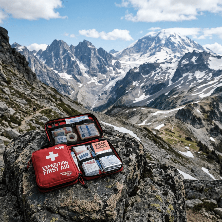
Open expedition first-aid kit on a rock overlooking snow-capped mountains

Medical Kits for Expeditions and Backcountry Travel

shutterstock_35627896I give credit for the information learned and presented here to Howard Donner, MD who is an expert in Wilderness Medicine. Those of us who enjoy traveling in the backcountry understand there are inherent dangers and risks for serious injury and even death.  Many people go out into the wilderness and travel far into remote areas and might be days away from the nearest medical clinic.  I think it’s important for everyone to carry a first aid kit, and someone in the group to carry more advanced supplies if needed. I realize that organizing medical equipment for an expedition into the backcountry requires a lot of planning.  You cannot be prepared for every possible illness and accident no matter how many supplies you bring with you.  There are many opinions on what should be brought and the opinions here are my own after my travels, adventures and taking multiple classes in Wilderness Medicine and Search and Rescue disciplines.  Remember that when the medical kits are too heavy they are more likely to be left behind and not be available when they are desperately needed.  I try to take into consideration size and weight constraints and I choose items for my kits with that in mind. The contents of the kits may depend on: 1)   Age of the hikers 2)   Distance from local medical care 3)   Number of people in the trip 4)   Environmental extremes of the trip 5)   Length of the expedition 6)   Diseases endemic to the area of travel 7)   Medical expertise of the medical officer 8)   Availability of rescue (helicopter for example) 9)   Medical experience of the other expedition members 10)  Pre-existing health problems of the participants I generally recommend that each hiker carry their own personal first aid kit, and then have a base camp kit for the entire group depending on the size. Personal first aid kits might have the following: 1)   Pain medications such as ibuprofen and Tylenol 2)   Sunscreen and lip protection 3)   Water disinfection supplies 4)   Blister care 5)   Minor wound care – band aids/ointment 6)   Insect repellent 7)   Personal medications (for pre-existing problems) 8)   Malaria prophylaxis (if there is risk) 9)   Throat lozenges/cough drops Base Camp Kits will depend on the factors described above, but may contain some of the following – note that I would pick and chose from the list below but usually not take everything here: 1)   Antibiotics – Preparation for common problems. i.e. upper and lower respiratory infections, skin and soft tissue infections and bacterial diarrheas.  I usually carry azithromycin (i.e. Z-Pack) for the respiratory illness.  Duricef (which has the advantage of dosing once to twice a day) is useful for skin infections but consider Bactrim for MRSA.  I usually carry Ciprofloxacin for bacterial diarrheas although there are some newer agents out there (see my blog on traveler’s diarrhea).  Also consider taking antibiotics for protozoan infections if going on longer or more remote trips (i.e. metronidazole or tinidazole). 2)   Analgesics – I like to have a strong oral narcotic medication in case of  a serious  medical problem such as a broken bone or worse.  Consider Percocet or Vicodin. 3)   Respiratory – I have asthma, so I carry albuterol, but it is also useful for exercise induced bronchospasm.  Prednisone is also useful to have for asthma exacerbations and to treat poison ivy, poison hemlock, or poison oak exposure. 4)   ENT (Ear/Nose/Throat) – Rhinorockets are essentially nasal tampons that are useful for nosebleeds.  They are lightweight and simple for anterior nasal packing.  Afrin nasal spray is a great topical decongestant and is useful for nosebleeds and Eustachian tube dysfunction during altitude changes.  Zyrtec or other antihistamine for allergies. Throat lozenges or hard candy are very useful for cough or sore throat.  Sudafed or other oral decongestants. 5)   Eye – Topical eye antibiotic such as Ciprofloxacin ophthalmic for the treatment of corneal ulcers.  Cyclopentolate HCL can be helpful for relieving the ciliary spasm of photokeratitis or uveitis.  A topical anesthetic such as Tetracaine is useful for corneal exam and to help get a climber off the mountain – but should not be abused.  Fluorescein for staining corneal defects.  Ketorolac Ophthalmic solution for minor irritation, Zaditor for allergic conjunctivitis and a penlight or blue LED. 6)   CNS/Brain – Ativan or Xanax for anxiety.  Anxiety can be common with individuals travelling far from home.  Caffeine (No-Doz) useful to relieve caffeine withdrawal headaches or for a little extra “get up and go.” Motion sickness medication – i.e. scopolamine or antihistamines. 7)   Cardiovascular – A beta-blocker such as Lopressor or Tenormin for chest pain or heart attack.  Nitroglycerine for chest pain/MI, Aspirin for chest pain or heart attack, Nifedipine for HAPE (High Altitude Pulmonary Edema) or hypertension. 8)   Gynecological – Urine pregnancy test – rule out an ectopic pregnancy in a female in reproductive age with pelvic pain or bleeding.  Oral birth control pill for hormonal cycling or dysfunctional uterine bleeding.  Fluconazole for yeast vaginitis 9)   Gastrointestinal – Pepto-Bismol for non-dysenteric diarrhea, Imodium for long bus rides, etc., Miralax for – “Freeze dried megacolon” i.e. constipation as a result of eating freeze-dried food.  An anti-nausea medicine such as Zofran – I prefer the oral disintegrating tablets.  An antacid such as Prilosec, oral rehydration solution (dehydrated packets) – i.e.. “Jeevan jal”, or other Sports Drink.  Annusol HC or Tucks pads to treat hemorrhoids. 10)  Altitude illness – Acetazolamide (Diamox), Dexamethasone (for cerebral edema), Compazine for altitude associated nausea and ibuprofen or naproxen for altitude associated headache.  Albuterol inhaler.  Nifedipine for field HAPE treatment. 11)  Topicals – Lamisil AT (or other antifungal cream), Lidex or other topical steroid cream (good for insect bites), Insect repellant with DEET, Silvadene cream for burns or Bactroban for topical antibiosis.  Aloe Vera Gel is excellent for minor burns and irritation.  Vaseline for chapped lips or fever blisters.  Labosan or other sun protection for lips.  Betadine solution (not the scrub). 12)  Blisters – Moleskin (lots of it), Adhesive foam for making donuts and padding for boots, Compeed is a very durable, product for painful blisters or Blistoban to reduce friction.  Duct Tape – good for preventing blisters (i.e. a new pair of boots on a long trip). 13)  Wound Care – Steristrips (multiple sizes), Tegaderm or Opsite for abrasions, Staplers (the small 15 shot  3M are great), Sutures – multiple sizes of both nylon and absorbable.  Superglue for instantaneous treatment of painful skin fissures.  Dermabond and misc. gauze and Band-Aids. 14)  Surgery – Angiocath for emergency tube thoracotomy, Uncle Bill’s tweezers, Sliver pickers (for foreign object removal), #11 scalpels for incision and drainage.  Instruments:  Bandage scissors, tissue scissors, needle drivers, and tissue forceps.  20 or 30cc syringes with 18 G. angiocath for wound irrigation and TB syringes for administration of lidocaine.  Gloves, Safety pins. 15)  Orthopedics – SAM splints, both full-length and finger sizes (great for improvising just about anything).  Kendrick Traction Device – very lightweight design for femoral traction.  Another newer splint is the Slishman Traction Splint (STS) for femoral fractures.  Adhesive tape for splinting ankles and fingers.  ACE Wraps, Fiberglass 3M One “One Step” splints, Air casts or gel casts are great for sprained ankles. 16)  Injectable Medications – Epinephrine 1:1000 (anaphylaxis treatment), Morphine sulfate or consider ketamine.  Dexamethasone, Ondansetron or Compazine, Benadryl, Ativan or Valium (consider rectal administration), Lidocaine, Toradol, Antibiotics such as Rocephin might be an option. 17)  IV’s – Multiple sizes of angiocaths (16, 18 & 20G).  Intraosseous needle for IO infusions, Heparin locks, Bacteriostatic normal saline or heparin flushes, IV fluid volume expanders 18)  Airway – Oral airways such as the LMA, pocket mask 19)  Dental Kit – Cavit (no mix temporary filling), Eugenol (topical analgesic), Dental floss 20)  Miscellaneous items to consider – Stethoscope, snakebite kit, paper/pencil, headlamp, tongue blades, sterile applicators, Foley catheter (16F with 30cc balloon).  A Foley can be used as a urinary catheter, improvised chest tube or posterior nasal pack for bloody nose.  Water disinfection system, spare sunglasses, urine chemstrips. If you are interested in learning more about wilderness medicine, a great resource for information is the wilderness medical society:  http://www.wms.org/   I hope that you have found this information useful.  Wishing you the best of health,

Scott Rennie, D.O.
Board Certified in Obesity Medicine and Family Medicine

This blog is for educational purposes only and does not constitute individual medical advice. Always consult your own physician before making changes to your health, medications, or treatment plan.
Golden retriever wearing a harness and leash in a sunny park

Animal Bites: What to Do if Your Neighbor’s Dog Bites You

shutterstock_54807220Quite a few patients come to the urgent care and see me for an animal bite.  Usually it’s a dog or cat bite, but sometimes I’ve seen patients who’ve been bit by a squirrel or raccoon and then there’s the rare person who is worried that a bat might have bit them.  In the US, two to five million people get bit each year and children are bitten much more often than adults.

Most people worry about the possibility of rabies, although an infection in the skin is much more common.  Bites on the hands can be serious because the skin surface is close to underlying bones and joints.

Dog bites:  Most dog bites are to boys between the ages of 5-9 years old, and the most common areas to be bit are on the head and neck for the younger children.  The right hand is the most frequent site of injury for older children and adults.  The complications of the bite range in severity from tearing away a body part to deep open cuts, crush injuries, to minor scratches.   In Washington State, we have not had a rabies case from a dog bite in many years and do not routinely give rabies treatments for individuals bitten by a dog unless it is known to have rabies infection.

Cat bites:  Claws or teeth can cause wounds from cats.  67% of cat bites are on the arms or hands and scratches are most often on the face.  Puncture wounds that are deep from a cat bite is concerning because bacteria from the cat’s mouth can go deep into the wound and cause infection to the underlying bones or joints.  Infections from cat bites generally cause increased redness to the skin, swelling and pain as early as 12-24 hours after the bite and progress quickly.

Rodent bites:  Rats are the most common rodents that cause bites, but squirrels, hamsters, rabbits and guinea pigs are generally all treated the same way as cat bites.

Other bites:  Bites from raccoons, skunks, fox, coyote or bats should be evaluated right away even if the bite is small and does not look like it’s infected.  These animals can carry rabies and medications are generally given to prevent rabies infection in individuals who are bit by these types of animals.

Treatment:  The first step is to clean the wound with soap and lots of water.  In the event of bleeding, gauze pads can be applied and pressure used to stop the bleeding once the wounds have been washed.

When to seek medical care:

1)   The bleeding has not stopped after applying pressure for 15 minutes

2)   There is a broken bone or serious injury such as large or deep laceration

3)   The bite victim has medical problems such as diabetes, cancer, HIV or takes medication that weaken the immune system

4)   The animal is from one of the animals listed above which have a high risk of rabies

Antibiotics:  Skin infections are the most common complication of an animal bite.  High-risk wounds such as on the face, or involving the bone or a joint are usually treated with an antibiotic.  Many experts also recommend antibiotics to treat people bitten by cats because of the high risk of infection from cat bites.  Most of the time oral antibiotics such as Augmentin are given to treat animal bites however in severe bites with infections that are already present, we will use IV antibiotics.

Tetanus immunization:  Tetanus is a serious, and potentially life threatening infection that can be transmitted by animal or human bite.  If the last vaccine was greater than 5 years ago, the patient should receive a tetanus vaccine.

Sutures (aka stiches):  Wounds on the face are usually closed to avoid developing a scar however due to the risk of infection, some bites may not be sutured immediately.  They may be flushed with a sterile solution and left open for 72 hours after injury and watched closely for signs of infection.

Infected bite wound:  If bite wounds are not treated appropriately right after the injury, an infection may develop.  Surgical treatment and antibiotics may be required at that point.

For more information on animal associated hazards, a useful resource is the centers for disease control article: http://wwwnc.cdc.gov/travel/yellowbook/2012/chapter-2-the-pre-travel-consultation/animal-associated-hazards.htm


I hope that you have found this information useful.  Wishing you the best of health,

Scott Rennie, D.O.
Board Certified in Obesity Medicine and Family Medicine

This blog is for educational purposes only and does not constitute individual medical advice. Always consult your own physician before making changes to your health, medications, or treatment plan.

Healthcare professional examining a baby with a stethoscope

Whooping Cough Epidemic Spreads Across Washington State

shutterstock_163366394From:  Washington Department of Health Pertussis (whooping cough) spreads easily by coughing and sneezing. The number of reported whooping cough cases continue to rise in our state. On April 3, 2012, the Secretary of Health declared a statewide epidemic of pertussis.

Symptoms of pertussis vary depending on age.

Infants: Infants with pertussis may have trouble feeding and breathing and may turn bluish. Many infants are unable to even cough. The disease is most serious in infants, especially those not fully protected or too young to get the vaccine. Babies and young kids: Babies older than six months and kids with pertussis can have severe coughing spells that make it hard to eat, drink, breathe, or sleep. At this age, the cough is often followed by a “whooping” sound, which is how the disease got its common name. Kids may also vomit after a long coughing spell. Whooping cough can cause pneumonia, seizures, brain damage, and death. Babies with whooping cough are often hospitalized. Older kids and adults: With older kids and adults, the disease can be quite mild or can cause several weeks of exhausting coughing. Babies usually get whooping cough from caregivers or family members who don’t realize they have the disease, like older brothers and sisters, parents, and grandparents. Research shows that it’s most common for moms to pass on the disease to babies. How to protect infants and young kids: Anyone with a persistent cough, especially if it includes fits of coughing or causes vomiting, should seek medical care. There is a vaccine to prevent pertussis in older kids and adults called Tdap. All pre-teens, teens, and adults should get the vaccine, especially those in contact with babies. The vaccine can help stop the spread of the disease to babies. Anyone with a cough should avoid being around infants. Not all coughs are whooping cough, but without testing, it’s better to avoid the possible spread. If it’s not possible to avoid being around infants, cough into a tissue, then wash your hands thoroughly, or wear a surgical mask to prevent the spread of bacteria. Age Groups at Risk:  All Fact Sheets: 
Vaccine Information:
    • Kids should be immunized in the first 18 months of life with a four-shot series of the combination vaccine, DTaP. It includes diphtheria, tetanus, and pertussis. Kids who get all four doses before their fourth birthday should get a fifth dose before starting kindergarten or elementary school. The fifth dose isn’t necessary if the fourth dose was given on or after the fourth birthday. This combination vaccine is not given to people over age seven.
DT vaccine is available for kids under seven who can’t tolerate the pertussis (whooping cough) component. Ages 7-10: Tdap Vaccine
  • Kids aged 7-10 years who aren’t fully immunized against pertussis (such as, those who didn’t complete a series of pertussis-containing vaccine before their seventh birthday) should get a one-time dose of Tdap vaccine.
Ages 10 and up: Tdap or Td Vaccine
  • One dose of Tdap vaccine is recommended for adolescents aged 11 or 12 years in place of one Td booster. Kids at least 11 years old are required to show proof of Tdap vaccination. One dose of Tdap vaccine is also recommended for older adolescents aged 13-18 years and adults aged 19 through 64 years.
Vaccine Information Statements:  There is no separate Vaccine Information Statement for combination vaccines.
Related Information from the Department of Health:
Centers for Disease Control and Prevention: Sounds of Pertussis For the latest information on infectious disease, I recommend checking out the website for the Centers of Disease Control: http://www.cdc.gov/

Scott Rennie, D.O.
Board Certified in Obesity Medicine and Family Medicine

This blog is for educational purposes only and does not constitute individual medical advice. Always consult your own physician before making changes to your health, medications, or treatment plan.
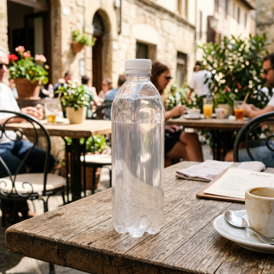
Clear water bottle on a wooden café table with coffee and menu

Traveler’s Diarrhea: What Every Traveler Should Know

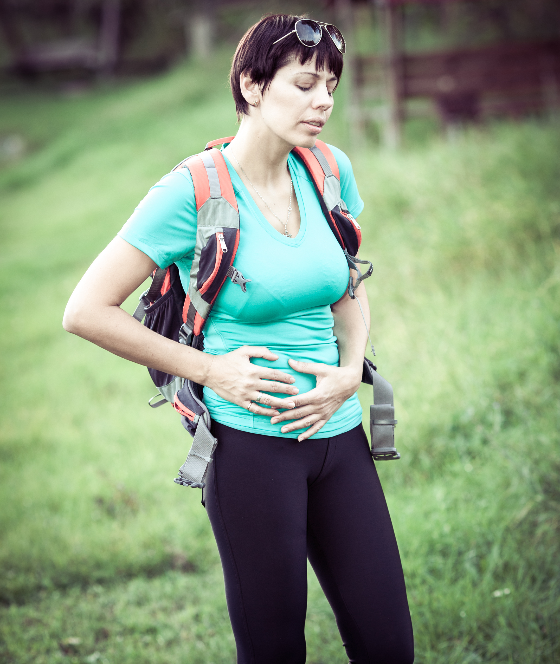
shutterstock_158924261I give credit to Dr. Gene Allred for the information that I’ve learned and posted here.  He is an expert in Wilderness and Travel Medicine. I enjoy travelling, but I am always concerned the possibility of developing diarrhea when I go the backcountry or remote areas.  The majority of cases of traveler’s diarrhea (TD) occur in the first 2 weeks of travel. Symptoms:  The “typical” symptoms are either 4 loose stools in 24 hours or 3+ loose stools per day x 4-5 days plus at least one of the following:  nausea, vomiting, abdominal pain, fever (10%), blood in the stools (15%).  Symptoms can begin as early as 8-10 hours after exposure to contaminated food or water.  With persistent diarrhea (>14 days) we usually consider a parasitic infection with Giardia, E. histolytica, Crypto, or Cyclospora more likely. The majority of diarrheal diseases improve on their own and specific tests to identify the pathogen may not be necessary.  Treatment typically involves ensuring adequate fluid replacement, and empiric antibiotic treatment.  If there is fever along with abdominal pain and dysentery we will obtain a stool culture to look for Salmonella, Shigella and Campylobacter as well for ova and parasites. We used to think that water contaminated with bacteria was the way the infection was spread from person-to-person but we now know that the flies spread the disease very effectively.  The travel from fecal samples and then land on food that’s been sitting out. Diarrhea can be caused from viruses, bacteria or protozoan.  The most common pathogen that causes diarrhea in travellers is E-Coli.   Bacteria cause 50-75% of TD with E-Coli causing 25-40%, Campylobacter – 0-30%, Salmonella 0-15%, and Shigella 0-15%.  Protozoan cause 0-5% of TD, and viruses cause 0-20%. I try to take meticulous drinking water precautions (including ice) but it’s often very challenging.  When I go out in the backcountry or to remote areas I take along water purification tablets such as chlorine dioxide.  Boiling water for at least 1 minute can be helpful, as can chemical disinfection, filtration and use of UV devices.  Remember to cook your food well, and peel all your fruits and vegetables – it’s easy to remember but impossible to do.  As the saying goes “Boil it, cook it, peel it, or forget it!” Diagnosis:  I’m not sure if there is any hard and fast rule for diagnosing traveler’s diarrhea when out in the field without access to a laboratory, but if I’m with a group of people in a developing country and one of the group members has at least one loose stool, I treat them for traveler’s diarrhea.  In the clinic, we can check the stool cultures and for ova & parasites, but we usually only need to do this if there are symptoms of fever, bloody stools or persistent abdominal pain. Treatment:  There is increasing resistance of campylobacter to the fluroquinolone medications such as Cipro.  Azithromycin has been found to be a better choice – 1000mg taken daily for 2-3 days usually works well. Rifaximin is a newer antibiotic with few side effects and has a broad spectrum of coverage – the dose is 200mg three times a day for three days.   PeptoBismol often improves diarrhea and cramps (2 tablets every 30 minutes for up to 8 doses).   If there is no fever, vomiting or blood in the stool, loperamide can help reduce the frequency of diarrhea and can be invaluable for long bus rides. If there is persistent infection, even after treatment with antibiotics we think about the cause being a protozoa.  Giardia is a protozoan that can be detected by a immunoassay and causes diarrhea that lasts for 10 days or longer.  Nitazoxanide (Alinia) 500mg twice a day for three days or Tinidazole (Tiniba) 2 grams in a single dose for adults. Cryptosporidiosis can be a concern, however it is very sensitive to heat.  Even 65 degrees C will kill it, so just boil the water. In third world countries, we wary of swimming in fresh water or going barefooted because there are infectious agents that can get through the skin and cause serious infections (Schistosomiasis, Leptospirosis and Crytopsoridium). Prevention when travelling:  Here are a few of my recommendations about preventing traveler’s diarrhea with water precautions: 1)   Drink carbonated water from a sealed container – the carbonation process kills many pathogens – beer is also acceptable 2)   If non-carbonated, bring beverages to a boil – you can bring them just up to boiling temperature 3)   Avoid ice cubes in your beverages unless you prepared them If you are interested in learning more about wilderness medicine, a great resource for information is the wilderness medical society:  http://www.wms.org/   Stay safe, and happy travels,

Scott Rennie, D.O.
Board Certified in Obesity Medicine and Family Medicine

This blog is for educational purposes only and does not constitute individual medical advice. Always consult your own physician before making changes to your health, medications, or treatment plan.
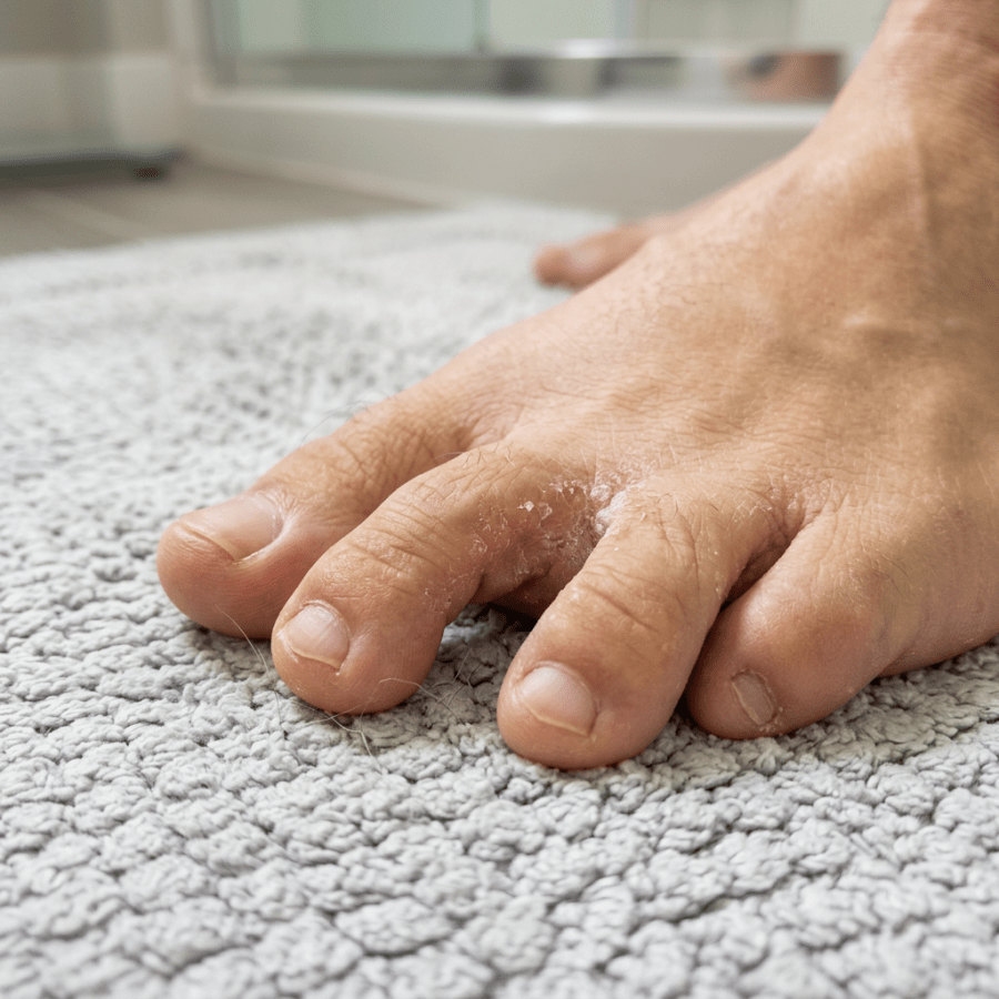
Close-up of toes with dry, peeling skin on a textured towel

Ringworm, Athlete’s Foot and Jock Itch and Fungal Nail Infections – “There Are Fungus Among Us”

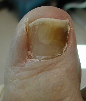
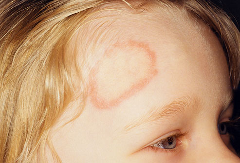
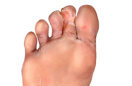
Photo credit:  http://fluconazole.biz/fluconazole-resistant-tinea/ People come to their doctor quite frequently due to fungal infections of the skin or nails.   One might think that because of the name, “Ringworm,” it would be caused by a worm, but it is actually caused by a fungus.  The shape of the rash looks like a ring and thus it got its name.  It’s a red, itchy rash on the skin and is also caused tinea. Four different type of ringworm infections are named for the body-part that is affected: 1)   Tinea capitis affects the head or scalp and is mostly seen in children 2)   Tinea pedis affects the feet and is called athlete’s food because athlete’s common acquire it in the locker room 3)   Tinea cruris affects the groin and is also called jock itch because it also commonly affects athletes or “jocks” presumably because of the moisture on the skin that the fungus prefers 4)   Tinea corporis is a term for fungal infections that affect other body surfaces Ringworm is a contagious infection and can be spread by someone else who is infected or even by an infected dog or cat.  It may be spread in a shower stall, locker room floor or pool area.  Often small skin shavings from the infected person are left behind in these areas and an unknowing person touches the infected area with their feet or other body part. Tinea capitis:  a fungal infection of the scalp may be related to cradle cap in children.  It causes a red scaly rash and can lead to bald patches.  It rarely affects adults.  We treat scalp infections with either oral or topical medications or a combination of both.  Children often improve with the use of gentle massage of the scalp during a bath to remove the scaly areas and then use of an antifungal medical on the scalp to kill the bacteria.  Adults and children are usually treated with oral medications and treatment can take 2-12 weeks depending on the severity of the infection. Tinea pedis (athlete’s foot):  the skin of the feet (often between the toes of the 3-5th toes) become itchy, red, tender, cracked and scaly.  We also see it on the soles of feet and in this area it is usually scaly and simply looks like dry skin.  Sometimes blisters form on the feet, especially between the toes.  It’s very important to treat athlete’s foot in diabetic patients because the fungal infection predisposes the patient even worse – a bacterial infection in the feet.  Diabetic patients can have a decreased sensation on their feet, so they can develop worsening infections without realizing it.  We usually start by removing the dead, scaling skin from the feet, and then treat athlete’s foot with a topical antifungal cream such as Lamisil AT twice a day for up to 12 weeks. The fungus can live on the skin even after the scaling skin is gone, so it’s important to treat the infection even for 1 week after the infection is totally gone.  This infection can be very difficult to treat because if you miss a day of applying the cream, the fungal infection can grow rapidly.  It’s important to keep the feet as dry as possible because fungal infections grow better in warm, damp areas. Tinea corporis (body infection):  often occurs when the fungal infection is transferred from one part of the body to another.  It is commonly seen in high school wrestlers.  It usually appears as a circular or oval scaly area with an outer red edge that is slightly raised while the center is usually flat and skin colored.  We treat this infection with a topical antifungal medication twice a day for 1-2 weeks.  Wrestlers may not be allowed to compete (by their school policy) until their infection clears. Onychomycosis (fungal nail infection):  a fugal infection of the nail that causes the nail to thicken, and change color such as white, yellow or brown.  This type of infection happens most commonly in the toenail but can also happen in fingernails. Fungal nail infections are common and don’t usually lead to serious long-term problems.  It is usually very easy to diagnose a fungal infection of the nails just by examining them.  Most over the counter medications don’t work for fungal nail infections.  In fact even prescription medications don’t work very well, and most fungal nail infections return after being treated with oral antifungal medications. In addition, the oral medications used to treat fungal infections of the nails can cause severe liver disease, so I usually don’t recommend treating this type of infection and instead recommend keeping the nails trimmed to prevent nail fracture or trauma. Preventing ringworm: 1)   Do not share clothing or sports equipment/towels with others 2)   Wear slippers or sandals at the gym, local pool or public shower 3)   Wash with soap and shampoo after skin-to-skin contact with others 4)   Avoid tight-fitting clothing to allow the skin to dry 5)   With athlete’s foot, put socks on before underwear to prevent spread of the fungal infection to other parts of the body including the groin 6)   Treat pet fungal infections to prevent spread to humans 7)   If a family member has a fungal infection, make sure they get treatment right away To find a Dermatologist in your area, the American Academy of Dermatology’s Website has a very useful locator:  http://www.aad.org/find-a-derm   I hope that you have found this information useful.  Wishing you the best of health,

Athlete’s foot and thickened fungal nails matter more than they look, because the cracks they leave between the toes are a common entry point for a deeper foot infection. That risk is highest in diabetes, which is why I wrote a separate post on how to check and protect your feet when you have diabetes.

Updated for 2026: There Is a Resistant Ringworm Now

When I wrote this in 2012, ringworm was a nuisance with a reliable answer. Put an antifungal cream on it, wait a couple of weeks, done. For most people that is still true. But a genuinely new problem has arrived and it is worth your knowing about, because it changes what to do when the cream does not work. Trichophyton indotineae emerged on the Indian subcontinent and has spread internationally, including to multiple US states. It is frequently resistant to terbinafine through mutations in the squalene epoxidase gene. One documented US case had a terbinafine minimum inhibitory concentration of 16 micrograms per milliliter, which is to say the drug was doing essentially nothing (1). It tends to look different. Extensive rather than a tidy ring. Often on the face, trunk, and groin at once. Sometimes a ring inside a ring. Intensely itchy, widespread, and stubborn in a way ordinary ringworm is not. It has also been reported as possibly sexually transmitted (2). The frustrating part is that an ordinary fungal culture cannot tell it apart from garden variety Trichophyton. Identifying it takes specialized gene sequencing available at only a handful of labs. Itraconazole usually works, but often needs more than three months. Terbinafine resistant Trichophyton rubrum, the far more common species, is also increasing in the United States (3).

What That Means Practically

Mostly it means stop assuming that treatment failure is your fault for not applying the cream long enough. If you have had an adequate course of a topical antifungal, or a proper course of oral terbinafine, and the rash is still spreading, that is not a reason to run the same drug again. That is a reason to have the diagnosis reconsidered and, if it still looks fungal, to change class rather than repeat. For ordinary tinea nothing has changed much. Body and groin ringworm respond to a topical antifungal over about two weeks, and the terbinafine class tends to clear it slightly faster than the azoles. Athlete’s foot takes longer, roughly four weeks with an azole. For toenails, oral terbinafine 250 milligrams daily remains first line, and current guidance is to confirm the diagnosis with testing before committing someone to months of an oral antifungal (3).

The Video Visit Version

Most of what I need for this one I can get from a photograph and a few questions, which makes it a reasonable fit for a virtual visit. What helps me is a well lit close up, plus a wider shot showing how far it extends. Tell me how long it has been there, what you have already tried and for how long, whether anyone else at home or any pet has it, and whether you have been treating it with a steroid cream, because a steroid on ringworm changes its appearance and makes it worse. Where I will send you elsewhere is toenails. If we are talking about months of an oral antifungal, I want the diagnosis confirmed rather than assumed, and roughly half of thickened toenails turn out not to be fungal at all.

When To Be Seen In Person

Extensive or rapidly spreading tinea that has already failed proper treatment, which raises the question of the resistant species and needs specialized testing. Scalp involvement, which needs oral treatment and cannot be handled with cream. Pain, warmth, or spreading redness suggesting a bacterial infection on top, particularly if you are diabetic. And any widespread fungal infection if your immune system is suppressed.

The Bottom Line

Ordinary ringworm is still ordinary and still responds to cream. What is new is that a failed course now means something. If two weeks of the right treatment has not touched it, do not just buy another tube. Get it looked at again.

Sources

1. Caplan AS, et al. Notes from the Field: First Reported US Cases of Tinea Caused by Trichophyton indotineae. https://www.ncbi.nlm.nih.gov/pmc/articles/PMC10208369/ 2. Potential Sexual Transmission of Antifungal-Resistant Trichophyton indotineae. Emerging Infectious Diseases. 2024;30(4). https://wwwnc.cdc.gov/eid/article/30/4/24-0115_article 3. Diagnosis and Management of Tinea Infections. American Family Physician. October 2025. https://www.aafp.org/afp/2025/1000/tinea-infections

Related Reading

Scabies Infection: The Mite Bite When to Use Telemedicine vs. Urgent Care: How I Spot the Sick One Ingrown toenails and paronychia A comparison of topical steroid medications Calluses and Corns: Causes, Prevention, and Treatment Newly Diagnosed With Type 2 Diabetes: What You Should Know Scott Rennie, D.O.

Board Certified in Obesity Medicine and Family Medicine

This blog is for educational purposes only and does not constitute individual medical advice. Always consult your own physician before making changes to your health, medications, or treatment plan.