
Vintage balance scale holding fresh tomatoes, carrots, broccoli, and greens

How Do You Actually Lose Weight? A Doctor Explains

Disclosures: I have no conflicts of interest with pharmaceutical companies or research groups, and I don’t make money from sharing this information. My purpose is to provide educational value for patients and colleagues. If you’re on medications for diabetes, hypertension, or heart disease, work with a physician who understands nutrition and diet so adjustments can be made safely.

Weight management is one of the biggest health challenges we face in the U.S. About 75% of Americans are overweight or obese, and that number keeps climbing. Obesity contributes to metabolic syndrome, diabetes, heart disease, stroke, arthritis, sleep apnea, and several cancers. It also lowers energy, reduces confidence, and limits mobility. Patients often tell me they can’t do the things they imagined doing in retirement, like traveling, because of joint pain, back problems, or poor stamina.

Measuring Weight and Risk

In medicine, we usually start with Body Mass Index (BMI), which compares weight to height. A BMI of 18.5-24.9 is considered normal, 25-29.9 overweight, 30-39.9 obese, and 40 or higher morbidly obese. BMI isn’t perfect, especially in athletes with high muscle mass; for them, body fat percentage is more accurate. Obesity is defined as 25% body fat or more for men and 32% or more for women.

Why Weight Loss Is So Hard

Patients often tell me they’ve tried diets, lost a few pounds, then stalled or regained. Many describe feeling cold, hungry, and irritable when cutting calories. Even when weight loss succeeds, it often rebounds, the familiar yo-yo effect.

Part of this is due to how the body regulates weight. Think of it as a “set point.” If you’ve been 230 pounds for years, your body may accept that as its new baseline. If you then cut calories, your metabolism adapts by slowing down. Instead of burning 1800 calories a day, your body may drop to 1300. You feel sluggish, chilled, and hungry. Weight loss slows, and sometimes you even gain despite eating less.

The Role of Insulin

Insulin is usually discussed in the context of diabetes, but it’s central to weight regulation. Our blood can only hold about one teaspoon of sugar, roughly 4 grams, at any time. When we eat carbohydrates, they break down into sugar, and insulin moves that sugar into cells. Excess gets stored as fat.

This mechanism is protective. In times of famine or illness, stored fat can be used for energy. But in modern life, where high-carb foods are constantly available, insulin is triggered frequently. That keeps the body in fat-storage mode and blocks fat breakdown (lipolysis).

Carbohydrates, Fat, and Protein

Carbohydrates include sugars, starches, and grains, but also fruits and vegetables. Foods like rice, pasta, potatoes, and bread raise blood sugar the most, leading to higher insulin release. Federal nutrition advice pushed hard in this direction: the 1977 Senate report Dietary Goals for the United States recommended that most calories come from carbohydrate, and the USDA Food Guide Pyramid that followed in 1992 carried the same message. Since then, obesity and diabetes rates have surged (Micha R, et al. JAMA. 2017;317(9):912-924).

Fat was once vilified, but the story is more nuanced. Eating fat doesn’t necessarily make you fat. Fat provides satiety and doesn’t cause the same insulin spikes that carbohydrates do. Traditional populations consuming higher-fat diets often have lower rates of obesity and diabetes. Fats that are liquid at room temperature, such as olive, avocado, or nut oils, are generally healthier than industrial seed oils like soybean or corn.

Protein is essential, but too much can be converted into sugar through gluconeogenesis. I usually recommend moderate protein intake, since overeating protein can work against weight loss by raising glucose levels.

A Practical Approach: Low Carbohydrate, High Fat (LCHF)

For many patients, shifting to a low-carb, higher-fat diet is effective. It reduces insulin spikes, promotes fat burning, and increases satiety. Foods that fit well include avocados, eggs, nuts, fatty fish, cheese, olive oil, and non-starchy vegetables. Processed foods, sugary drinks, cereals, pastries, and high-carb snacks usually derail progress.

One example I often share with patients: compare a 300-calorie soda to 300 calories of eggs and avocado. The soda doesn’t trigger satiety hormones like leptin and ghrelin, so you don’t feel full. The eggs and avocado, higher in fat and protein, send stronger signals of fullness and reduce the urge to keep eating.

The Role of Exercise

Exercise is valuable for mood, cardiovascular health, and strength. But weight loss depends more on diet. A single cookie may contain 200 calories. You can eat it in seconds, but it takes a 20-minute run to burn it off. Most people can’t out-exercise a high-calorie, high-carb diet.

Alcohol and Weight

Alcohol can complicate weight loss. Beer and sugary cocktails are carb-heavy. Wine and spirits without mixers are lower, but alcohol lowers inhibitions, making it easy to overeat. Patients trying to lose weight need to be mindful about drinking.

Safety in Diabetes

Low-carb, high-fat diets can be safe and beneficial in both type 1 and type 2 diabetes, but medication adjustments are often required. Many patients with type 2 diabetes reduce or even stop insulin once carbs are lowered. Patients with type 1 often report more stable blood sugars and improved A1c. Close monitoring with a physician is critical.

Side Effects and Adjustments

Some patients experience “keto flu” when transitioning from carbs to fat metabolism. Symptoms include fatigue, nausea, irritability, and cramps. They usually resolve in a few days with hydration and adequate electrolytes. Because ketosis increases sodium loss, patients may need to add salt, about 5 grams a day total, from foods or broth, unless restricted for heart failure or severe hypertension. Magnesium, potassium, and calcium should also be maintained.

A rare but important issue is cholesterol. About 10% of patients on a low-carb, high-fat diet may see an increase in LDL cholesterol. Most improve, but it’s worth monitoring.

Tools for Tracking

Some patients like evidence that they’re in ketosis. Urine test strips are unreliable. Blood ketone meters, similar to glucose meters, are more accurate. Even patients without diabetes can use glucose meters to see how food choices affect their blood sugar. Unfortunately, there’s no simple home test for insulin levels, but labs can measure it when needed.

Intermittent Fasting

Intermittent fasting fits naturally with low-carb eating. Eating once or twice a day allows insulin levels to fall further, encouraging fat breakdown. Patients may consume the same total calories, but spacing meals differently can improve insulin sensitivity and fat loss. Early research also suggests fasting may reduce cancer risk and extend lifespan, though more studies are needed (Longo VD, et al. Cell Metab. 2014;19:181-192).

The Bottom Line

Weight gain is complex, but insulin, carbohydrate intake, and the body’s set point all play key roles. Shifting to a low-carb, higher-fat diet can help reset metabolism, improve diabetes, and reduce reliance on medications. Adding intermittent fasting may enhance those effects.

Patients should approach these strategies carefully, ideally with medical guidance, especially if they take insulin or blood pressure medications. Done properly, this approach helps with far more than weight. It improves energy, mood, and long-term health.

Scott Rennie, D.O.

Sources

  • Sumithran P, et al. Long-term persistence of hormonal adaptations to weight loss. N Engl J Med. 2011;365:1597-1604.
  • Micha R, et al. Association Between Dietary Factors and Mortality From Heart Disease, Stroke, and Type 2 Diabetes in the United States. JAMA. 2017;317(9):912-924.
  • Longo VD, Mattson MP. Fasting: Molecular Mechanisms and Clinical Applications. Cell Metab. 2014;19:181-192.

Board Certified in Obesity Medicine and Family Medicine

This blog is for educational purposes only and does not constitute individual medical advice. Always consult your own physician before making changes to your health, medications, or treatment plan.


Kratom: A New Drug Growing Popular in Washington State

Through 2016 I saw a growing number of patients at Urgent Care dealing with the effects of Kratom. Some came in sick; others called worried about withdrawal or side effects. Because it came up so often, I think it’s worth sharing what I’ve learned.

Kratom comes from the leaves of a tree native to Southeast Asia. People there have used it for a long time as a painkiller, for diarrhea, or recreationally. In Washington State, it’s sold in capsules filled with powdered leaf material. Some people also brew it into tea or smoke it.

The United Nations Office on Drugs and Crime places Kratom in a group called “New Psychoactive Substances.” That same group includes Khat from East Africa, Salvia divinorum, and certain synthetic drugs such as ketamine analogs and mephedrone. As of 2016, Kratom wasn’t illegal at the federal level in the U.S., though it was illegal in countries like Thailand, where thousands of people had been arrested for related offenses (United Nations Office on Drugs and Crime, 2013). Despite that, it could be found for sale in some recreational marijuana shops here.

Kratom’s effects show up quickly and can last about five to seven hours. People use it for both stimulation and sedation. At lower doses, it may make someone feel more energetic, talkative, or sociable. At higher doses, it often causes lethargy and a sense of euphoria. Not every user finds it pleasant, and the side effects can be significant.

Some patients report nausea and vomiting, sometimes severe enough to bring them to Urgent Care. Others describe nervousness, sweating, itching, constipation, or tremors. I’ve also seen reports of hallucinations, paranoia, and even aggressive behavior.

With longer-term use, addiction becomes a real concern. People can develop cravings, lose weight, notice skin changes on the face, and report a loss of sexual desire. When they stop, withdrawal can be difficult. Symptoms may include diarrhea, muscle pain, tremors, trouble sleeping, depression, anxiety, and sudden mood swings (Drug Enforcement Administration, 2016).

In 2016, the Drug Enforcement Administration announced plans to classify Kratom as a Schedule I drug, the same category as heroin or LSD. That decision was paused after a public comment period, and at the time of writing Kratom hadn’t been rescheduled. The DEA had called it a potential “imminent hazard to public safety” (Federal Register, 2016).

If you or someone you know is using Kratom and experiencing health problems, talk with a healthcare provider.

Scott Rennie, D.O.

Board Certified in Obesity Medicine and Family Medicine

This blog is for educational purposes only and does not constitute individual medical advice. Always consult your own physician before making changes to your health, medications, or treatment plan.
Glowing neural network with brain at top surrounded by a golden shield containing various emblem icons.

Acute Flaccid Myelitis: A Mystery Illness in Washington State

shutterstock_142857034

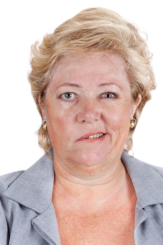
Recently, eight children in Washington State were diagnosed with acute flaccid myelitis (AFM). It is a rare condition that affects the central nervous system, specifically the spinal cord. It causes weakness in the arms or legs, and it can also produce facial drooping or weakness, difficulty moving the eyes, drooping eyelids, and trouble with speech or swallowing. As of September 2016, 89 people in 33 states had been confirmed with the illness, according to the CDC.

Diagnosis: If you think you or a family member has this condition, seek consultation with a medical provider. But how do you know whether AFM is causing the symptoms? A doctor may be able to make the diagnosis through a careful examination, and an MRI is sometimes helpful as well. Spinal fluid, which surrounds the brain and spinal cord, may be collected by a spinal tap (lumbar puncture) and can also aid in the diagnosis.

There are nerve tests that may help confirm the diagnosis, though they need to be done 7 to 10 days after the onset of illness.

Causes: A number of viruses have been thought to be possible causal agents, including enteroviruses (polio among them), West Nile virus, Japanese encephalitis, St. Louis encephalitis, and various adenoviruses.

AFM is not the only cause of weakness in the arms or legs. Other causes include viral infections, environmental toxins, genetic disorders, and Guillain-Barré syndrome (GBS). Neurological conditions such as stroke (cerebrovascular accident) can also cause weakness in an arm or leg, or facial drooping, so it’s important to seek medical attention immediately and call 911 if you or someone you know develops these symptoms.

Treatment: No specific treatment exists for AFM. A neurologist may be consulted to help with the diagnosis and to make recommendations.

If you or your child is having problems walking or standing, or develops sudden weakness in an arm or leg, contact a medical provider right away.

I hope you have found this information useful. Wishing you the best of health,

Scott Rennie, D.O.

Board Certified in Obesity Medicine and Family Medicine

This blog is for educational purposes only and does not constitute individual medical advice. Always consult your own physician before making changes to your health, medications, or treatment plan.
Blog: https://doctorrennie.wordpress.com

This information comes from the CDC website, About Acute Flaccid Myelitis.

Trigger Finger: What Is Stenosing Tenosynovitis Exactly?

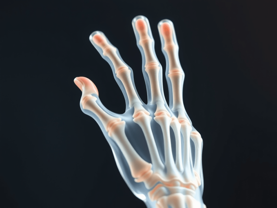
In my urgent care years I often saw patients complaining that one of their fingers got stuck when they tried to bend or straighten it. They would describe how the finger locked in a bent position, and when they forced it straight, it popped painfully back into place instead of moving smoothly. This is what we call trigger finger. It’s a type of tenosynovitis, which means inflammation of a tendon and the protective sheath around it.

Tendons are the strong cords that connect the muscles in your hand to your finger bones. When the tendon or its sheath gets inflamed, movement becomes restricted. Most of the time this happens in the hand or wrist, though it can also occur in the ankle.

The causes vary. Repeated use of the hand with the same motion, like gripping tools or typing, is a common reason. Infection is another possibility, especially if bacteria spread into tissues under the skin.

Symptoms usually start with pain and stiffness, often worse in the morning. Many patients notice a popping or catching sensation when trying to straighten the finger. In some cases the finger locks completely in a bent position. A small, tender lump can sometimes be felt at the base of the affected finger on the palm side. Swelling is also possible, along with trouble gripping or holding objects. Trigger finger most often affects the thumb and middle fingers.

Diagnosis is usually made by physical exam, and most of the time testing isn’t needed. If there’s a history of trauma or concern for infection, your doctor may order x-rays, an ultrasound, or blood work to rule out other issues.

Treatment depends on the severity and the underlying cause. Rest and ice are often the first steps. A cold pack, or even a bag of frozen vegetables, applied to the swollen area for 15 minutes every few hours can help calm inflammation. Nonsteroidal medications like ibuprofen or naproxen may ease discomfort, though they don’t cure the condition.

For more persistent cases, a steroid injection directly into the tendon sheath can reduce swelling and improve movement. If infection is the culprit, antibiotics are required. Surgery is an option for severe or stubborn cases where conservative measures don’t help. Once the symptoms improve, gentle finger stretches can restore motion and prevent stiffness from returning.

Scott Rennie, D.O.

Board Certified in Obesity Medicine and Family Medicine

This blog is for educational purposes only and does not constitute individual medical advice. Always consult your own physician before making changes to your health, medications, or treatment plan.
doctorrennie.wordpress.com

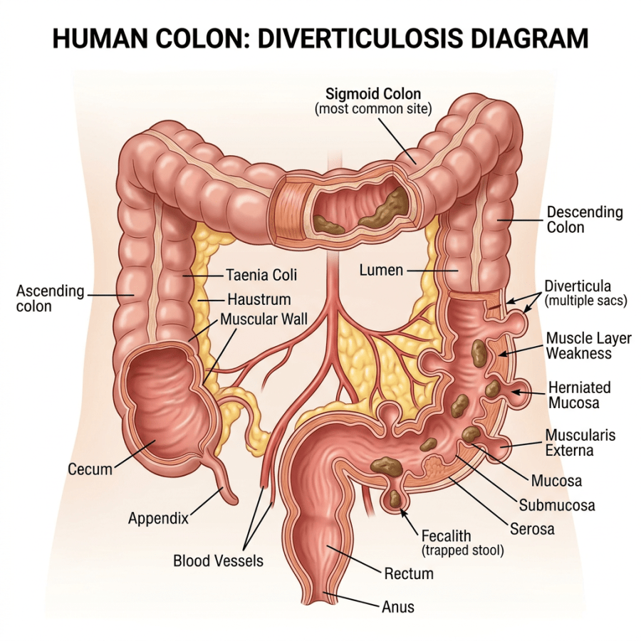
Diagram of the human colon highlighting diverticulosis features including diverticula, muscle layer weakness, herniated mucosa, and fecalith.

Diverticular Disease: Diverticulosis vs Diverticulitis Explained

shutterstock_75923548

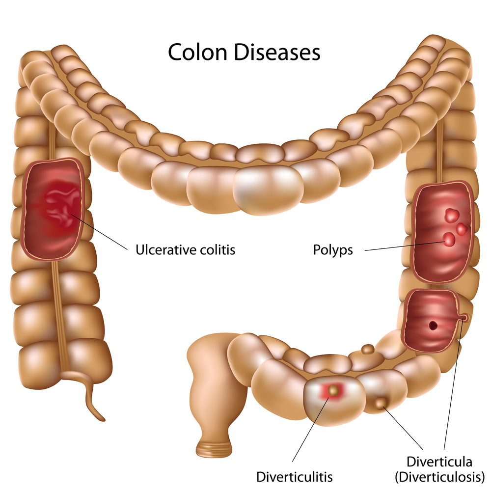
When a patient came in with abdominal pain or blood in their stool, one of the conditions we considered was diverticulitis. You may not have heard much about diverticulitis, so this post will try to answer some common questions about diverticular disease.

A diverticulum is a small pouch-like structure that sometimes forms in the muscular wall of the colon. These little pouches often cause no pain, and we usually become aware of them only after a procedure such as a colonoscopy, flexible sigmoidoscopy, a barium enema or a CT scan.

What is diverticulosis? Diverticulosis simply means that diverticula are present. Most people have no symptoms and will remain free of symptoms throughout their lives. (Roughly 15 to 25 percent go on to develop diverticulitis, which is more severe.) I usually think of a diverticulum as a small area where the colon balloons out. These are potentially weak spots that might become inflamed or may rupture under pressure.

What is diverticulitis? Diverticulitis is inflammation of a diverticulum, which occurs when there is thinning or breakdown of the wall of the colon. It is more severe than diverticulosis because the pouches have become irritated, inflamed or stretched, either from pressure within the colon or from hard particles of stool lodged inside them. The diverticulum becomes painful and may rupture, meaning the wall of the colon breaks down and allows stool and bacteria to enter the abdomen. Diverticulitis may also cause bleeding within the colon, because blood vessels often run through the area where diverticula form and they can bleed when the area gets inflamed.

Symptoms of diverticulitis: The symptoms depend on how severe the inflammation inside the colon is. The most common symptom is pain in the left lower abdomen. Patients may also have blood in the stool, nausea, vomiting, constipation, or diarrhea.

Classification of diverticulitis: Simple diverticulitis is more common and usually responds well to medical treatment without surgery. Complicated diverticulitis, about 25 percent of cases, usually requires surgery to remove the section of colon that has become inflamed or ruptured.

Treatment: Diverticulosis itself usually needs no treatment, since diverticula that are not inflamed are generally painless and cause no problems. Many providers do recommend increasing dietary fiber to reduce the chance of constipation, since constipation raises pressure within the colon, which can in turn lead to diverticulitis. Increasing fruits, vegetables, hydration and fiber has not been proven to prevent diverticulitis, however. We used to tell patients with diverticulosis to avoid seeds and nuts, on the theory that they increased the risk of diverticulitis. Research has since shown that advice to be wrong.

Diverticulitis does require treatment, and what you receive depends on the severity of your symptoms. Mild abdominal pain from diverticulitis can usually be managed at home with a clear liquid diet and oral antibiotics. If a patient develops a fever greater than 100.4 degrees F, worsening or severe abdominal pain, or an inability to keep fluids down, hospital treatment is usually recommended.

Hospitalization: If you need to be in the hospital for diverticulitis, you usually will not be allowed to eat or drink until you start feeling better, and antibiotics and IV fluids are given. If an abscess forms, a surgeon may need to drain it by placing a tube through the abdominal wall.

Complications: Peritonitis is a generalized infection within the abdomen that occurs if the colon ruptures. An emergency operation is often required in these cases to remove the diseased section of colon and reconnect the unaffected ends. Sometimes those two steps are done at separate times, with the removal first, so the colon is allowed to heal before the pieces are reattached.

Surgical treatment: Patients who don’t respond well to medical treatment, or who have repeated attacks of diverticulitis, may benefit from surgery to remove the diseased section of colon.

Bleeding from diverticulitis: Most of the time the bleeding stops on its own, but sometimes a colonoscopy or another procedure is necessary to get it under control.

Scott Rennie, D.O.

Board Certified in Obesity Medicine and Family Medicine

This blog is for educational purposes only and does not constitute individual medical advice. Always consult your own physician before making changes to your health, medications, or treatment plan.
Blog: https://doctorrennie.wordpress.com

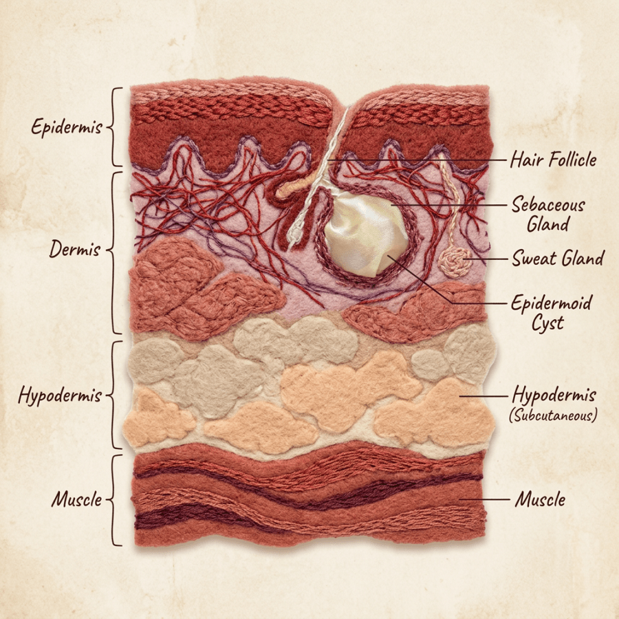
Cross-section of skin layers showing epidermis, dermis, hypodermis, muscle, hair follicle, sebaceous gland, sweat gland, and epidermoid cyst

Epidermal Inclusion Cysts Explained: What Are They?

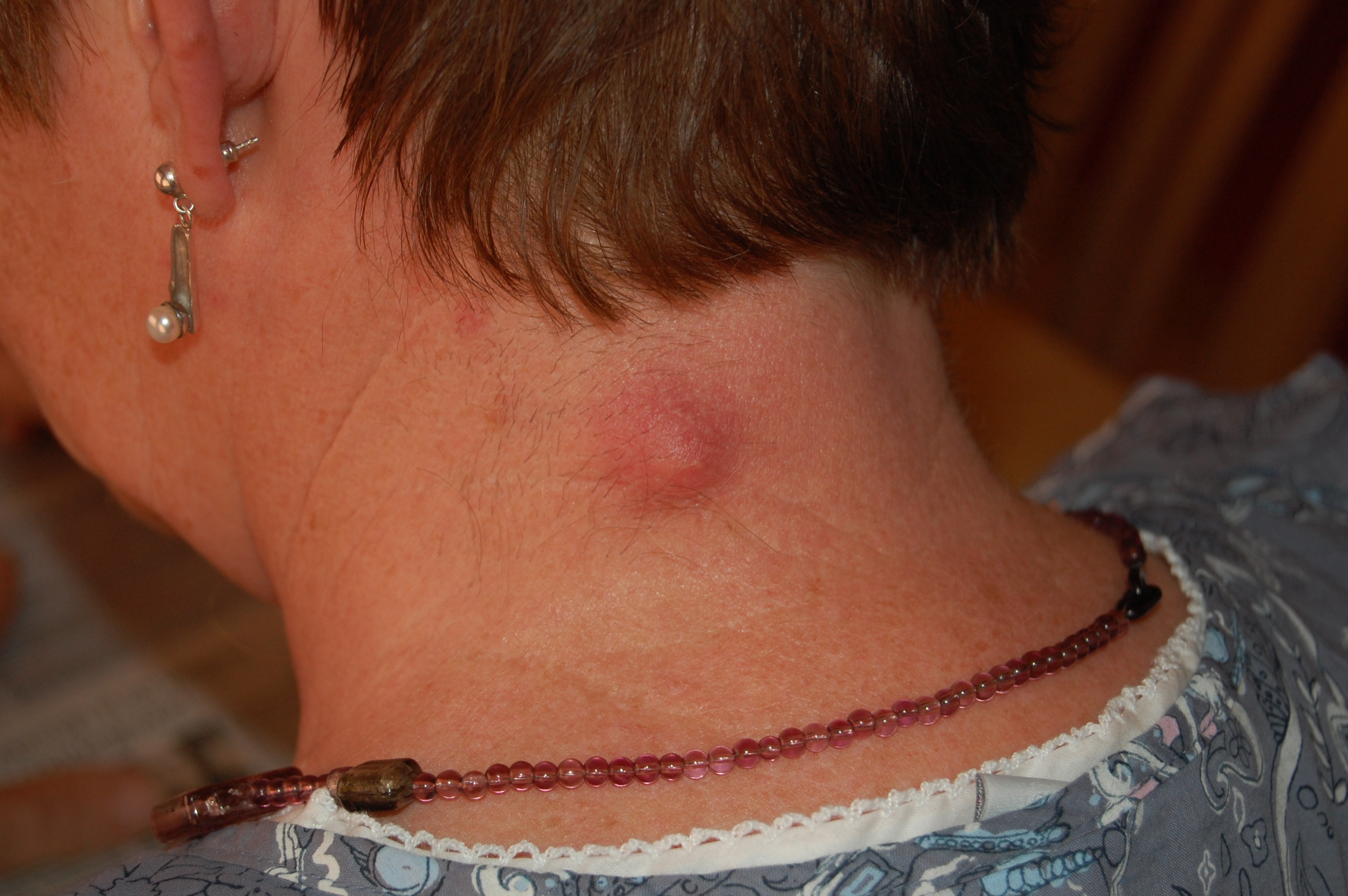
Inflamed_epidermal_inclusion_cyst

Photo credit: http://en.wikipedia.org/wiki/Sebaceous_cyst

Patients often came into the urgent care with a small skin lump that had become red or painful. Frequently, thinking back, they would recall feeling a small nodule under the skin in that same spot months or even years before it became swollen and red. The epidermal inclusion cyst is one of the most common skin cysts. It can occur anywhere on the body, though it is more common on the face and upper body. Most of the time these cysts cause no problems, but they can be cosmetically unwelcome.

Other names for epidermal cysts:

1) Epidermoid cyst

2) Sebaceous cyst

3) Keratin cyst

4) Epidermal inclusion cyst

5) Infundibular cyst

Appearance: Epidermal cysts have a wall made of skin cells from the outer layer of the skin, called the epidermis. The cyst wall is like a balloon that extends down into the second layer of skin, the dermis. That wall produces keratin, a protein found in skin and nails, which is usually white and either cheesy or firm in consistency. It is often foul smelling as well.

Cyst Rupture: If the cyst wall ruptures underneath the skin, usually from trauma or from bumping the area unintentionally, the keratin escapes into the surrounding tissue and is very irritating. The skin becomes red, swollen and painful. It’s best to see your doctor rather than trying to “pop” or drain the cyst yourself. Your doctor may recommend treating you with an oral antibiotic before opening the cyst if he or she thinks it has become infected.

How epidermal cysts are removed: If the cyst needs to come out, your doctor will try to remove the entire cyst including the wall. Remember, the cyst wall is what makes the keratin. If the wall is left behind under the skin, it may start producing keratin again and the cyst can come back.

Usually we make an incision over the cyst, separate the overlying skin from the cyst wall, and try to remove it in one piece. If the cyst has ruptured, which is most likely what brought the patient in, it is removed piecemeal, with an effort to get all of the keratin and all of the wall out. The doctor may irrigate the wound with sterile solution afterward. The skin is usually left open rather than stitched. The doctor may place a small piece of packing gauze under the skin where the cyst was, and then cover it with a bandage. This allows the wound to drain while it heals. The wound is usually examined by a medical provider every two to three days to check on healing, and part or all of the packing gauze is removed at those visits.

The reason your provider may pack the wound and ask you to come back, instead of simply putting stitches over it, is that these areas are sometimes contaminated with bacteria. If the skin is closed right away with sutures, the bacteria have a small sealed pocket in which to grow and form an abscess. Letting the wound heal from the inside out lowers that risk and often speeds healing.

When to have a cyst removed: If it’s small, painless, and not red or swollen, it probably doesn’t need to be removed. I might recommend removing a cyst if:

1) It keeps getting red and irritated or infected

2) It’s getting larger quickly

3) It’s in a place that rubs against clothing or jewelry and gets irritated

4) It becomes red, inflamed or painful

Scott Rennie, D.O.

Board Certified in Obesity Medicine and Family Medicine

This blog is for educational purposes only and does not constitute individual medical advice. Always consult your own physician before making changes to your health, medications, or treatment plan.
Blog: https://doctorrennie.wordpress.com

Abstract swirling blue and orange shapes with dynamic flow and light accents

Google Glass: Is It Really Ready for Prime Time Yet?

Yes, I admit it, I’m a technophile, aka “nerd,” “geek,” or perhaps a “neek.” I picked up the Explorer Edition of Google Glass when it became available to the public. I thought it might have some interesting applications in the medical field. I liked the idea of using it for navigation, and I was curious to see what you could do with it. It’s a great concept and in many ways a great device. Here are some of my observations.

  1. If you wear Google Glass in public, people notice the device and worry that you’re recording them. There are perhaps hundreds of devices out now that can record or take photos, and people carry them everywhere: smartphones, watches, pens, glasses, clip-on cameras and plenty of others. I think people feel uncomfortable around Google Glass because it’s so visibly recognizable to everyone who isn’t wearing it. It’s the opposite of covert.
  2. People will stop and ask you about it. You might get a small group gathered around you wanting to know more, so if you’re already late for work, you might not want to wear Glass on your walk, or you could find yourself running further behind schedule.
Image

3) You need to pair it with your smartphone, not only over Bluetooth but also Wi-Fi. I found it challenging to pair with my iPhone’s personal Wi-Fi network. It seems to lose the connection and then have trouble re-establishing it. Worse, you don’t even know it’s disconnected from your phone, or from another Wi-Fi network, until you try to use it. I’m not sure whether that’s a problem inherent to Glass or to the iPhone personal hotspot feature. It may work better with Android devices, but I can’t say.

If you’d like to connect to a different Wi-Fi network, it isn’t easy. You enter the settings into Glass, then create a QR code in the Google Glass application on your phone, and then Glass photographs the QR code to store your network information.

4) It seems to lock up or stop responding at times. One of the supposedly great things about wearable tech is that it’s ready whenever you want it, without having to pull anything out of your pocket. If Google Glass would wake up whenever I wanted it to, that would be a good start. Sometimes it also decides to update itself without asking, and during that time it’s completely unusable.

5) Google advertises it as a device you can use all day, but I find the battery dies quickly. With regular use it lasts about three hours. The battery also gets very warm.

6) At least at this point, you’re limited to Google Calendar, with no other options. If you’d like to add all of your phone contacts to Glass, that isn’t easy or intuitive either.

7) Scanning through the Glass timeline to find your photos, tweets, missed calls, text messages or other data can be time consuming and difficult.

8) The audio quality is very poor. Google has tried to remedy this by supplying a small earbud you can plug in, but in my opinion that isn’t a great solution.

9) The screen, the glass that projects in front of your eye, has very low resolution and can be difficult to see in bright sunlight.

10) There are a very limited number of applications available right now. Applications that interact with Facebook or Twitter only let you post an update or a tweet. Be careful taking a photo, because it’s very easy to accidentally share it to your Facebook timeline or send it out as a tweet.

11) Perhaps the best feature is navigation. Having directions appear right in front of you is great, but you need the Google Glass application already open on your iPhone before you use it, or it won’t work. I can’t tell you how many times I asked Glass for directions only to be told to open the Glass application on my phone. So much for hands free.

12) There’s a beta feature that lets you take a picture by winking. This is genuinely cool, if a bit creepy, but it doesn’t work all the time. That feature is in beta along with the rest of the device.

13) At $1,500, Google Glass costs more than most people will want to pay. If the device worked 90 percent of the time, and there were more you could do with it, and better application support, meaning Facebook, Twitter, medical applications such as Epocrates, better audio, and music apps, I would pay $500 for it.

Is Google Glass ready for prime time? I think it’s a great device with real potential. You be the judge. What do you think about Google Glass? Post a comment and share your opinion.

Scott Rennie, D.O.

Board Certified in Obesity Medicine and Family Medicine

Bottle of scalp care shampoo labeled Renew & Refresh with hairbrush and towel

Seborrheic Dermatitis – Dandruff, Cradle Cap and Adult Seborrhea

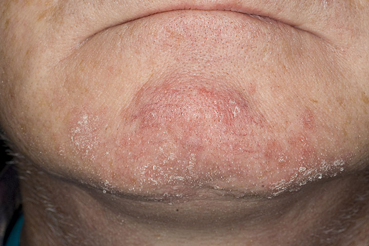
??????????????????????????????????????????????????????????????????????
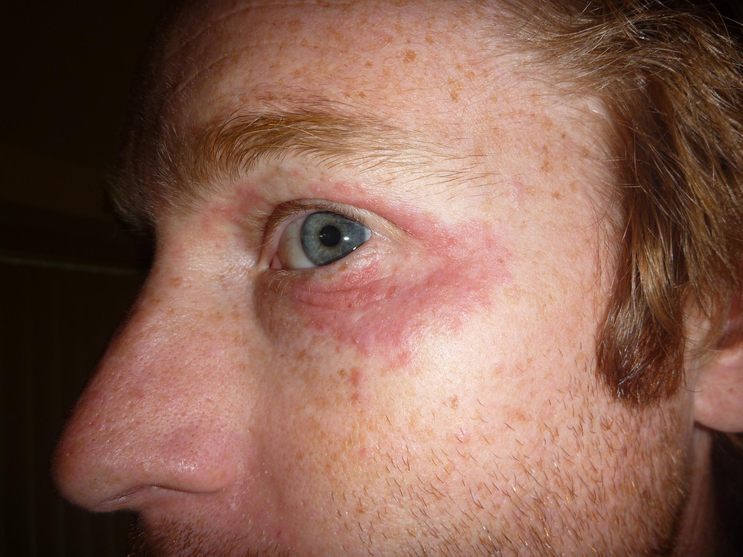
seborrheic-dermatitis-eyes
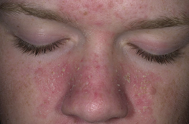
??????????????????????????????????????????????????????????????????????

Photo credit: http://hardinmd.lib.uiowa.edu/dermnet/seborrheicdermatitis22.html

Photo credit: http://siklusair.com/seborrheic-dermatitis-eyes

Photo credit: http://medicalcontent.hubpages.com/hub/Seborrheic-Dermatitis-Pictures-on-Face-Scalp-Hair-loss-Causes-Treatment

Seborrheic dermatitis is a common skin condition affecting both infants and adults. It causes redness, greasy scales, flaking along the eyebrows, and itching. It was one of the most common skin conditions I saw as an urgent care physician.

Seborrheic dermatitis, also called seborrhea, is an inflammation of the skin that usually occurs in areas with an abundance of oil glands: the scalp, face, around the eyes, ears, neck, and even the diaper area. In men it is more common where there is facial hair. Infants often have seborrhea on the scalp, where it is known as cradle cap. A mild form confined to the scalp, in children or adults, is what we call dandruff. Symptoms may include:

  1. Skin scales, white or yellow, usually oily or greasy
  2. Itching
  3. Mild redness
  4. Skin lesions or plaques

The exact cause is unknown, but there is evidence that it flares in times of stress, with hormonal changes, and during particular seasons, especially in extreme cold or warm weather. It is more common in people with weakened immune systems, in those with oily skin, and in certain neurological conditions such as Parkinson’s disease.

Diagnosis: Your healthcare provider can usually diagnose seborrheic dermatitis by examining you and taking a history. A biopsy, a sample of skin removed surgically and examined under a microscope, is rarely needed.

Treatment: Treatment is tailored to the individual patient and to which part of the body is affected. Options include:

  1. Medicated shampoos such as Neutrogena T-Gel or T-Sal, Head and Shoulders, or Nizoral. These usually contain salicylic acid, coal tar, zinc, selenium sulfide, ketoconazole, or resorcinol. Even if you do not have dandruff, these shampoos can be used on the face and other affected areas, and they work best left in place for five to ten minutes before rinsing.
  2. Topical steroid creams, lotions, foams or shampoos. Low, medium or even high potency steroids are sometimes prescribed to reduce inflammation, depending on severity and location. Lower potency preparations are used on the face, such as desonide 0.05% lotion.
  3. Antibacterial creams. Topical agents such as sodium sulfacetamide with sulfur are sometimes prescribed.
  4. Antifungal creams. Ketoconazole 2% cream and ciclopirox 1% cream are commonly used.
  5. Other anti-inflammatory medications such as pimecrolimus cream or tacrolimus ointment, sometimes prescribed for facial seborrhea.
  6. Dermatologists sometimes recommend a compounded mixture combining a steroid with an antifungal or antibacterial agent.

Cure: Unfortunately there is no cure for seborrheic dermatitis. It is a chronic, lifelong condition. It may disappear for months or years and then relapse. We focus on controlling it, usually by combining the treatments above with efforts to reduce known triggers such as emotional stress, extreme cold, and excess body weight.

Blog: https://doctorrennie.wordpress.com

This one is from 2014. I am board certified in obesity medicine and most of what I write now sits there, down to narrow questions like whether you can do intermittent fasting while you are on Ozempic or Wegovy.

Updated for 2026: A New Drug, and the Same Old Shampoo

Most of what I wrote here in 2014 still stands. Ketoconazole 2 percent or ciclopirox 1 percent shampoo, used a couple of times a week for at least a month, is still the backbone. A short run of a mild steroid for a flare. A calcineurin inhibitor like tacrolimus for the face, where I would rather not leave someone on steroids. What is new is roflumilast foam 0.3 percent, sold as Zoryve. It is a topical PDE4 inhibitor, approved in December 2023 for seborrheic dermatitis in anyone nine and older, applied once a day. It is the first genuinely different mechanism approved for this condition in more than twenty years (1). The trial numbers are good. In the phase 3 STRATUM study, 79.5 percent of patients on roflumilast reached clear or almost clear skin at eight weeks against 58 percent on vehicle, with about half completely clear. Itch improved within two days (2). My honest read: it works, and it is a real option when shampoo and a steroid have not been enough or when the face is involved and you want off steroids. It is also expensive, and for a lot of people ketoconazole shampoo bought over the counter handles the problem for a few dollars. I would still start there.

Cradle Cap Has Not Changed

Worth saying because parents ask constantly. It resolves on its own over weeks to months. Baby shampoo, soften the scale with mineral oil or petroleum jelly, lift what comes away easily, leave what does not. Do not use medicated dandruff shampoo on an infant. If it is genuinely inflamed and persistent, a low potency steroid or topical antifungal briefly, and that is a conversation to have rather than something to start yourself (1).

What a Video Visit Handles Well Here

This is a good fit for the format. Seborrheic dermatitis has a recognizable look and a recognizable distribution, scalp, eyebrows, the creases beside the nose, behind the ears, sometimes mid chest, and a photograph in decent light usually tells me what I need. The questions matter as much as the picture. How long, whether it comes and goes with stress or season, what you have already tried and for how long, and whether you have been using a steroid cream continuously, which causes its own problems on facial skin. Where I want a closer look is when the distribution is not typical, because psoriasis, tinea, and contact dermatitis all get called seborrhea and are treated differently.

When To Be Seen In Person

Seborrheic dermatitis that is unusually widespread or severe deserves a conversation about HIV testing, because that association is real and longstanding. Signs of bacterial infection on top, meaning spreading redness, warmth, pus, or fever. Failure to respond to standard treatment, which usually means the diagnosis is wrong rather than the drug. And in an infant, scale spreading well beyond the scalp along with poor feeding or poor weight gain needs a pediatrician.

The Bottom Line

Start cheap. Antifungal shampoo, used properly and for long enough, still fixes most of this. Roflumilast is a genuine addition for the cases it does not, particularly on the face. Cradle cap needs patience and mineral oil, not medicine.

Sources

1. Vidal SI, Menta N, Green L. Child and Adult Seborrheic Dermatitis: A Narrative Review of the Current Treatment Landscape. Dermatol Ther (Heidelb). 2025;15(3):599-613. https://pmc.ncbi.nlm.nih.gov/articles/PMC11909311/ 2. Blauvelt A, et al. Roflumilast foam 0.3% for adolescent and adult patients with seborrheic dermatitis: a randomized, double-blinded, vehicle-controlled, phase 3 trial. J Am Acad Dermatol. 2024;90(5):986-993. https://pubmed.ncbi.nlm.nih.gov/38253129/ 3. American Academy of Dermatology. Seborrheic dermatitis: Diagnosis and treatment. https://www.aad.org/public/diseases/a-z/seborrheic-dermatitis-treatment

Related Reading

Ringworm, Athlete’s Foot and Fungal Nail Infections When to Use Telemedicine vs. Urgent Care: How I Spot the Sick One What Is Psoriasis? An Overview of This Skin Condition A comparison of topical steroid medications Dry, Itchy Skin: Could It Be Eczema or Dermatitis? Scott Rennie, D.O.

Board Certified in Obesity Medicine and Family Medicine

This blog is for educational purposes only and does not constitute individual medical advice. Always consult your own physician before making changes to your health, medications, or treatment plan.
Abstract artistic illustration of the human digestive system with surrounding microbiome cells

Rectal Bleeding: What Are the Possible Causes of It?

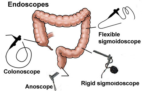
Colon-Endoscopes
anat_colon_1.0

Photo credit: http://www.naturalhealingsolutionsllc.com/learn-about-colon-hydrotherapy.html

Photo credit: http://www.thenurseslockerroom.com/2013/03/sigmoidoscopy-screening-test-for.html

One of the more common problems that brought patients in to see me, believe it or not, was seeing blood in the toilet, on the stool, or on the toilet paper after a bowel movement. Since I had seen several patients about this, I thought I would write about some of the possible causes.

Healthcare providers take this seriously, because blood noticed after a bowel movement can sometimes be a sign of colon or rectal cancer. Fortunately, most causes of rectal bleeding are not cancer.

Causes of rectal bleeding:

  1. Hemorrhoids: Swollen blood vessels in the rectum or anus can cause itching, pain, and bleeding. Patients usually describe hemorrhoidal blood as bright red. It may coat the surface of the stool, drip into the toilet and turn the water red, or show up on the toilet paper. Hemorrhoids do not have to hurt. Painless rectal bleeding during a bowel movement is a common presentation.
  2. Anal fissure: A tear in the lining of the anus can bleed, and there may be pain with a bowel movement.
  3. Other causes include infection, colitis (which may be due to an autoimmune disease such as ulcerative colitis or Crohn’s disease), colon polyps, and colon cancer. If the bleeding comes from higher in the digestive tract, such as in or above the stomach, the blood may look dark black or tarry.

Diagnosis and testing: To find the cause of bloody stools, your provider may perform tests or refer you to a specialist. They will take into account the information you give them, your history, your symptoms, and your age.

1) Rectal exam: Your provider will usually examine the rectum and look for a source of bleeding such as a hemorrhoid or fissure. This may include a digital rectal exam, where the doctor inserts a gloved and lubricated finger into the rectum to feel for possible rectal cancers.

2) Anoscopy: Your doctor may use a small plastic device with an attached light to get a better look at the source of bleeding. Most of the time this is not painful, though it can be a bit uncomfortable, and it can be done in the office.

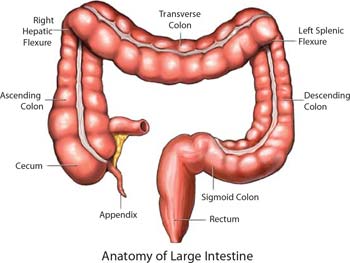
3) Sigmoidoscopy: This is usually done in an outpatient center, and the patient is usually not sedated. Sigmoidoscopes come in rigid and flexible forms, and the flexible one is used most often. A flexible tube, roughly 70 cm long and 1 cm wide, carrying a tiny video camera and a light, is inserted through the anus and gently into the colon while air is introduced to open the area and improve the view. A biopsy is often taken with a small tool. Sigmoidoscopy allows visualization of the anus, rectum, sigmoid colon and the top of the descending colon. It does not reach the entire colon, so it can miss cancers, polyps or bleeding sources in other areas.

4) Colonoscopy: Similar to sigmoidoscopy, but it allows the doctor to examine the entire colon with a longer flexible tube. The colonoscope, about 140 cm long, reaches everything the sigmoidoscope can and also allows visualization of the transverse colon, ascending colon and cecum. The patient is usually sedated.

When to seek help: It is impossible to know the cause of rectal bleeding without an examination. Everyone with rectal bleeding should talk to their healthcare provider about what evaluation they need. Even though there are common causes that are not cancerous, bleeding can be caused by cancer or by precancerous conditions.

Precancerous polyps may sit in the colon for years before turning cancerous, and they can be removed safely, which prevents them from ever becoming cancer. These polyps can produce symptoms very similar to an innocent hemorrhoid.

I’ve met patients who have ignored rectal bleeding for years because they thought it was because they had a hemorrhoid and it turned out to be cancer. With increasing age comes increasing risk of polyps and colon cancer.

Colon cancer screening: For patients at average risk, screening was started by checking the stool for small amounts of blood, which may be hidden, each year beginning at age 50. A first colonoscopy at age 50 was also recommended, unless other risk factors were present, and every 10 years thereafter unless a person was at increased risk based on family history or a previous diagnosis or biopsy result.

Update, 2026: The starting age has changed since this post was written. The American Cancer Society lowered it to 45 for people at average risk in 2018, the U.S. Preventive Services Task Force adopted 45 in 2021, and the ACS reaffirmed age 45 in its 2026 guideline update, with screening continuing through age 75 for those with a life expectancy over 10 years. If you are 45 or older and have not been screened, talk to your provider. See https://www.cancer.org/cancer/types/colon-rectal-cancer/detection-diagnosis-staging/acs-recommendations.html

People at increased or high risk:

If you are at increased or high risk of colorectal cancer, you should begin screening before the standard starting age, be screened more often, or both. The following raise your risk above average:

  1. A personal history of colorectal cancer or adenomatous polyps
  2. A personal history of inflammatory bowel disease (ulcerative colitis or Crohn’s)
  3. A strong family history of colorectal cancer or polyps
  4. A known family history of a hereditary colorectal cancer syndrome such as familial adenomatous polyposis (FAP) or hereditary non-polyposis colon cancer (HNPCC)

Scott Rennie, D.O.

Board Certified in Obesity Medicine and Family Medicine

This blog is for educational purposes only and does not constitute individual medical advice. Always consult your own physician before making changes to your health, medications, or treatment plan.

Blog: https://doctorrennie.wordpress.com

Hospital bed with medical equipment and patient repositioning guide on wall

What is a Pressure Ulcer (AKA Pressure sore)?

shutterstock_89421025
bed-sores

Photo credit: http://diseasespictures.com/bedsores/

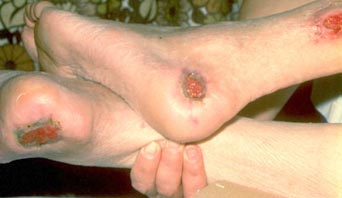
A patient came in to see me today with a sore on his heel that’s been bothering him for the past few months. He’s diabetic and has lost feeling in the bottom of his feet. He’s had these pressure sores in the past but has trouble getting them to heal up.

Pressure sore: An area of skin damaged by pressure, such as sitting or lying in one position for a long period. They are also called bedsores. They are most common where bone sits near the surface of the skin: the hips, elbows, ankles, and back or buttocks. The skin and soft tissue become damaged because not enough oxygenated blood reaches the area to support healing, usually because the soft tissue is compressed between the surface and the hard bone beneath.

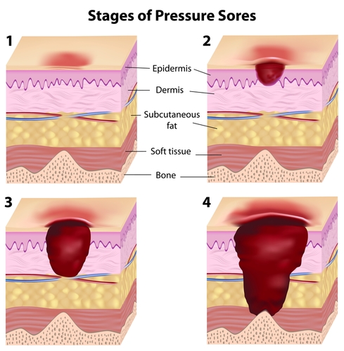
Appearance: The sores change in appearance depending on how long they have been present and how much damage has been done. At the start, the sore looks like a small red patch of skin. If it is not treated, the skin breaks down and a hole or crater forms, which is what we call an ulcer.

Stage 1: The skin is intact without ulceration, but when you press on it the color does not change the way it should. Healthy tissue is pink, and pressing on it with a finger blanches it, with the pinkness returning within a couple of seconds. That does not happen in damaged skin at this stage, and the area may look darkly pigmented.

Stage 2: There is an open, shallow ulcer with a red-pink base. Blisters may be present, either intact or ruptured.

Stage 3: Structures beneath the skin such as fat may be exposed, but at this stage you should not see bone, tendon or muscle.

Stage 4: Bone, tendon and muscle may be visible in the base of the ulcer.

People at risk: Some patients are far more likely than others to develop pressure sores.

  1. Patients who cannot move well because of a medical problem. They may sit or lie in one position for a long time and need help repositioning so the skin does not break down.
  2. Older people, who often move less and whose skin is thinner and more fragile.
  3. Patients with diabetes or nerve problems in their feet, who may not feel a pebble in a shoe or pressure building against the foot.
  4. Patients in the hospital or a nursing home, who are at especially high risk because several of these factors stack: increased age, reduced mobility, and other complicated medical problems.

Prevention: Several things lower the chance of developing pressure sores.

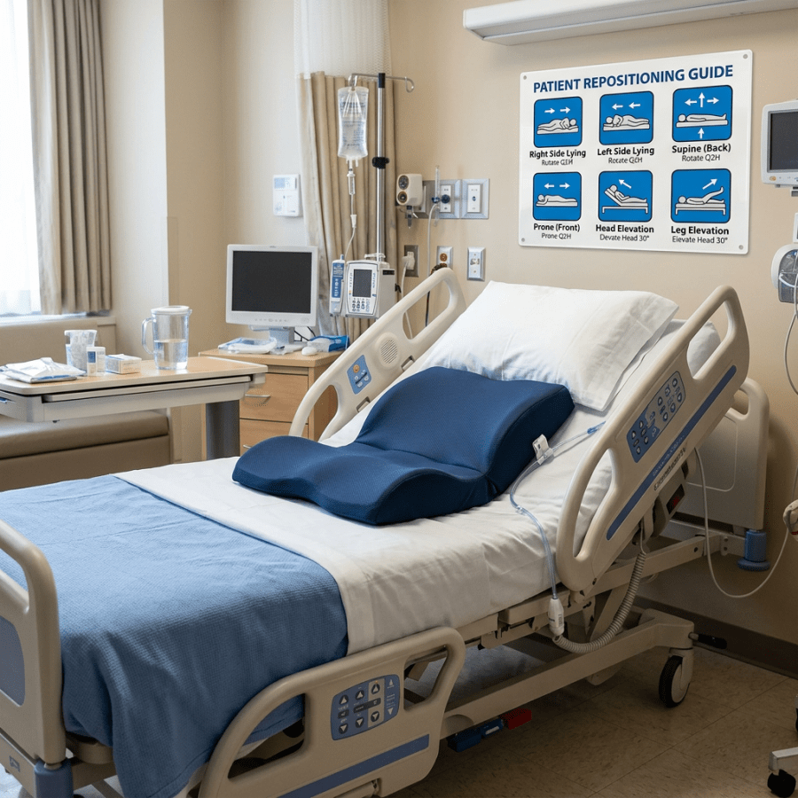
  1. Repositioning the patient’s body every two hours so no one area of skin stays crushed, pinched or under pressure.
  2. Putting pillows between the ankles and knees to reduce pressure on the skin over those bony areas.
  3. Raising the head of the bed when the patient is lying on their side to reduce pressure on the hip bone.
  4. Using special foam or soft mattresses that reduce pressure over the areas that bear the most load.

For patients in wheelchairs:

  1. Use a cushioned seat if possible to prevent pressure on the sacrum.
  2. Tilt forward or to the side every hour to release pressure on the seat.
  3. If the ankles or heels press against the chair, use foam padding to protect against sores.
  4. Check the skin regularly for signs of pressure or ulceration.

Treatment: Pressure sores are treated differently depending on the stage and how severe the skin damage is.

  1. If there is mild erythema, treatment is generally offloading the area by reducing the time it spends compressed, through repositioning and pillows to cushion it. We also use transparent films over the ulcers to protect them.
  2. In patients with diabetes, keeping blood sugars under good control is very important. Elevated blood sugars impede wound healing.
  3. If there is dead or dying skin or soft tissue, it often needs to be removed to help prevent infection.
  4. Special bandages may be needed to keep the healing tissue moist while preventing maceration from being too moist. The dressings we use can be expensive.
  5. Antibiotics may be prescribed if there is a wound infection.
  6. Medication for pain may also be prescribed.

There are scoring tools to grade pressure and track healing. These help for patients who return for repeat visits to a wound care clinic or their primary care provider and need their progress graded. Clinical features examined include:

  1. Amount of exudate
  2. Skin color surrounding the wound
  3. Peripheral tissue swelling
  4. Peripheral tissue firmness around the wound
  5. Amount of granulation (healing) tissue
  6. How much epithelialization is present

It’s important to optimize the nutritional status of patients with wounds. Patients with stage 3 and stage 4 ulcers in particular need enough protein and calories to heal.

Related Reading

Diabetic Foot Care: How to Check and Protect Your Feet Basic Wound Care Tips for Non-Medical Professionals Cellulitis – a soft tissue/skin infection – is it MRSA? Calluses and Corns: Causes, Prevention, and Treatment

Scott Rennie, D.O.

Board Certified in Obesity Medicine and Family Medicine

This blog is for educational purposes only and does not constitute individual medical advice. Always consult your own physician before making changes to your health, medications, or treatment plan.

Blog: https://doctorrennie.wordpress.com