Three blank white squeeze tubes on a laboratory countertop

A comparison of topical steroid medications

shutterstock_47533036Many patients come into the clinic and are confused about steroid creams, ointments, lotions, sprays, foams, and gels.  They come in many different forms and vary in their degree of potency.  They are generally grouped into classes according to their strength.  The medical provider’s choice in prescribing  a particular topical steroid medication depends on many factors including the medical condition (ie. eczema, psoriasis, bee sting, allergic reaction, etc.) the location of the body that is being treated and of course the patient’s ability to afford the prescription. There are some potentially harmful consequences that can occur if a steroid cream is used inappropriately.  For example, applying a high potency steroid cream to an area of the body such as the face or groin can cause permanent thinning of the skin. Here is a list of the different classes or categories that topical steroids are grouped into: Class 1:  Superpotent Class 2:  Potent Class 3:  Upper Mid-Strength Class 4:  Mid-Strength Class 5:  Lower Mid-Strength Class 6:  Mild Class 7:  Least Potent Over the counter topical steroids are generally in the class 7 or least potent category. Some examples of topical steroids and their respective classes include:
CLASS 1—Superpotent
Clobex Lotion/Spray/Shampoo, 0.05% Clobetasol propionate
Cormax Cream/Solution, 0.05% Clobetasol propionate
Diprolene Ointment, 0.05% Betamethasone dipropionate
Olux E Foam, 0.05% Clobetasol propionate
Olux Foam, 0.05% Clobetasol propionate
Temovate Cream/Ointment/Solution, 0.05% Clobetasol propionate
Ultravate Cream/Ointment, 0.05% Halobetasol propionate
Vanos Cream, 0.1% Fluocinonide
Psorcon Ointment, 0.05% Diflorasone diacetate
Psorcon E Ointment, 0.05% Diflorasone diacetate
CLASS 2—Potent
Diprolene Cream AF, 0.05% Betamethasone dipropionate
Elocon Ointment, 0.1% Mometasone furoate
Florone Ointment, 0.05% Diflorasone diacetate
Halog Ointment/Cream, 0.1% Halcinonide
Lidex Cream/Gel/Ointment, 0.05% Fluocinonide
Psorcon Cream, 0.05% Diflorasone diacetate
Topicort Cream/Ointment, 0.25% Desoximetasone
Topicort Gel, 0.05% Desoximetasone
CLASS 3—Upper Mid-Strength
Cutivate Ointment, 0.005% Fluticasone propionate
Lidex-E Cream, 0.05% Fluocinonide
Luxiq Foam, 0.12% Betamethasone valerate
Topicort LP Cream, 0.05% Desoximetasone
CLASS 4—Mid-Strength
Cordran Ointment, 0.05% Flurandrenolide
Elocon Cream, 0.1% Mometasone furoate
Kenalog Cream/Spray, 0.1% Triamcinolone acetonide
Synalar Ointment, 0.03% Fluocinolone acetonide
Westcort Ointment, 0.2% Hydrocortisone valerate
CLASS 5—Lower Mid-Strength
Capex Shampoo, 0.01% Fluocinolone acetonide
Cordran Cream/Lotion/Tape, 0.05% Flurandrenolide
Cutivate Cream/Lotion, 0.05% Fluticasone propionate
DermAtop Cream, 0.1% Prednicarbate
DesOwen Lotion, 0.05% Desonide
Locoid Cream/Lotion/Ointment/Solution, 0.1% Hydrocortisone
Pandel Cream, 0.1% Hydrocortisone
Synalar Cream, 0.03%/0.01% Fluocinolone acetonide
Westcort Cream, 0.2% Hydrocortisone valerate
CLASS 6—Mild
Aclovate Cream/Ointment, 0.05% Alclometasone dipropionate
Derma-Smoothe/FS Oil, 0.01% Fluocinolone acetonide
Desonate Gel, 0.05% Desonide
Synalar Cream/Solution, 0.01% Fluocinolone acetonide
Verdeso Foam, 0.05% Desonide
CLASS 7—Least Potent
Cetacort Lotion, 0.5%/1% Hydrocortisone
Cortaid Cream/Spray/Ointment Hydrocortisone
Hytone Cream/Lotion, 1%/2.5% Hydrocortisone
Micort-HC Cream, 2%/2.5% Hydrocortisone
Nutracort Lotion, 1%/2.5% Hydrocortisone
Synacort Cream, 1%/2.5% Hydrocortisone
  I hope that you have found this information useful.  Wishing you the best of health,

Scott Rennie, D.O.
Board Certified in Obesity Medicine and Family Medicine

This blog is for educational purposes only and does not constitute individual medical advice. Always consult your own physician before making changes to your health, medications, or treatment plan.
Gloved medical professional arranging surgical instruments on a sterile operating table

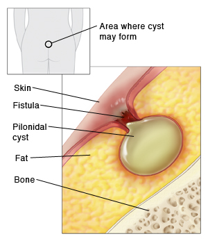
Pilonidal Cysts – A pain in the rear

Photo credit:  http://www.pilonidalcystcure.com

A pilonidal cyst tends to announce itself in a particular way.  They usually have complaints of difficulty sitting down or laying on their back because of pain.

A pilonidal cyst is a fluid-filled sac that forms above the crease of the buttocks.  The cysts are usually red, inflamed and most often infected when I see them.  In many cases the cysts are hard, but sometimes they have a small hole in them where some fluid or blood has been draining.  Pus (milky yellow/green fluid) is sometimes draining from them as well.  The drainage often has a foul smell.

Cause:  The cause of these cysts is not clear. They may form when a hair is forced under the skin or when a hair follicle ruptures. Or, a person may be born with the cyst. These cysts are often diagnosed in people between the ages of 16 and 26. But people of any age can have a pilonidal cyst. And they affect both men and women.

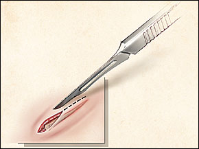
Treatment:  We usually refer patients to a surgeon who can drain the cyst or remove it with surgery.  They are often very painful, so performing an incision and drainage in the urgent care or clinic is often not possible because the patient requires a higher level of pain control and possibly conscious sedation to tolerate the procedure.  To drain a cyst, the medical provider will first numb the area and then cut open the cyst with a scalpel and push the fluid out.  They will then wash the area to clean it thoroughly and sometimes they might pack the empty cyst with gauze or leave a drain in place so that it doesn’t fill up with blood or pus again immediately.  The drain or gauze can then be removed in a few days.  We usually try to provide the patient with medication for pain, and often recommend sitting on a doughnut-like pillow with a hole in the center to help with pain.

Removing the cyst entirely often requires a more extensive surgical procedure because pilonidal cysts often have a sinus (small channel) that goes deep under the skin and can lead to another cyst or be deep enough to almost touch the sacrum or tailbone area.  Patients with multiple cysts often need an even larger surgical procedure and consequently the recovery time can be much longer for multiple or larger cysts.


I hope that you have found this information useful.  Wishing you the best of health,

Carrying extra weight is one of the recognized risk factors for pilonidal disease and for it coming back after surgery. Weight is the other half of my practice now, so if that is on your list, here is what Wegovy and Zepbound cost without insurance.

Related Reading

Abscesses and MRSA: What To Do About a Skin Infection

When to Use Telemedicine vs. Urgent Care: How I Spot the Sick One What is Hidradenitis suppurativa? Hemorrhoids Explained: Why They’re a Real Pain in the Rear

Scott Rennie, D.O.
Board Certified in Obesity Medicine and Family Medicine

This blog is for educational purposes only and does not constitute individual medical advice. Always consult your own physician before making changes to your health, medications, or treatment plan.
Tweezers removing debris from beneath a person’s fingernail

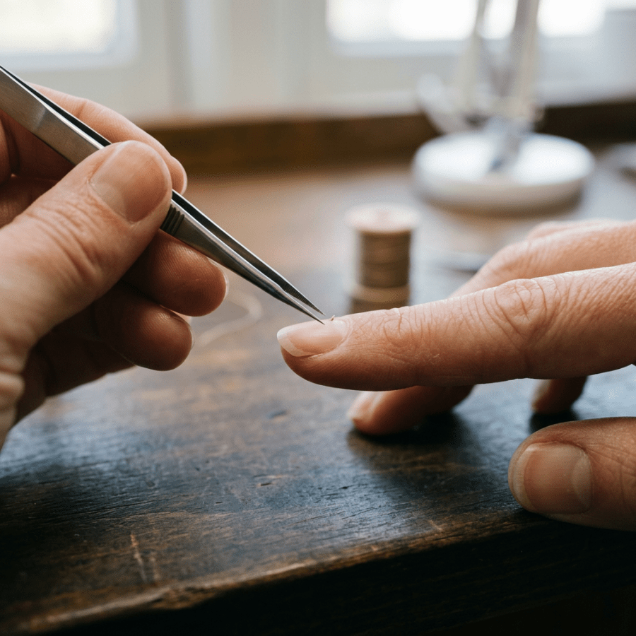
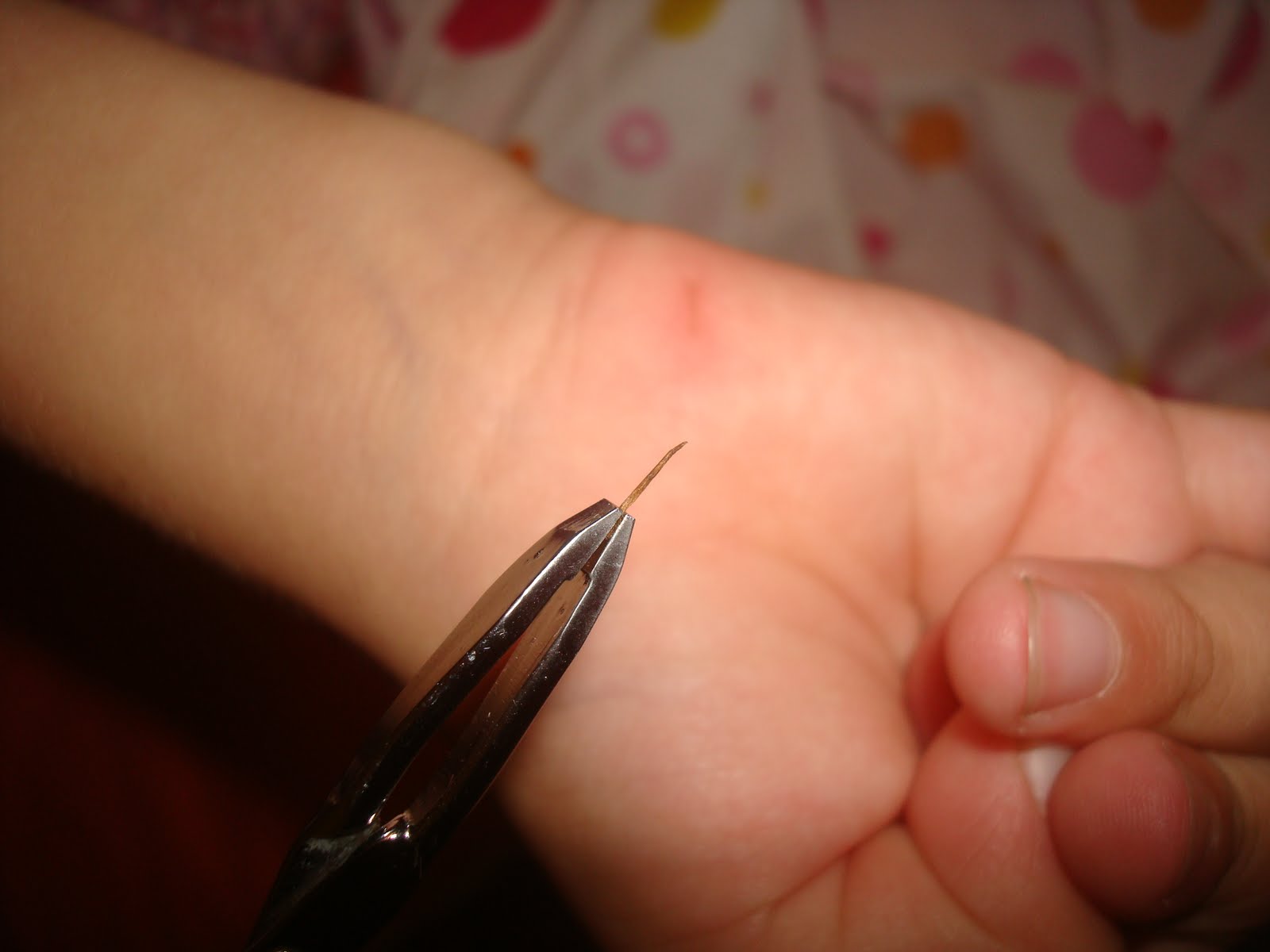
Splinter (Sliver) Removal


Photo credits: http://www.lemamme.it/sos-cosa-fare-quando-il-bambino-si-fa-male/

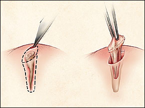
http://www.aafp.org/afp/2003/0615/p2557.html

Splinters look like the simplest possible problem, right up until one of them is not.  I thought I’d discuss some different types of splinters and some techniques that you can use at home to remove them.  In addition, it’s important to know when to have it taken out in person for help with that process.

Splinters are actually very common, and children and adults need it taken out in person usually with a small foreign body embedded into the superficial or subcutaneous soft tissues of the hands or feet.  Wood, glass and metallic splinters are the most common types.  Many splinters can be removed at home by our patients, which leaves the physicians only the deeper and larger splinters or retained splinters that have been broken down during an attempt at removal.  If splinters are not removed completely, they may cause complications such as infection and inflammation.

When to see a medical provider:  I recommend that you see a medical provider instead of trying to remove the splinter yourself if there is lots of redness around the splinter, increased swelling or pus draining from the wound.

The severity of the reaction and type of reaction that the body has to a particular splinter.  Some examples of reactions to retained foreign materials include:

1)   Glass – Mild reaction and the body attempts encapsulation

2)   Blackthorns – Severe reaction with inflammation from the plant alkaloids

3)   Wood – Severe reaction with inflammation from the oils and resins

4)   Cactus Spines – Moderate to severe reaction from the fungal coating on the plant and possibly an allergic reaction

5)   Rose thorns – Moderate to severe reaction with inflammation from the fungal coating on the plant

6)   Sea urchins – Moderate to severe reaction with inflammation and possible infection

7)   Metal – Mild reaction and the body attempts encapsulation

8)   Plant spines – Mild to severe reaction from the toxins in the plant

9)   Animal spines – Mild to severe reaction from the toxins in the animal spines

10)  Plastic – Mild reaction and the body attempts encapsulation

Sometimes splinters can be difficult to detect, especially if they are deep or very small.  Clues that a splinter is present might be swelling, tenderness, a mass, or soft tissue infection such as cellulitis, or an abscess.  Sometimes we might use a diagnostic test such as a x-ray, ultrasound, CT scan or MRI to detect a splinter if it’s deep and difficult to see.

Removal:  We try to remove splinters quickly before inflammation or infection occurs.  There are several ways to remove a splinter, some of those might include:

1)   Using tweezers: Make sure you have the right kind of tweezers.  The sharp tipped splinter tweezers work best most of the time.  If the sliver is sticking out of the skin, clean the pair of tweezers with alcohol and carefully pinch the sliver as close to the skin as possible and gently slide it out.

2)   Use a small needle:  If the sliver isn’t sticking out of the skin, often a needle can help push the sliver out.  First clean the needle with alcohol and then push the sliver from the bottom (the pointed end that entered the skin first) and angle upward toward the puncture hold in the skin’s surface.  Try to start at the bottom of the sliver so you don’t break it and leave sliver shreds in the skin.

3)   Use a razor blade to make a small slice in the skin over the sliver:  If the sliver is deep and using tweezers or a needle isn’t working well, you can clean a razor blade in alcohol and make a shallow incision parallel to the sliver, just above it.  I know this sounds scary, but the top layer of skin is made of dead skin cells that do not have nerves, so this is not likely to hurt unless you cut too deeply.  Once the incision is made, gently part the skin and pick out the sliver.

4)   Clean the wound:   Whatever method you use to remove the sliver, make sure you clean the wound with soap and water after the sliver is removed.  Puncture wounds are often laced with bacteria and are very likely to become infected.

5)   Antibiotics:  Apply a small amount of antibiotic ointment such as bacitracin over the puncture wound.

6)   Examine the area at least once a day for the next several days to look for increasing redness, swelling or pain.  If any of these occur, I recommend that you see a medical provider because this might be the start of an infection.


I hope that you have found this information useful.  Wishing you the best of health,

A splinter you cannot reach is one of the few things a video visit genuinely cannot fix. I wrote about the conditions that still need an in person visit.

Related Reading

Abscesses and MRSA: What To Do About a Skin Infection

When to Use Telemedicine vs. Urgent Care: How I Spot the Sick One Seborrheic Keratosis: The Skin Growth That Looks Stuck On Skin Tags (Acrochordons): What They Are and Look Like Calluses and Corns: Causes, Prevention, and Treatment HPV (Human Papillomavirus) Vaccine: What You Need to Know

Scott Rennie, D.O.
Board Certified in Obesity Medicine and Family Medicine

This blog is for educational purposes only and does not constitute individual medical advice. Always consult your own physician before making changes to your health, medications, or treatment plan.
Woman grimacing and holding her lower back in a living room

Kidney Stones: Pain That Can Rival Childbirth, Explained

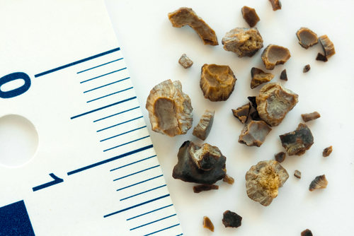
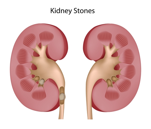
shutterstock_116071294shutterstock_77325922I’ve had quite a few female patients who’ve told me that the pain of having a kidney stone is similar to the intensity of pain during childbirth.  A patient came in the other night with intense abdominal/side pain associated with nausea and vomiting.  At first I thought he might have had appendicitis but after we were able to give him enough fluid by IV to rehydrate him and get a urine sample I saw he had blood in his urine and the CT scan showed a 3mm stone in his kidney. Kidney stones (also called nephrolithiasis or urolithiasis) are small solid fragments or crystals with sharp barbs on them that form within the urinary tract.  They can be made up of calcium, oxalate, cysteine, uric acid or a combination of these and other substances.  When these crystals form within the kidney or urinary tract they can become anchored or stuck and get larger.  Sometimes additional kidney stones are also formed while one is already lodged within the kidney or ureter.  The ureter is a small tube that drains the urine from the kidney down to the bladder. When a kidney stone travels down the ureter it can cause pain that radiates from the kidney all the way down the side and abdomen to the urinary bladder. Risks for Developing Kidney Stones:  Certain factors can increase your chances of developing a kidney stone including: 1)   Dehydration 2)   Diet with high level of sugars 3)   High sodium diet 4)   Low calcium in your diet 5)   Diet with lots of animal protein 6)   Diet with low levels of phytate (phosphorus that we get mostly from plants) 7)   Patients with gout 8)   Obesity 9)   Crohn’s disease 10)  History of bariatric surgery such as gastric or intestinal bypass surgery 11)  Diabetes 12)  Hyperparathyroidism Symptoms: 1)  Pain – most patients complain of pain either in the back, flank or abdomen.  The pain seems to come on in waves (called renal colic) and sometimes it may be barely noticeable and then become so intense that it requires treatment in the hospital. 2)  Bloody urine 3)  Pass small stones in the urine 4)  Nausea 5)  Vomiting 6)  Pain with urination Diagnosis:  Sometimes the patient’s symptoms and history can be very suggestive of kidney stones and making the diagnosis is easy.  Other times it can be more challenging to diagnose kidney stones and imaging studies such as a CT scan or ultrasound.  Plain x-rays can also sometimes show kidney stones. Treatment:  For patients who come to the medical clinic we try to ensure that they are well hydrated to prevent the formation of more stones and help the current stones to pass.  Some patients need IV fluids to help with the hydration process, especially if they have been vomiting and aren’t keeping fluids down.  Depending on the size of the stone and location, the patient may be able to treat their symptoms at home with over the counter pain medications such as Advil or Aleve.  In addition, we may prescribe medications that help the stone to pass more easily.  If the pain becomes severe, the patient may need to be treated with stronger pain medications and sometimes hospitalization is necessary.  If the stones are larger than 5mm, they don’t pass on their own very easily and generally require a procedure to break them up or remove them.  Some treatments to remove the stones may include: 1)   Shock wave lithotripsy (SWL):  Ultrasonic energy is used to break up the larger stone so that it can be passed more easily 2)   Percutaneous nephrolithotomy (PNL):  A surgical procedure that is minimally invasive may be required to remove a stone that is extremely large or complex, especially if SWL is not effective.  Small instruments are inserted through the skin in the operating room to remove the stone. 3)   Ureteroscopy:  A small scope is passed through the urethra up into the bladder and then up into the ureter.  This scope has a small camera and instruments that are used to remove the stone or break it up into small pieces.  This procedure is used more often if a stone is blocking the ureter and causing swelling of the kidney.   I hope that you have found this information useful.  Wishing you the best of health,

Scott Rennie, D.O.
Board Certified in Obesity Medicine and Family Medicine

This blog is for educational purposes only and does not constitute individual medical advice. Always consult your own physician before making changes to your health, medications, or treatment plan.
Woman sleeping on folded arms beside a glass of water

What Really Causes a Hangover? The Truth Explained

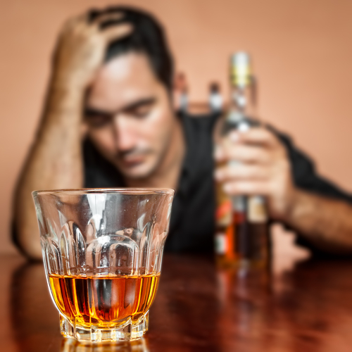
shutterstock_137241881It’s the 4th of July and it is a big night for parties for many people.  People often drink on holidays so I thought it was important to discuss hangovers. A hangover is the unpleasant set of symptoms that come with drinking too much alcohol too quickly.  The particular set of symptoms experienced and their intensity may vary from person to person and depend on the occasion.  The characteristics may also depend on the type of alcohol consumed and the amount that a person drinks.  Typically, a hangover begins within several hours after the individual stops drinking.  Many people are probably familiar with these, but here is a list of some of the symptoms that may be present with hangover: 1)   Headache 2)   Dizziness 3)   Nausea & Vomiting 4)   Fatigue 5)   Disrupted sleep – it may be easy to fall asleep but as the alcohol wears off, people often wake up frequently and complain of feeling tired the next day. 6)   Increased thirst 7)   Sensitivity to light and sound 8)   Muscle aches 9)   Rapid heart rate 10)  Tremor 11)  Sweating Dehydration is common because alcohol causes the body to increase urination.  Increased sweating, vomiting and diarrhea are also common during a hangover and that can lead to dehydration and increase and the symptoms of hangover. Here are several tips to prevent a hangover: 1)   Have something to eat before you drink alcohol – fat slows your body’s absorption of alcohol – eat steak, sausage, fried foods or pizza before going to the party 2)   Drink slowly 3)   Drink in moderation – The National Institute on Alcohol Abuse and Alcoholism recommend that women have no more than 1 drink per day and men no more than 2 drinks per day.  Definition of one drink:  12 ounces of beer, 5 ounces of wine or 1 ½ ounces of 80 proof liquor 4)   Drink a glass of water between alcoholic drinks 5)   Avoid alcohol completely if you want to be sure to prevent a hangover Home treatment tips for hangover: 1)   Dehydration is common with a hangover, so replace fluids by drinking  sports drinks such as Gatorade.  I suggest making a mixture of ½ sports drink and ½ water. 2)   Eat a meal as soon as you are able to. 3)   Rest.  Most hangovers are gone within 24 hours. 4)   Remember that even if you feel well the morning after drinking alcohol, the lasting effects of alcohol will reduce your ability to perform at your best. 5)   Ibuprofen or aspirin may be helpful for headache or muscle aches A Few Facts About Hangover Myths: 1)   A wake-up cocktail will only delay, not prevent your hangover. 2)   The order in which you drink alcoholic drinks does not determine the magnitude of the hangover, it’s the amount of alcohol that you drink 3)   Diet drinks will not help with hangovers Hangover pills don’t do much to help with hangovers and may actually make your symptoms worse   I hope that you have found this information useful.  Wishing you the best of health,

Scott Rennie, D.O.
Board Certified in Obesity Medicine and Family Medicine

This blog is for educational purposes only and does not constitute individual medical advice. Always consult your own physician before making changes to your health, medications, or treatment plan.
Smiling woman holding two sparklers in a flower-filled garden at dusk

11 Fourth of July Safety Tips to Know Before You Celebrate

shutterstock_142548190I thought this article had some particularly great tips for people regarding being safe during the July 4th holiday. Medical Author: Melissa Conrad Stöppler, MD Medical Editor: William C. Shiel Jr., MD, FACP, FACR 1)  Be a safe swimmer. Water sports and fireworks are two of the biggest pastimes for Fourth of July celebrations, and these are both linked to numerous deaths and injuries each year. Never swim alone, and make sure that kids’ water play is adequately supervised at all times. Many drownings occur when parents and other adults are nearby, so always have a designated chaperone for water play and don’t assume that others are watching the kids. Statistics show that most young children who drown in pools have been out of sight for less than five minutes. 2)  If fireworks are legal in your community and are a part of your celebration, be sure to store and use them safely. Keep the kids away from the fireworks at all times, and keep spectators at a safe distance. Attending fireworks displays organized by professionals is always safer than trying to put on your own show. 3) Use alcohol responsibly.  Alcohol and fireworks can be a hazardous and dangerous combination. Also, have a designated driver to bring partygoers home from the festivities. Remember also that alcohol and swimming can be as dangerous as drinking and driving. 4)  Lakes, waterways, and seas will be crowded with boats. Review safe boating practices, and don’t drink and drive your boat. Alcohol consumption while operating boats or other motorized water vessels is illegal, and you can be arrested for a BWI (boating under the influence!). Be sure that you have an adequate number of life preservers on hand for extra guests. Become familiar with the boating laws in your area. 5)  Cover food and beverages outdoors to discourage bees and wasps from attending your party. If someone is allergic to insect stings, you should have an emergency anaphylaxis kit on hand. Wearing shoes, long sleeves, and long pants outdoors and avoiding fragranced body products, bright colors, and sugary drinks can also help prevent bee stings. 6)  Apply sunscreen both before and during an outdoor party. Ultraviolet rays from the sun can cause both premature aging and skin cancer in the long term, and a painful burn the next day. Even those with darker skin should use a sunscreen with a minimum sun protection factor (SPF) of 15, according to recommendations from the American Academy of Dermatology. 7)  Check prescription medications you are taking to assure you will not have a sun-sensitizing drug reaction to the medication. Check prescription medications you are taking to assure you will not have a sun-sensitizing drug reaction to the medication. 8)  If you’ll be hiking or camping in an area where ticks are abundant, wear long-sleeved, light-colored shirts and long pants tucked into socks or boots to protect yourself from tick-borne diseases. For your skin, you can use a tick repellent with no more than 30% DEET according to the manufacturer’s instructions. Products containing DEET should not be used on children less than 2 months of age and should not be applied to the hands or face of young children. Check yourself (and your pets) for ticks at the end of the day. 9)  Spend adequate time indoors or in the shade and drink plenty of fluids to avoid heat illness in extremely hot climates. The risk of heat illness is increased when participating in strenuous activity or sports, and those with chronic medical conditions and the elderly are also at an increased risk of heat exhaustion and/or heat stroke. Alcohol consumption can also promote dehydration and increase the risk. 10)  Keep children away from campfires and grills. Gas leaks, blocked tubes, and overfilled propane tanks can be a cause of grill fires and explosions. 11)  Don’t leave the picnic spread out all day. Allowing food to sit in outdoor temperatures can invite foodborne illness. The U.S. FDA suggests never leaving food out for more than one hour when the temperature is above 90 F and not more than two hours at other times. Foods that need to be kept cold should be placed in a cooler with plenty of ice or freezing packs and held at a maximum temperature of 40 F. While mayonnaise and other egg dishes are often associated with food poisoning, any food can potentially become contaminated. Adequate hand washing and food preparation can also help prevent food poisoning.

Scott Rennie, D.O.
Board Certified in Obesity Medicine and Family Medicine

This blog is for educational purposes only and does not constitute individual medical advice. Always consult your own physician before making changes to your health, medications, or treatment plan.

Golden retriever sleeping on a patterned rug beside a sofa

How to Keep Your Pets Safe and Calm This 4th of July

My dogs hate fireworks and I was just discussing what we are going to do with our dogs tomorrow night during all the fireworks.  I will be working in the Urgent Care and my wife will be attending some 4th of July festivities with friends, so our dogs would be home alone.  We will make accommodations so that they will not be alone.  I came across an article that I thought was useful and timely regarding 4th of July safety tips for pet owners. By:
Denver Pets Examiner
When celebrating with family and friends this 4th of July, remember that fireworks and some common barbecue foods can be hazardous to the well-being of pets. Despite most firework shows being banned this year in Colorado due to fire dangers, fireworks will still be the biggest perpetrators of 4th of July issues for pets, the most common being pet noise phobias. The loud noises can cause fear and anxiety for pets of all kinds, including dogs and cats and even horses. It is best to keep dogs and cats a safe distance from the activity – indoors is best. For pets with severe noise phobias, a veterinarian can prescribe anti-anxiety drugs or sedatives to help ease the stress. Larger pets, like horses and other livestock, are extremely susceptible to noise phobias. While most pet owners are aware of noise phobias, many are unaware that unused fireworks can be poisonous if ingested by curious dogs or cats. Many contain hazardous chemicals like chlorates, potent oxidizing agents that are harmful to red blood cells and kidneys; soluble barium salts that cause a life-threatening drop in potassium; sulfur; and coloring agents, which can contain dangerous heavy metals. Gastrointestinal issues like vomiting, a painful abdomen and bloody diarrhea can result. The severity of the reaction will depend on the type of firework, the amount ingested and what type of coloring agents it contains. In severe cases, pets can suffer tremors or seizures, along with acute kidney failure, bone marrow changes, shallow breathing and jaundice (yellowing of the skin).

Scott Rennie, D.O.
Board Certified in Obesity Medicine and Family Medicine

This blog is for educational purposes only and does not constitute individual medical advice. Always consult your own physician before making changes to your health, medications, or treatment plan.
Clinician reviewing a chest CT scan on a computer monitor

Blood Clot in the Lung: What Is a Pulmonary Embolism?

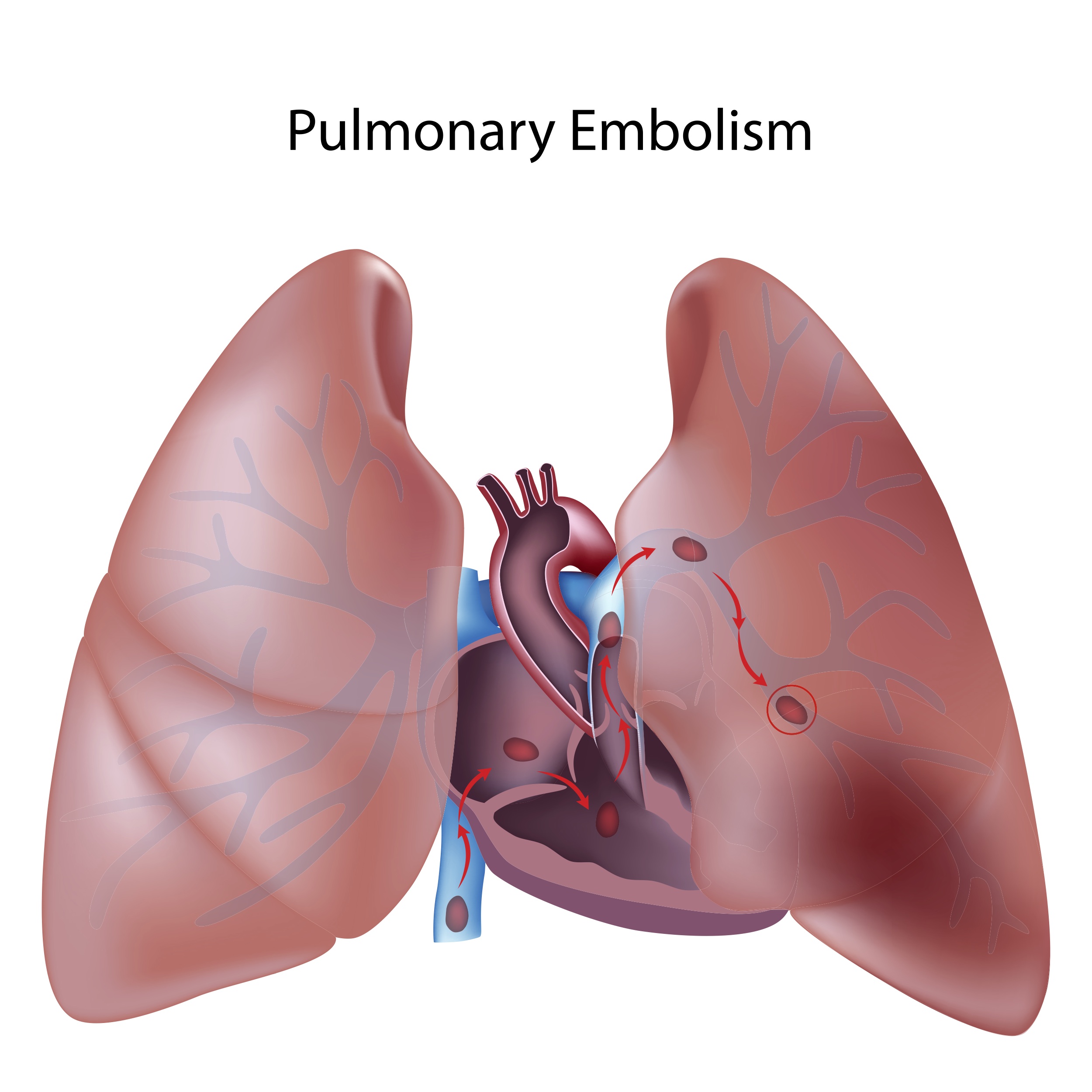
shutterstock_94626226A patient who regularly competes in Iron Man Triathlons came into the clinic tonight with complaints of pain with taking a deep breath.  She had coughed up some blood once today and complained that it hurts when she takes a big breath in.  She is an athlete and remembered running in a race about three weeks ago and developed pain in the right calf and behind the right knee.  That pain has been improving over the last few weeks.  As you can probably guess by the title of this blog entry, she had a blood clot in her lungs.  Actually she had large blood clots called pulmonary emboli in both lungs.  She required transport emergently to the hospital for treatment. Definition:  A pulmonary embolism (PE) is a blockage of one of the blood vessels to the lungs.  These are usually caused by a blood clot that travels from somewhere else in the body, most commonly from the legs.  Other causes of blockages to the blood vessels in the lungs are air bubbles, or from tiny pieces of fat. A pulmonary embolism is a serious condition that is a medical emergency.  A blocked artery or vein in the lung can make it hard to breath and can lead to damage to the lungs and can even cause death. Most of the time the blood clots start in the legs, or in the pelvic area and then move along the veins and travel to the lungs. Symptoms:  People who have blood clots in the lungs might complain of: 1)   Sharp, stabbing pain when they take a breath in 2)   Coughing up blood 3)   Rapid heart rate 4)   Trouble breathing or feeling short of breath If you have any of these symptoms, it is important to call 9-1-1 and be evaluated in the hospital.  Do not try to drive yourself to the urgent care or to the hospital yourself. Risk Factors:  There are some factors that can increase the chance of having a blood clot in your lungs including: 1)   Having a family history of blood clots or blood clotting disorders 2)   Having recent surgery – especially on the knees, lower legs or pelvis 3)   Smoking 4)   Taking birth control pills or estrogen replacement 5)   Having a previous history of DVT (Deep Venous Thrombosis) or PE 6)   Having a history of heart failure 7)   Patients with cancer 8)   History of kidney problems such as nephrotic syndrome 9)   Having a recent trauma or injury with a large clot (hematoma) – more likely to cause DVT if it’s in the legs Diagnosis:  To diagnose a pulmonary embolism, special tests such as a special x-ray called a CT scan (Cat-Scan) with contrast.  During this test, the patient has special a detailed x-ray of the lungs while contrast (a solution of medication that can be seen under x-ray) is put into a vein (usually in the arm) and circulates through the lungs and rest of the body.  The CT scan is used to see the blood vessels and determine if a blood clot is present.  There are other tests to determine if a pulmonary embolism is present as well such as a ventilation/perfusion lung scan, but we usually use CT scan instead because it is easier and less time consuming.  If we suspect that there might be a blood clot in a patient’s leg, we can perform a vascular ultrasound to look for blood clots in the vessels of the legs. Treatment:  We start treatment of a pulmonary embolism without delay to prevent a new blood clot from forming and to reduce the chance of enlargement of the existing clot.  Usually your own body’s enzymes dissolve the clot.  We treat patients by anticoagulation.  That means that we give medications to reduce the body’s abilities to form new blood clots – this is also sometimes called using “blood thinners.”  It isn’t actually thinning the blood however.  For patients who are at high risk for forming new blood clots, we sometimes put small screens or “filters” in a major blood vessel such as the inferior vena cava to prevent a new blood clot from reaching the lungs. Most of the time we use a combination of oral anticoagulation medications such as Coumdin and also use an injectable medication such as heparin.  Patients who have pulmonary embolisms often stay in the hospital for a few days and receive intravenous heparin to prevent more blood clots from forming.  It takes a few days for the medication that is taken orally to become effective, so during this time the patient is giving the heparin either through an IV or the heparin is injected under the patients skin. Patients who have had a PE usually take the Coumadin (also called Warfarin) for a minimum of three months.  Some doctors recommend that patients with a PE take Coumadin or something similar for the rest of their lives.  Other medications to prevent blood clots might be used if the patient is pregnant or cannot tolerate Coumadin. Prevention:  Patients who are in the hospital and confined to the bed for extended periods of time are usually started on a prophylactic dose of heparin each day or special compression devices on their legs to prevent blood clots.  People who are on long air plane flights, car, bus or train rides should get up regularly and stretch their legs to help prevent the development of the blood clot in their legs (DVT).  Patients who have had recent surgery often use heparin injections even at home for a few days to help prevent the development of a blood clot in their leg. If you or someone you know if having symptoms of a pulmonary embolism (PE), they should dial 9-1-1 and go to the hospital immediately.  They should not attempt to drive to the hospital or urgent care.   I hope that you have found this information useful.  Wishing you the best of health,

Scott Rennie, D.O.
Board Certified in Obesity Medicine and Family Medicine

This blog is for educational purposes only and does not constitute individual medical advice. Always consult your own physician before making changes to your health, medications, or treatment plan.
Sole of foot with dry, peeling skin and calluses

Calluses and Corns: Causes, Prevention, and Treatment

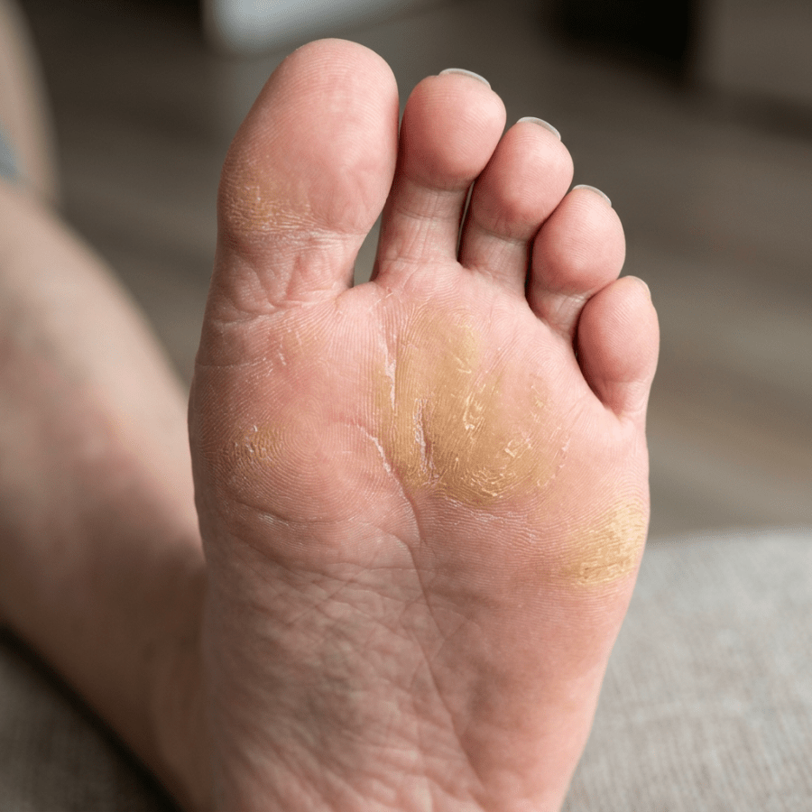
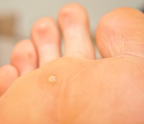
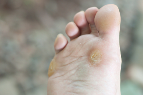
shutterstock_113610118shutterstock_93910513I can’t tell you how many patients come in to the clinic because of a callus or corn that’s bothering them – too many to count!  Corns and calluses can cause significant pain, especially when they’re on the feet because we often get them in areas over pressure areas.  When they get thickened they can re-distribute your weight onto other areas of your feet and that may lead to foot instability or worsening pain. Calluses are usually on the hands and feet and basically just thickened areas of skin that form when something rubs or presses on these areas over prolonged periods of time. Corns are thickened areas of skin that are often on the soles of the feet or sides of the toes and look like a small nodule that has a hard center.  Corns are usually more painful because they are often over a smaller area and press harder on a more specific area of the foot. Causes:  Possible reasons that calluses or corns develop include: 1)   Wearing shoes that don’t fit properly for your feet – either too loose or too tight 2)   Walking barefoot 3)   Wearing shoes without socks 4)   Calluses on the hands can be caused by repetitive sports such as rowing, golfing, tennis or biking without gloves Prevention: 1)   Get shoes that fit properly!  It’s important to be proactive and to prevent calluses or corns if they bother you.  There are special shoe stores that work with foot doctors to help you select shoes that fit your feet properly.  Some people are born with narrow or wide feet and that makes it more difficult to find shoes that fit properly.  If you are getting painful corns or calluses, take time and visit a store where someone can help you to pick out shoes that fit you well 2)   Avoid going barefoot or wearing shoes without socks 3)   If you have spots on your feet that rub inside your shoes, you can get special pads that prevent rubbing Treatment:  I often work with patients who come into the clinic to trim the corn or callus down so that it reduces the pressure in the affected area.  I use a scalpel to carefully trim away the thickened skin.  A foot doctor (podiatrist or orthopedic surgeon who specializes in treating conditions of the foot/ankle) can make special orthotic devices that can help reduce the pain and help prevent recurrence of corns/calluses.  Some people treat calluses themselves by purchasing special pads that contain medications to burn or dissolve the thick skin.  I highly recommend that patients with diabetes be seen and treated by a medical provider to reduce the risk of infection.  Diabetic patients are at increased risk of foot infections and should be seen for yearly foot examinations even if they don’t have calluses or corns.   I hope that you have found this information useful.  Wishing you the best of health,

Scott Rennie, D.O.
Board Certified in Obesity Medicine and Family Medicine

This blog is for educational purposes only and does not constitute individual medical advice. Always consult your own physician before making changes to your health, medications, or treatment plan.

Clinician examining the side of a patient’s face during a clinic visit

Why is the Side of My face Drooping? All about Bell’s Palsy

shutterstock_142857034A few weeks ago a family brought their son in to the clinic because one side of his face including his eye and lips were not moving symmetrically with the other side.  Of course they were worried about the possibility of him having a stroke.  He’d had an upper respiratory infection that started about one week before and had a slight fever with runny nose.  He’d never had any neurological problems before.  He had a condition called Bell’s Palsy. Bell’s Palsy is a problem with the nerves on one side of the face and it causes the muscles of the face to have decreased ability to move.  The muscles of the face can become weak or even paralyzed.  Patients often complain that one of their eyelids starts drooping or they drool out one side of their mouth.  When they smile, one half of the mouth doesn’t seem to move. Most people who get Bell’s palsy recover entirely but a small number of patients have symptoms for the rest of their life. Causes:  Inflammation of the facial nerve on one side of the face is the cause of Bell’s palsy.  A virus is the cause and there is some evidence that it’s the virus that causes cold sores (Herpes Simplex Virus – HSV) that causes the condition.  Other viruses may cause Bell’s palsy however including the viruses that cause Chicken Pox and Mononucleosis. Symptoms:  When the facial nerve because inflamed from the virus, and the nerve may swell and get pinched as it travel’s through some tight spaces in the face.  If this happens it can cause weakness and even paralysis of the muscles of the face so you may see: 1)   Drooping of one eyelid 2)   Eyebrow that sags 3)   Corner of the mouth that does not move or sags 4)   One eye might not close completely 5)   Loss of taste in the front of the tongue 6)   Loud noises may cause pain on the side of the dysfunction If your eye is not able to fully close, this can lead to dryness of the eye, so it’s important to seek treatment to prevent eye damage. Treatment:  We don’t have any specific treatments for Bell’s palsy, but seeing a medical provider may be helpful to: 1)   Ensure the proper diagnosis, because the symptoms may be confused with a stroke or other neurological problems which can be dangerous if not treated appropriately 2)   Prevent damage to the cornea of your eye from dryness 3)   Steroids such as prednisone may be given to reduce the swelling of the affected facial nerve – this works better when started within the first 2-3 days of symptoms 4)   Antiviral medications such as acyclovir are sometimes given in hopes that they will help the body overcome the virus more quickly, however studies have not found any added benefits from using antiviral medications for Bell’s palsy Recovery/Prognosis:  People who have less severe symptoms seem to recover more rapidly and have a better chance of full recovery.  If you are getting better within the first three weeks, the chances are better that you will totally recover.  A small group of people however have permanent moderate to severe muscle weakness in their face from Bell’s palsy. Rare effects:  If there is severe damage to the facial nerve, it may heal in a disorganized fashion.  I have one patient who was bothered by tears coming from their eye when they salivate (before eating).   I’ve also had a patient whose eye would close whenever he smiled.  Fortunately this is not common with Bell’s palsy.   I hope that you have found this information useful.  Wishing you the best of health,

Scott Rennie, D.O.
Board Certified in Obesity Medicine and Family Medicine

This blog is for educational purposes only and does not constitute individual medical advice. Always consult your own physician before making changes to your health, medications, or treatment plan.