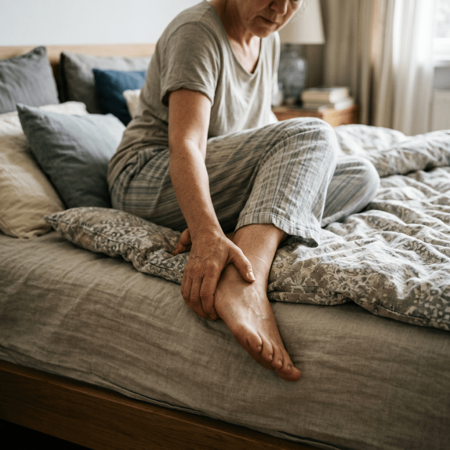
Person sitting on bed and holding their lower leg

Plantar Fasciitis Explained: A Real Pain in the Foot

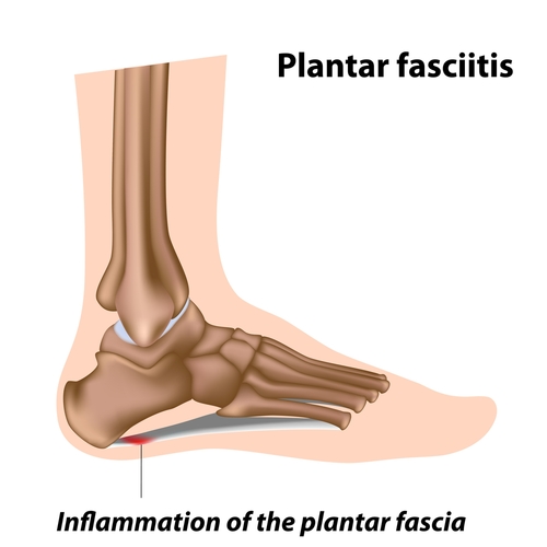
shutterstock_90181162One of the more common reasons that I see patients in the urgent care for is plantar fasciitis.  It causes foot pain, mostly in adults.  Patients are more prone to this kind of foot problem if they jump or stand for prolonged periods of time.  These motions stretch the plantar fascia.  Most people who have an episode of plantar fasciitis have no pain within one year. Causes:  A strain in the ligaments in the fascia which is a thick, white pearly tissue with long fibers that starts at the heal bone and fans out along the under surface of the foot and extends to the toes.  The plantar fascia provides support as the toes bear the weight of the body when the heal rises while walking. Symptoms:  pain beneath the heel and sole of the foot.  The pain is often worst when stepping out of bed early in the morning or getting up after being seated for a period of time. Risk factors: 1)   Long distance running 2)   Standing for long periods of time (grocery store cashiers for example) 3)   Dancing 4)   Repeated squatting or standing on the toes 5)   Poor fitting shoes 6)   Obesity Diagnosis:  The diagnosis is usually made by a healthcare provider who takes the history of the patient and performs a physical examination. Treatment:  Several treatment options are used to help with plantar fasciitis.  They may include: 1)   Rest – Limit jumping or standing for extended periods of time.  A complete lack of physical activity is not recommended however 2)   Ice – Putting ice on the area 4x/day for 20 minutes at a time may reduce pain 3)   Exercise – Home exercises including calf-plantar stretches, foot and ankle circles, toe curls, and toe towels often help reduce pain.  Careful attention not to overdo the stretches is important 4)   Pain medication – Ibuprofen or Naproxen may be helpful to reduce the swelling and pain 5)   Shoes with good arch support or metal shanks take the pressure off the plantar fascia and are one of the most important parts of treatment. 6)   Steroid Injection – a steroid shot may be given in the foot to relieve pain but usually the effect wears off after a few weeks.  There is also a risk of worsening the foot pain due to small crystals of the steroid forming in the plantar fascia 7)   Casting – a short walking cast that has a rocker-shaped bottom decreases the flexibility of the sole of the foot and can provide relief from pain. 8)   Surgery  – this is rarely recommended for plantar fasciitis.  If symptoms persist for at least 6-12 months and all other treatments have failed, it may be an option for some people   I hope that you have found this information useful.  Wishing you the best of health,

Scott Rennie, D.O.
Board Certified in Obesity Medicine and Family Medicine

This blog is for educational purposes only and does not constitute individual medical advice. Always consult your own physician before making changes to your health, medications, or treatment plan.

Office worker holding his neck while seated at a desk

Cervicalgia – A Pain in the Neck

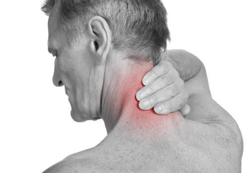
shutterstock_142402684Patients frequently come into the Urgent Care clinic for problems related to neck pain.  It can be caused by a number of different factors including ligament or muscle strain, arthritis or a “pinched nerve.” It is quite common, and in fact 10% of adults have neck pain at any one time.  Most patients recover with conservative therapy regardless of the cause of the pain.  Understanding neck pain is much easier if you have a good knowledge of the structural anatomy of the neck. Brief description of the anatomy is outlined below: 1)   Cervical vertebrae:  Seven small bones the make up the cervical spine of the neck 2)   Spinal canal:  The structure through which the spinal cord (nerves) flow which is made from the cervical vertebrae as well as the supporting ligaments and overlying neck muscles. 3)   Cervical discs:  Between the neck bones, these tiny shock absorbers cushion one bone from another.  The inner part of the disc contains a gelatin-like material and when excessive pressure on the disc occurs, this gelatin-like material can protrude and cause what we call a “herniated disc.” Causes:  There are several causes for neck pain, some of which are mentioned here: 1)   Whiplash injury:  A traumatic event that causes sudden forward/backward movement of the cervical spine.  A motor vehicle accident is the most common cause. 2)   Cervical strain:  Injury to muscles of the neck that cause spasm of the neck and upper back muscles causes this type of pain.  It can be a result of physical stresses of everyday life including poor sleep habits, muscle tension from psychological stress, or poor posture. 3)    Diffuse skeletal hyperostosis:  Also called (DISH) is when there are abnormal calcifications in the ligaments and tendons along the cervical spine. 4)   Cervical spondylosis:  Abnormal wear and tear causes gradual narrowing of the disk space and loss of normal bone structure which often leads to bone spurs.  These spurs can increase the pressure on surroundings areas. 5)   Cervical discogenic pain:  The intervertebral discs of the neck function as shock absorbers that cushion the neck bones from one another.  If there are structural changes in these discs, it can cause pain. 6)   Cervical facet syndrome:  Pain involving the facet joints is common in people who repeatedly extend the neck (tilt the head backwards).  The facet joints are on the left and right of the vertebrae. 7)   Cervical radiculopathy:  When disk or neck pass pushes on or irritates a nerve root, this can cause pain, weakness or numbness/tingling of the neck and/or arm. 8)   Cervical spondylotic myelopathy:  This is narrowing of the spinal canal inside the bones of the neck, and is usually caused by either damage to the disks or degeneration from arthritis. Testing:  In order to determine the cause of the pain, your healthcare provider will examine your neck and look at the movement or range of motion to the neck and observe posture of the neck and shoulders.  In some cases there may be a radiological study such as an x-ray, MRI (magnetic resonance imaging) or CT scan (computed tomography) ordered.  The need for on of these tests depends on the patient’s history and physical examination. Treatment:  The individual treatment is tailored to the patient to help treat the underlying cause of pain.  Some possible treatments may include: 1)   Medications:  Ibuprofen, naproxen or Tylenol may be prescribed.  Other medications such as muscle relaxants or narcotics may also be prescribed depending on the circumstances. 2)   Heat:  Can be helpful for decreasing the muscle spasm in the neck.  Moist heat (from a shower, hot tub, or moist towel warmed in a microwave or with warm water) seams to work best 3)   Ice:  Can reduce the pain in many people.  Ice is applied directly to the painful area.  A bag of ice, frozen peas or a ice cubes in a plastic bag is often used, but avoid applying the blue ice that is used for coolers/camping to the skin as this can cause freezer burn. 4)   Massage:  By applying pressure on both sides of the neck and upper back, the neck muscles may be relaxed.  This is usually most helpful if done by a professional massage therapist. 5)   Stretching exercises:  Do not attempt exercises without being evaluated by a healthcare provider as they can actually make the problem worse if performed incorrectly.  Exercises can be performed to relieve stiffness and improve range of function. 6)   Stress reduction:  Neck tension can be increased due to emotional stress and can delay the recovery process.  To help with stress reduction, breathing exercises, meditation, progressive muscle relaxation, biofeedback, prayer or self-hypnosis are helpful for some patients. 7)   Posture:  It’s important to avoid extreme ranges of motion or positions that cause constant tension.  Avoid sitting in the same position for extended periods of time.  Also avoid placing backpacks, over-the-shoulder purses, or children on the shoulders.  Do not perform overhead work for prolonged periods of time.  Hold your head up and keep shoulders back and down to maintain a good posture.  Sleep with your neck in a neutral position by sleeping with a small pillow under the nape of your neck (while laying on your back).  Carry heavy objects close to your body rather than with outstretched arms. Other treatments: 1)   Osteopathic manipulative therapy (OMT)– physically manipulating the muscles, soft tissues and joints can help with neck pain 2)   Trigger Point Injection:  A local anesthetic such as lidocaine can be injected into an area of muscle spasm 3)   Accupuncture:  A needle placed into the proper body area by a healthcare professional who is trained in accupunture therapy can be helpful 4)   Electrical stimulation:  Transcutaneous electrical nerve stimulation (TENS) is a treatment that uses a mild electric current that is applied to the skin to decrease pain and increase mobility and strength.  Some people have found TENS helpful. 5)   Cervical traction:  The use of weights to pull the spinal column into alignment have been helpful for some people in the short term, however clinical studies have shown no long term benefits. 6)   Surgery:  Surgery has a role in relieving symptoms related to a pinched nerve in some cases.  Usually these patients have tried all prior treatment options before proceeding with surgery. To find an Osteopathic physician in your area, the American Osteopathic Association has a useful website:  http://www.osteopathic.org/osteopathic-health/find-a-do/Pages/default.aspx   I hope that you have found this information useful.  Wishing you the best of health,

Scott Rennie, D.O.
Board Certified in Obesity Medicine and Family Medicine

This blog is for educational purposes only and does not constitute individual medical advice. Always consult your own physician before making changes to your health, medications, or treatment plan.
Woman seated on a sofa holding her knee with both hands

All About Knee Pain: What Every Patient Should Know

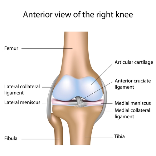
shutterstock_94626553I often have people come to the Urgent Care who have problems due to knee pain. Pain that originates in the knee can be complex because there are quite a few areas within the knee that can cause pain. The knee is a “hinge” type joint that is at the greatest risk of injury, age-related wear and tear, arthritis as well as infection related arthritis. Causes: As I mentioned above, there are many causes of knee pain. Several are listed below: 1) Patellofemoral pain syndrome – Group of symptoms that is sometimes caused from overuse of the knee. It can affect running athletes, and is more common in women. The pain is usually in the front of the knee and is made worse with squatting, running, prolonged sitting or when climbing or going down stairs. We also call this Patellar tracking syndrome because it is related to the knee cap (patella) sliding out of the groove that it normally sits in. 2) Meniscal tears – The meniscus is a specialized shock absorber that provides cushion on both sides of the knee. They can become damaged or torn from an acute knee injury or from overuse that comes with age. They have a very poor blood supply, so they don’t heal very quickly 3) Bursitis – The knee is lubricated by joint fluid that is produced by a lubricating bursa (or sac). These bursa sacs can become irritated as a result of injury or even overuse. Inflammation of the bursa is called bursitis 4) Arthritis – This refers to inflammation of the cartilage covering the ends of the bones and undersurface of the knee cap. When it gets worn down, irritated or irregular, it can become painful and is known as arthritis. 5) Tearing of a ligament – The knee is held together by a combination of ligaments including the anterior and posterior cruciate ligaments as well as the medial and collateral lateral ligaments. These ligaments function to hold the bones together and prevent side-to-side or back and forth motion. 6) Muscle strain – If the muscles on the front or back of the knee area become injured, or in spasm they can cause pain around the knee joint. These muscle groups are the quadriceps (located on the front of the top of the knee) and the hamstrings (located on the back part of the leg). These muscle groups work to give support to and move the knee joint 7) Fractures – Broken bones around the knee can obviously cause pain 8) Infection – If bacteria get into the knee joint, this can cause pain, swelling and decreased range of motion 9) Gout – This is caused by the formation of uric acid crystals that build up within the knee joint and is less common in the knee than some other joints 10) Baker’s Cyst – a cyst in the back of the knee can cause pain Diagnosis: Since there are so many different possible causes of knee pain, it is important to make an accurate diagnosis to treat the underlying problem. In addition to getting a good history from the patient about their pain, a physical evaluation will be performed and sometimes knee x-rays, an ultrasound, or an MRI will be ordered. Additionally, a small sample of synovial fluid is sometimes removed from the knee using a needle. This fluid can be examined under a microscope and/or sent for culture to look for bacteria, crystals or signs of inflammation. Treatment: The individual patients injury will dictate the kind of treatment that they need to recover the fastest. Physical therapy is often used because it speeds recovery and regain motion. We also often recommend ice, elevation of the leg and muscle-toning exercises when appropriate. Sometimes a knee brace or immobilizer may be appropriate. Medications such as ibuprofen, Aleve or Tylenol may be appropriate for knee injuries. If infection is present, antibiotics may also be prescribed. If injury to the bone or ligaments is the cause, surgery can be helpful in some patients. Limiting certain activities: Speeding the recovery and helping prevent further injury sometimes involve limiting activities temporarily. Excessive pressure on the knee joint by the following activities should be avoided to help recovery: 1) Kneeling 2) Jogging 3) Squatting 4) Twisting and pivoting 5) Aerobics/Dancing 6) Playing stop and go sports such as basketball or racquet sports 7) Swimming with frog leg or whip kick techniques 8) Rowing machine 9) Stationary bike 10) Stair stepper 11) Leg extensions with weights   I hope that you have found this information useful.  Wishing you the best of health,

Scott Rennie, D.O.
Board Certified in Obesity Medicine and Family Medicine

This blog is for educational purposes only and does not constitute individual medical advice. Always consult your own physician before making changes to your health, medications, or treatment plan.

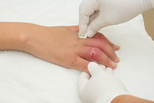
Healthcare worker bandaging a patient’s injured hand

I Hurt My Finger or Toe: How Do I Know if It’s Fractured?

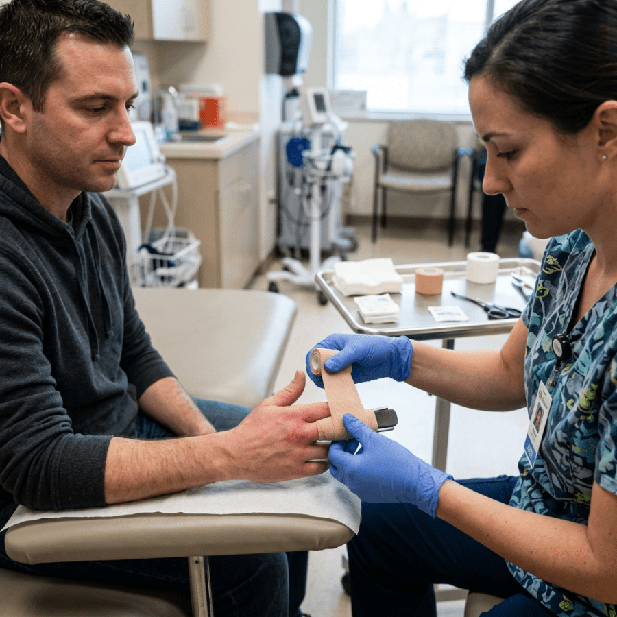
shutterstock_136665947Patients frequently come into the urgent care after injuring a finger or toe and are concerned about the possibility of a fracture. A finger or toe fracture is when a person breaks a bone within their digit. There are different types of fractures, and they are described based on how the bone breaks and whether the broken bone actually sticks out of the skin or not. Symptoms of fracture: 1) Pain 2) Swelling 3) Bruising 4) Stiffness 5) Weakness of the finger/hand or toe 6) The finger or toe looks bent in an abnormal position Testing: You healthcare provider will examine your finger or toe and obtain an X-ray Treatment: The treatment depends on the type of fracture and where it is. Some possible treatments may include: 1) Medication for pain such as Ibuprofen or Tylenol 2) Splinting or “buddy taping” 3) If the fracture is severe with a bone sticking out of the skin, you will need antibiotics and referral to a bone specialist (orthopedic physician). 3) After the splint can come off, your healthcare provider may recommend you work with a physical or occupational therapist. The therapist can show you exercises to strengthen your finger muscles and keep the fingers from getting stiff. Length of time to health: Finger/Toe fractures can take weeks to months to heal, depending on the type of fracture. Home therapy: to help the bone heal rapidly we generally recommend the following: 1) No smoking 2) Keep the cast or splint from getting wet, and keep it clean 3) Keep the finger splinted and immobile to decrease movement   I hope that you have found this information useful.  Wishing you the best of health,
Scott Rennie, D.O.
Board Certified in Obesity Medicine and Family Medicine

This blog is for educational purposes only and does not constitute individual medical advice. Always consult your own physician before making changes to your health, medications, or treatment plan.

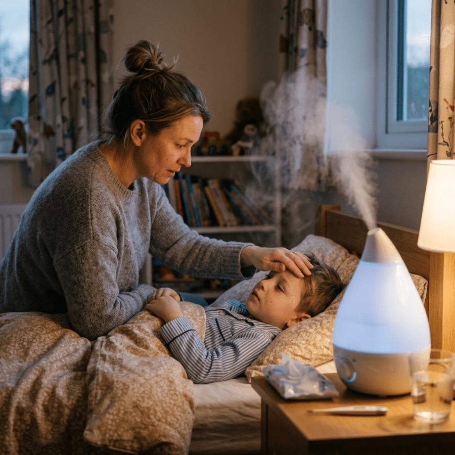
Mother comforting a child in bed beside a running humidifier

My Child Has a Barking Seal-like Cough: Is It Croup?

shutterstock_109784702A variety of respiratory illness in children can cause what we cause croup.  We see children with croup in the urgent care frequently, mostly during the winter months.  It occurs mostly in infants and young children between 6 months and 3 years of age.  We don’t see it often in kids that are older than 6 years of age.  Most of the time croup is mild and can be treated at home, but sometimes it can become serious and need immediate medical care. When to seek help:  A child with severe or worsening croup should be evaluated immediately because croup can be a life-threatening illness .  Some features of worsening or severe croup include: 1)   Drooling or difficulty swallowing 2)   Difficulty breathing 3)   Inability to speak or cry due to difficulty taking a breath 4)   A whistling sound when breathing, or noisy-high pitched breathing while sitting or resting 5)   Sucking in of the skin around the ribs or abdomen with breathing 6)   Pale or blue-tinged skin It’s important for parents not to attempt to drive their children to their medical provider if they are struggling to breath or are having severe croup symptoms.  Instead, calling 911 and having emergency medical care assist in the treatment on the scene where the child is located and help with transport is critical in this potentially life-threatening situation. Causes:  A viral infection with influenza or para-influenza are the most frequent causes although most of the time these viruses people with these viruses don’t get croup.  The infection can lead to swelling of the voice box and windpipe and as the infection progresses, this can cause the windpipe to become swollen and narrow. Bacterial infection can also cause croup or trachiitis but this is usually more severe and requires a different treatment than a viral infection. Symptoms:  Usually it starts with congestion and runny nose and then can progress to a characteristic “barking cough,” and hoarseness.  Symptoms seem to be worse at night in most children.  Fever can develop in patients as well as a rash and redness to the eyes.  Swollen lymph nodes are common.  As the upper airway narrows due to swelling from the infection, high-pitched, noisy breathing (called stridor) can develop.  If the child becomes anxious, the breathing often becomes more difficult because the agitation can increase the narrowing.  The effort to breathe faster and harder is tiring and some children become exhausted and in severe cases, they might not be able to breath on their own. A blue-tinge to the skin (called cyanosis) can develop if airflow to the lungs is restricted.  Cyanosis may first be noticed in the fingers and toenail, ear lobes, tip of the nose, lips and tongue.  This is uncommon in croup, but can happen in severe cases. Severity:  Croup is often graded in terms of severity as mild, moderate or severe.  A brief description of these grades is listed below: 1)   Mild:  Child is alert and may have a barking cough.  Stridor (high-pitched noisy breathing) is not present at rest, but may be notable as the child coughs or cries.  There are not retractions (severe abdominal or chest movements with breathing) 2)   Moderate:  Children with moderate croup may have stridor at rest.  They may also have retractions (severe abdominal or chest movement as they breath).  They may look uncomfortable. 3)   Severe:  Child with this grade of croup has stridor and chest/abdominal retractions at rest.  The child struggles to take each breath and may appear anxious, agitated or fatigued.  A patient with severe croup should generally not be seen in the primary care or urgent care and be seen in the emergency room.  These patients may be transported to the hospital by ambulance. Diagnosis:  Usually the diagnosis is made clinically and is based on the child’s symptoms including barking cough, and stridor.  X-ray or other laboratory work is rarely needed. Treatment:  The type of treatment generally depends on the severity of symptoms.  Generally moderate to severe symptoms suggest the child should be seen by a healthcare provider. Mild croup: most often is able to be treated at home without difficulty.  Use of a mist humidifier or sitting in the bathroom with parents with the shower running (to produce steam) may be helpful.  Other treatments such as allowing the child to breathe cold air during the night by opening a window or door, treatment of fever with Tylenol or Ibuprofen, and elevating the child’s head slightly may also be helpful. Moderate to severe croup:  Usually moderate to severe croup should be evaluated by a healthcare provider who is able to handle an urgent respiratory illness.  Severe croup can be life-threatening, and treatment should not be delayed.   We will use humidified air or oxygen as needed, intravenous fluids if there are signs of dehydration, and even place a breathing tube or assist in oxygenation by applying a non-rebreather mask over the child’s mouth so the oxygen is delivered in a higher concentration.  We also monitor the child’s oxygen levels, breathing rate, heart rate, skin color and the alertness of the child closely. Medications for croup:  In the clinic, we often prescribe a single to multiple doses of dexamethasone which is a steroid medication that decreases the inflammation of the airway (windpipe and voice box area).  If there is stridor or the croup is severe, we will sometimes give nebulized treatments of racemic epinephrine (aka adrenaline).  This also reduces swelling in the airway and actually starts working faster than dexamethasone.  Racemic epinephrine works for a short period of time (two hours or less) and may be given every 15-20 minutes for severe symptoms.   I hope that you have found this information useful.  Wishing you the best of health,

Scott Rennie, D.O.
Board Certified in Obesity Medicine and Family Medicine

This blog is for educational purposes only and does not constitute individual medical advice. Always consult your own physician before making changes to your health, medications, or treatment plan.

Woman holding her wrist beside a laptop at a home office desk

Finger and Hand Pain: Is It Carpal Tunnel Syndrome?

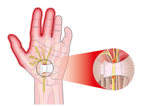
shutterstock_86604217Many patients come to the Urgent or their primary care office with complaints of hand/wrist pain, numbness, tingling or a combination of these symptoms.  They often wonder if it could be due to carpal tunnel syndrome. Carpal Tunnel Syndrome:  characterized by pain and numbness in the fingers and hands, and sometimes in the arms.  It happens when the median nerve in the wrist becomes pinched or squeezed.  The median nerve travels through a small “tunnel” in the wrist that is formed by bones and a ligament.  It’s a setup for this nerve to become pinched due to our normal anatomy, but may be worse in some people or due to certain conditions.  There is some thought that the nerve gets pinched possibly due to one or more of the following: 1)   Tissues that surround the surrounding tends in the tunnel harden 2)   Tendons that go through the tunnel get swollen 3)   People hold their hands in a position that causes the tunnel to get smaller. Parts of the hand affected by the median nerve: 1)   Thumb 2)   Index finger 3)   Middle finger 4)   Half of the ring finger 5)   The parts of the palm closest to the thumb Symptoms:  Pain, and tingling in the thumb, index, middle and ring fingers.  These symptoms may be present in one or both hands.  Rarely, the pain can travel up the wrist and forearm and even cause tingling past the elbow to the shoulder. The symptoms are usually worse at night.  Activities that may trigger carpal tunnel syndrome include: 1)   Typing 2)   Reading 3)   Driving 4)   Holding a phone 5)   Sleeping at night – many people bend their wrist while sleeping Testing:  Nerve conduction studies or Electromyography can measure the speed of the electrical nerve conduction of the median nerve or show whether muscles of the hand and wrist are responding appropriately to the electrical signals.  Most of the time the surgeons want these tests to be performed before they will consider surgical treatments for carpal tunnel syndrome. Treatment:  they are tailored to the individual patient and may include: 1)   Wrist splints keep the hands in a neutral position, where the wrists are not bent forward to backward 2)   Surgery is offered to patients who have severe symptoms and that involves cutting the ligament that stretches across the wrist to form the carpal tunnel. 3)   Steroid shots or pills:  The steroid medications that we use short-term are a group of medications that control inflammation and swelling.  Sometimes we will inject a steroid directly into the carpal tunnel, but this is usually done by a hand specialist because of the risks of getting the steroid directly into the median nerve. 4)   Osteopathic manipulation: There are techniques that an Osteopathic Physician (D.O.) may perform that can actually help increase the space inside the carpal tunnel ie. “carpal bone mobilization.”   I hope that you have found this information useful.  Wishing you the best of health,
Scott Rennie, D.O.
Board Certified in Obesity Medicine and Family Medicine

This blog is for educational purposes only and does not constitute individual medical advice. Always consult your own physician before making changes to your health, medications, or treatment plan.

Mother examines a rash on a child’s abdomen while seated indoors

The Viral Rash – Exanthem

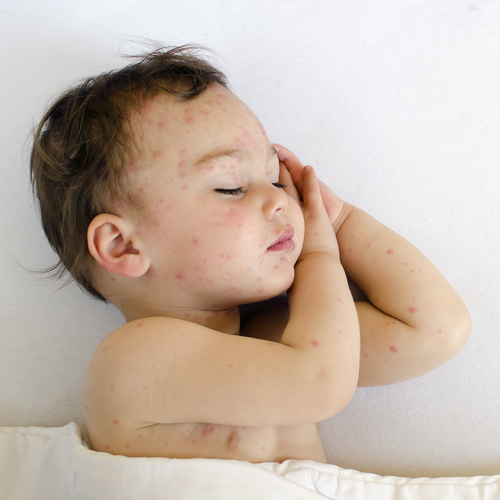
shutterstock_137048072A rash caused from a virus that is widespread and usually occurring in children is called a exanthem.  The first four listed below are the “classic” childhood viral rashes, but we recognize others that can also cause virus, some of those are also listed below (see 5-7): 1)   Rubeola is also known as the measles and is caused from the measles virus and produces reddish macules and papules behind the ears and at the anterior hairline, coalescing and spreading over the neck and trunk and finally affecting the arms/hands and legs/feet. 2)   Rubella is also known as German measles and is caused by the togavirus.  The eruption presents with pink-red macules and papules on the face and spreads down the body over 24 hours.  The rash begins to fade after 1-2 days in the order of appearance and disappears completely in 2-3 days. 3)   Erythema infectiosum is caused by parvovirus B19 and has confluent reddish and swollen patches on the cheeks, with sparing of the bridge of the nose and areas around the eyes.  These so-called slapped cheeks fade over 1-4 days.  The rash may spread to the trunk, arms and legs and may change to look like a lacy reticular pattern.  This rash may be itchy. 4)   Roseola infantum is caused by HHV-6 and HHV-7 and has non-itchy, rose-pink 2-3mm discrete macules and papules that blanch on pressure and are surrounded by white halos.  The eruption is usually first seen on the trunk and then spreads to the arms and legs. 5)   Chicken pox or shingles is caused by the Varicella zoster virus 6)   Mumps is caused by the mumps virus 7)   Rhinovirus which also causes the common cold can cause rash 8)   Hand-foot-mouth disease caused by Coxsackie virus can cause painful ulcers in the mouth, and the rash in the mouth begins as 2-8mm reddish macules and papules that progress through a short vesicular stage to form a yellow-grey ulcer with a reddish halo. Oral lesions usually resolve in 5-7 days.  The skin rash is characterized by 2-3mm reddish macules or papules with a central gray vesicle that usually appear shortly after oral lesions.  The hands are more commonly involved than the feet. Immunizations have decreased the numbers of measles, mumps and rubella but we still see these infections in the medical clinic. Description:  Usually pink or red rash without a typical pattern. It may have red spots that are slightly raised.  It usually isn’t very itchy. They may be faint pink or more extensive, and usually blanches (goes white) with pressure.  If you place a drinking glass against the rash, you may see it disappear through the glass. Some other symptoms that may be present along with a viral rash include low-grade fever, headache, sore throat, malaise, nausea, diarrhea or joint pain. Treatment:  There is no treatment for a viral rash.  The rash will disappear as the body recovers from the infection. When to get medical help: 1)   The rash does not blanch with pressure 2)   The rash is extremely itchy 3)   The patient is very sick or you are concerned about a serious illness   I hope that you have found this information useful.  Wishing you the best of health,
Related Reading

Measles is the one on this list that stopped being historical. If that is what brought you here, start with the current post rather than this one.
Measles Is Back: What Clinicians Need To Watch For When to Use Telemedicine vs. Urgent Care: How I Spot the Sick One Scott Rennie, D.O.

Board Certified in Obesity Medicine and Family Medicine

This blog is for educational purposes only and does not constitute individual medical advice. Always consult your own physician before making changes to your health, medications, or treatment plan.

Doctor talking with a patient across a desk in a clinic office

Bacterial Vaginosis: Most Common Cause of Vaginal Discharge

Bacterial Vaginosis (BV) is not a sexually transmitted infection.  It is the most frequent cause of vaginal discharge in women, but it can be difficult to know if the discharge is caused by BV or other types of vaginal infections. Definition:  Discharge caused by a large change in the number and types of bacteria in the vagina.  For some reason, the number Lactobacilli (which is a normal bacteria found in the vagina) are actually decreased while other bacteria are increased. Risk factors:  Multiple or new sexual partners, douching, cigarette smoking.  Again, BV is not a sexually transmitted infection, as it can occur in women who are not sexually active. Symptoms:  50-75% of women with BV do not have symptoms.  Those with symptoms may note an unpleasant, “fishy smelling” vaginal discharge that is more noticeable after vaginal intercourse.  The discharge is usually a thin, yellow-white color.  Most of the time it does not cause pain with urination or sex, vaginal itching or intercourse.  Self-treatment with over-the-counter medications such as yeast creams or deodorants are not recommended. Diagnosis:  Physical examination, which usually includes pelvic examination and laboratory testing may be used to test the vaginal secretions to determine if BV is present. Complications:  Bacterial vaginosis is usually not considered harmful but has been associated with some health problems such as: 1)   Increases risks for becoming infected with HIV, genital herpes, gonorrhea or chlamydia 2)   Pregnant women with BV are at higher risk of preterm delivery 3)   Untreated BV in woman who have had hysterectomy or abortion can lead to infection at the surgery site Treatment:  We commonly use one of two different treatment options for treating BV.  Either Metronidazole or Clindamycin can be taken in pill form or with a gel or cream that is inserted into the vagina.   There are more side effects possible if taken orally, but it is more convenient. Sexual partners:  There is no need to treat sexual partners of those infected with BV as it doesn’t decrease the risk of infection coming back.  BV is not a sexually transmitted infection, remember? Relapse:  Within 3 months after resolution of symptoms, 30% of women have recurrence.  More than 50% have recurrence after 12 months.  The reasons for recurrence are unknown.  Relapse may be treated with a more prolonged course of antibiotics and the CDC suggests a treatment regimen different from the initial treatment if recurrence occurs. Some patients use a preventative treatment with metronidazole vaginal gel twice weekly for 3-6 months if they get more than 3 episodes in 12 months. Prevention:  Some recommendations may include the following: 1)   Finish the entire course of antibiotics for the treatment of BV even if symptoms resolve rapidly 2)   Limit the number of sexual partners 3)   Do not douche.  There is no proven benefit to douching and the solution used to rinse the vagina may upset the balance of bacteria and actually flush other bacteria up into the uterus or fallopian tubes and cause other types of infections If you have vaginal discharge or questions, please contact your medical provider.  Do not attempt to treat yourself as this can actually make the situation worse.   I hope that you have found this information useful.  Wishing you the best of health,
Related Reading

Vaginal Yeast Infections: Symptoms, Causes, Treatment STI Testing: Should You Get Tested for Everything? FDA Removes Black Box Warning From Menopause Hormone Therapy Vaginal Dryness After Menopause: Why It Doesn’t Go Away

Scott Rennie, D.O.
Board Certified in Obesity Medicine and Family Medicine

This blog is for educational purposes only and does not constitute individual medical advice. Always consult your own physician before making changes to your health, medications, or treatment plan.

Pharmacist discussing medication with a patient at a pharmacy counter

Vaginal Yeast Infections: Symptoms, Causes, Treatment

shutterstock_128350532Patients frequently present to their health care provider with complaints of yeast infection symptoms.  Other terms that are used to describe yeast infections include yeast vaginitis or vaginal candidiasis.  The symptoms of this type of infection usually include itching and irritation to the vulva around the opening of the vagina. Yeast infections are more common during times of menstruation (having monthly periods) and also after taking antibiotics. There is no evidence that vaginal yeast infections are related to poor hygiene or tight synthetic clothing. Other symptoms of yeast infections: 1)   Pain with urination – can be confused with a urinary tract infection 2)   Itching or irritation around the opening of the vagina 3)   Pain with intercourse 4)   Watery or white colored vaginal discharge 5)   Red or swollen vaginal tissues The symptoms of yeast infection can be confusing because they can be similar to a number of other disorders such as a bacterial infection of the vagina, urinary tract infection, trichomoniasis, or common dermatitis (inflammation of the skin).  Most of the time the patient may not know if the itching or irritation is caused from yeast or another cause and this leads them to see a medical provider. Causes of yeast infection:  Candida is the name of the yeast that causes the infection and it normally lives in the intestines and vagina.  Most of the time Candida causes no symptoms.  If it grows out of control (which can be due to increased stress, antibiotic use, or stress to the immune system), it can overgrow and cause symptoms. Risk factors for yeast infection:  Often there are no underlying health problems that lead to yeast infections in women, but several factors may increase the chances of developing an infection including: 1)   Antibiotics – most antibiotics kill lots of bacteria including those that normally live in the vaginal area.  The bacteria that are normally found in the vagina help protect this area from being overgrown with yeast.  Antibiotics can kill off these bacteria and lead to an increase of the normal flora of yeast in the vagina 2)   Pregnancy can increase the chances of yeast infection because there is a normal increase in the amount of vaginal discharge. 3)   Diabetes causes an elevation of blood sugars which can lead to an increase in the likelihood of a yeast infection 4)   Hormonal contraceptives such as birth control pills, vaginal rings or patches that contain estrogen can sometimes increase the chance of yeast infection in some people. 5)   Contraceptive devices such as diaphragms, vaginal sponges and IUDs can also increase the chance of a yeast infection.  Spermicides usually don’t cause yeast infections, but can lead to vaginal irritation. 6)   Increased sexual activity can lead to a yeast infection.  The reason for this may be related to changes in the concentration of bacteria within the vagina that happen with sexual intercourse.  Yeast infections are not considered a sexually transmitted infection. Diagnosis:  Sometimes women who have symptoms of a yeast infection will diagnose themselves and seek treatment with over the counter medications.  One study showed that only 11% of women accurately diagnosed their infection.  Women who previously had a yeast infection were slightly more accurate (35% correct).  The general recommendation is for women who have symptoms consistent with yeast infection be be evaluated by a medical provider.  The exam usually consists of examination of the vulva and vagina and sometimes a swab is used to collect a sample of the discharge to look for yeast. Treatment:  You may be treated with oral or vaginal medication for yeast infection (or both). Vaginal treatment:  Most of the time the treatment includes a cream or tablet that is placed into the vagina at bedtime with an applicator. There are several different treatment options some of which are 1, 3 or 7 days in duration.  The vaginal treatment seems to be more effective in reducing the vaginal irritation quicker than the oral medications. Oral treatment:  Diflucan is a very common oral medication for treating yeast infections.  Most of the time only one dose is needed, however a second dose given 3 days or so after the first dose may be recommended for some patients. If you’re not feeling better within a few days after starting the treatment, make sure you call the office of your medical provider to let them know. Recurrent vaginal yeast infections:  There are about 5-8% of women who have vaginal yeast infections that come back repeatedly.  If the patient has more than 4 infections in one year, we call this recurrent vaginal yeast infection. There is no evidence that eating yogurt or other products containing live lactobacillus acidophilus or applying these products vaginally will benefit women with recurrent vaginal yeast infections. A persistent infection can be cause from a less common species of Candida called Candida glabrata or Candida krusei and in these less common species, we use different medications to treat them. Treatment of recurrent vaginal yeast infections:  Most of the time we use a longer course of anti-yeast medication in patients who have recurrent infections, often between 7 and 14 days for a topical cream or suppository or if the oral medication is used, it is taken with a 2nd dose 3 days later and possibly a 3rd dose 6 days later.  Some patients are started on preventative treatment with oral or vaginal creams once per week. Treatment of a sexual partner:  Vaginal yeast infections are not a sexually transmitted infection and most experts do not recommend treating a sexual partner. Prevention: 1)   Diabetes:  Keep blood sugars under good control in diabetic patients 2)   Antibiotics:  For patients who frequently get yeast infections after taking antibiotics, we often will prescribe a dose of fluconazole at the start and end of antibiotic therapy to prevent post-antibiotic vulvovaginitis. 3)   Patient awareness:  Patients who are using hormone birth control products or contraceptive devices should be aware of the increased risk of yeast infections with these products.   I hope that you have found this information useful.  Wishing you the best of health,
Related Reading

Bacterial Vaginosis: Most Common Cause of Vaginal Discharge Perimenopause and Menopause Symptoms and How to Manage Them Newly Diagnosed With Type 2 Diabetes: What You Should Know I hear it almost every day now. “I think I just need a Z-Pak.” Vaginal Dryness After Menopause: Why It Doesn’t Go Away

Scott Rennie, D.O.
Board Certified in Obesity Medicine and Family Medicine

This blog is for educational purposes only and does not constitute individual medical advice. Always consult your own physician before making changes to your health, medications, or treatment plan.

Honeybee collecting nectar from a purple wildflower

Bee and Insect Stings

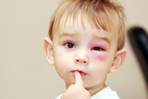
shutterstock_116816458Patients often come into the urgent care clinic due to bee or insect stings as the weather improves.  Being stung is often painful and can also be anxiety provoking because some people have serious or life-threatening allergic reactions to stings and require quick treatment. Sting Reaction:  Immediately after being stung, most people have a sharp or burning pain as well as redness or swelling at the site of the sting.  The swelling and pain usually improve within a few hours.  About 10% of people will develop severe redness and swelling after a sting, and this is called a large localized reaction.  These large red areas may reach up to 4 inches in diameter over 1-2 days, and then slowly resolve over 5-10 days. If you have a large area of inflammation after a sting, this does not mean that you will have an anaphylactic reaction if stung again. Rarely, some individuals will develop a severe allergic reaction, called anaphylaxis.  Only 5-10% of people with a large localized reaction will have an anaphylactic reaction. Symptoms of severe allergic reaction (usually develop quickly): 1)   Hives, redness or swelling of skin away from the area that was stung – ie. face or lips if being stung on the hand 2)   Shortness of breath, hoarse voice or difficulty breathing 3)   Abdominal pain, nausea, vomiting or diarrhea 4)   Feeling lightheaded, dizzy or passing out Treatment for local skin reaction: 1)    If the insect has left a stinger, remove it as soon as possible after being stung.  Flicking or scraping the stinger out is sufficient. 2)    Apply a cold or damp washcloth wrapped around ice to the area 3)    Take an antihistamine such as Benadryl or Zyrtec if you develop itching 4)    A pain medicine such as Ibuprofen or Aleve may be helpful Treatment for severe allergic reactions:  These are a medical emergency that can lead to death if not treated quickly.  Do not drive yourself to the hospital – call 911.  A shot of epinephrine (adrenaline) may be helpful to prevent more severe allergic reactions in some patients.  Epinephrine is prescription only and should only be used if there is concern about a possible life threatening reaction.  Training on how and when to use epinephrine should be given at your medical provider or pharmacy. Prevention:  Usually bees or wasps are not aggressive when they are away from their nests.  The usually only sting after being hit, stepped on or swatted.  Wearing a white or light-colored clothing may help reduce the chance of being attacked if you are near a nest.  If you are eating outside, keep food and drinks covered and clean up spills quickly.  Watch for yellow jackets inside of drink containers.  If you find a nest near your home, do not try to get rid of it yourself.  Call a pest control professional. If you are being swarmed or stung, cover your mouth and nose with your hand and retreat to inside a building or an enclosed vehicle.   I hope that you have found this information useful.  Wishing you the best of health,

Related Reading

Hives and Angioedema: Signs of an Allergic Reaction Hives – What am I allergic to? Animal Bites: What to Do if Your Neighbor’s Dog Bites You Cellulitis – a soft tissue/skin infection – is it MRSA?

Scott Rennie, D.O.
Board Certified in Obesity Medicine and Family Medicine

This blog is for educational purposes only and does not constitute individual medical advice. Always consult your own physician before making changes to your health, medications, or treatment plan.