

A plate of boiled eggs and cottage cheese beside a rolled gauze bandage and a glass of water on a sunlit wooden table

Why Won’t My Wound Heal?

The question comes up on almost every wound follow-up, and it is nearly always phrased the same way. What should I be eating to make this heal faster?

It is a good question. The answer patients get is usually a list of vitamins, which is mostly wrong, or a shrug, which is worse. There is real evidence here. It just does not point where the supplement aisle points.

A healing wound is a construction project

Closing a wound means building tissue. Collagen synthesis, angiogenesis, epithelial migration, immune cell proliferation. All of that runs on amino acids and calories, and the demand is above baseline for as long as the wound is open. A large or chronic wound raises resting energy expenditure meaningfully.

Which means the first question is not which supplement. It is whether the person is eating enough of anything at all. In older adults with a chronic wound, the answer is frequently no, and no capsule fixes an 1,100 calorie a day intake.

What glucose does to the machinery

Hyperglycemia interferes with wound healing at several points at once. Glycosylation impairs neutrophil and macrophage function, which stretches out the inflammatory phase instead of letting the wound progress to proliferation. Fibroblast activity falls. VEGF expression falls, so new vessels are slower to arrive. Matrix metalloproteinase activity rises, which means the collagen that does get laid down is broken down faster than it should be. Collagen density ends up lower.

Put underlying peripheral arterial disease on top and the wound has a supply problem as well as a manufacturing problem.

This is why I care about the A1c on a wound visit and not only on a diabetes visit. The same laceration on a person running an A1c of 11 and a person running a 6 is two different clinical situations, and only one of them is going to close on schedule.

An aside that patients find more persuasive than any of the biochemistry: the glucose control that matters for a wound is the control over the next several weeks, not the number on the day of the injury. That is actionable. It gives someone something to do about a wound that is already there.

The protein number

For adults with a pressure injury who are malnourished or at risk of it, the 2019 international guideline from EPUAP, NPIAP and the Pan Pacific alliance recommends 1.25 to 1.5 grams of protein per kilogram of body weight per day, with 30 to 35 kilocalories per kilogram per day for energy (1).

Those are the best-supported numbers in wound nutrition, and I use them as the floor for any significant open wound, not only pressure injuries. Most people are nowhere near them. A 70 kilogram adult needs roughly 88 to 105 grams of protein a day on that recommendation, and the typical intake I hear described on a video visit is half that.

Worth noting the overlap with the rest of my practice. When I start a patient on an incretin I ask for 1.6 grams per kilogram per day and strength training two to three times a week, because the risk on those drugs is lean mass loss. A patient on an incretin who also has a wound is being asked for the same thing for two different reasons, which makes the conversation easier rather than harder.

Arginine, and how good the evidence actually is

Arginine gets singled out because it is a substrate for nitric oxide and for proline, and proline goes into collagen. The 2019 guideline recommends a high-calorie, high-protein oral nutrition supplement fortified with arginine, zinc and antioxidants for adults with a stage 2 or greater pressure injury who are malnourished or at risk of malnutrition (1).

Read that recommendation carefully, because it is narrower than how it gets quoted. It is for a specific wound severity, in a specific nutritional context, and the arginine comes inside a fortified supplement rather than as arginine on its own (2). The trials behind it tested the whole formulation. Nobody has shown that arginine by itself, added to an adequate diet in a well-nourished person, does anything for a wound.

I have no objection to those supplements in the patients the guideline describes. I do object to a well-fed person with a healing surgical incision spending money on arginine capsules.

Vitamin C and zinc

Both are genuinely required for wound healing. Vitamin C is a cofactor for the hydroxylation steps in collagen synthesis, and frank scurvy produces wounds that will not close. Zinc deficiency impairs epithelialization.

None of that means supplementing someone who is not deficient helps. Reviews of vitamin and mineral supplementation in wound care keep landing in the same place: unclear benefit unless there is a confirmed or suspected deficiency, and no well-powered randomized trial showing that routine vitamin C or zinc speeds healing in replete patients (3).

So I correct documented deficiencies and I do not supplement reflexively. High-dose zinc in particular is not harmless; it interferes with copper absorption over time.

This is the part of the conversation patients like least, and I say it anyway.

What matters more than any of it

Smoking. Sørensen’s meta-analysis of surgical patients found substantially higher rates of wound complications and infection in smokers, and improvement with cessation before surgery (4). If a patient with a slow wound smokes, that is the highest-yield thing on the list, and it is not close.

Then perfusion. A wound on a limb without a decent pulse is a vascular problem wearing a dressing, and no amount of protein fixes it.

Then offloading and moisture balance, which are mechanical rather than nutritional and are where most home wound care goes wrong.

Nutrition belongs on the list. It belongs below those.

For patients

Eat more protein than you think you need while a wound is open. Aim for something in the range of 1.25 to 1.5 grams per kilogram of your body weight per day, which for most adults means adding a protein source to every meal rather than eating one large dinner. If you are diabetic, the wound is a reason to tighten control now, not later.

Skip the wound-healing supplement stack unless your clinician has found an actual deficiency. If you smoke, stopping will do more for that wound than everything else on this page combined.

For colleagues

Ask what people are eating before you reach for a supplement, and ask in grams rather than in general terms. The 1.25 to 1.5 g/kg recommendation is easy to quote and rarely translated into food for the patient, which is where it fails.

Check a zinc or vitamin C level if the history suggests deficiency, then treat what you find. Do not treat the wound with a multivitamin on principle.

And put smoking cessation at the top of the plan for any wound that is behind schedule, ahead of the dressing change conversation.

The Bottom Line

Wounds heal on protein, calories and blood flow, in something like that order, and they stall on high glucose and tobacco. The guideline numbers worth remembering are 1.25 to 1.5 grams of protein per kilogram per day and 30 to 35 kilocalories per kilogram per day. Arginine and antioxidant supplements have a real place, in malnourished patients with stage 2 or worse pressure injuries, and almost nowhere else. Vitamin C and zinc are worth correcting when they are low and worth skipping when they are not. If you fix only one thing on this list, fix the smoking.

Related Reading

Basic Wound Care Tips for Non-Medical Professionals

Diabetic Foot Care: How to Check and Protect Your Feet

What is a Pressure Ulcer (AKA Pressure sore)?

Cellulitis: A Soft Tissue and Skin Infection, Is It MRSA?

Newly Diagnosed With Type 2 Diabetes: What You Should Know

Doctor Supervised Weight Loss: What Works Long Term

Scott Rennie, D.O.
Board Certified in Obesity Medicine and Family Medicine

This blog is for educational purposes only and does not constitute individual medical advice. Always consult your own physician before making changes to your health, medications, or treatment plan.

Sources

1. European Pressure Ulcer Advisory Panel, National Pressure Injury Advisory Panel and Pan Pacific Pressure Injury Alliance. The Role of Nutrition for Pressure Injury Prevention and Healing: 2019 International Clinical Practice Guideline recommendations. https://cdn.ymaws.com/npiap.com/resource/resmgr/The_Role_of_Nutrition_for_Pr.pdf

2. Chu AS, Delmore B. Arginine: What You Need to Know for Pressure Injury Healing. Adv Skin Wound Care. 2021;34(12):630-636. PMID 34807894. https://pubmed.ncbi.nlm.nih.gov/34807894/

3. Indian Health Service National Pharmacy and Therapeutics Committee. Formulary Brief: Nutritional Supplements in Wound Healing. https://www.ihs.gov/sites/nptc/themes/responsive2017/display_objects/documents/guidance/NPTC-Formulary-Brief-NutritionalSupplementsinWoundHealing.pdf

4. Sørensen LT. Wound healing and infection in surgery. The clinical impact of smoking and smoking cessation: a systematic review and meta-analysis. Arch Surg. 2012;147(4):373-383. PMID 22508785. https://pubmed.ncbi.nlm.nih.gov/22508785/

5. Armstrong DG, Boulton AJM, Bus SA. Diabetic Foot Ulcers and Their Recurrence. N Engl J Med. 2017;376(24):2367-2375. PMID 28614678. https://www.nejm.org/doi/10.1056/NEJMra1615439
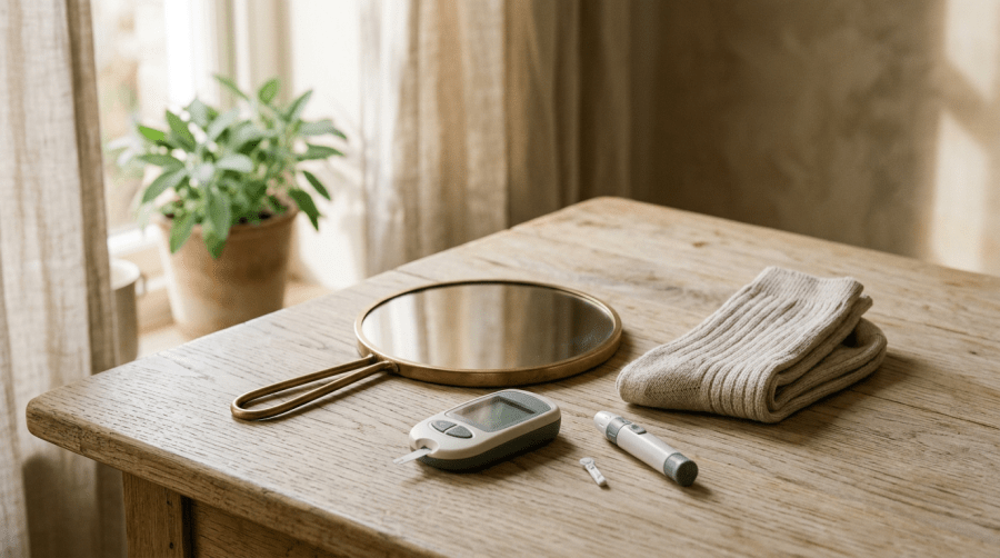
A hand mirror, a pair of folded cotton socks and a home glucose meter with a lancet on a sunlit wooden table

What Does Diabetes Do to Your Skin?

Skin is the one organ I can actually see on a video visit. No stethoscope, no palpation, no reaching across the desk. Just a camera, whatever light the patient happens to have, and a screen. That limitation has made me pay closer attention to skin than I did when I worked in urgent care, because on video it is often the first physical evidence that something metabolic is going on.

Between 30 and 70 percent of people with diabetes develop a skin problem attributable to the disease at some point (1). That is an enormous range, which tells you the studies are measuring different populations and different definitions rather than anything precise. Take the range as what it is, a signal that this is common, not a statistic to quote back to anyone.

What follows is what I look for, why it matters, and where I think the usual advice is weaker than it sounds.

The velvet patch on the back of the neck

Acanthosis nigricans is the one I care about most, because it is visible, it is early, and patients almost never bring it up. Dark, thickened, velvety skin in the folds. Back of the neck, armpits, groin, under the breasts. Patients describe it as dirt that will not wash off, and some have been scrubbing at it for years.

The mechanism is insulin. Hyperinsulinemia drives IGF-1 receptor activation on keratinocytes and fibroblasts, which proliferate (2). So the plaque is a rough readout of how much insulin the pancreas has been pushing out to keep glucose in range. It shows up before the fasting glucose does. Sometimes years before.

Skin tags travel with it and share the same driver (2). A patient with acanthosis nigricans and a crop of acrochordons on the neck and axillae is telling you about their insulin resistance without a single lab drawn.

Here is where I differ from what patients are usually told. Dermatology handles acanthosis nigricans as a cosmetic complaint and offers topical retinoids, urea, sometimes laser. Those do something to the appearance. None of them touch the reason it is there. I follow the ADA rule and screen every adult from 35 regardless of BMI, and when I see that patch I do not wait for the birthday. I check an A1c.

It also improves. Not overnight, and not completely in everyone, but weight loss and improved insulin sensitivity soften the plaques over months. That is worth telling someone who has been scrubbing their neck since high school.

Skin tags carry information

I have a whole post on what skin tags are and what they look like, and it gets steady traffic from people worried about a lesion. What that post does not do is explain why someone in their thirties suddenly has fifteen of them.

Friction is part of it. Insulin is the rest.

What high glucose actually does to a wound

This one deserves its own post and is getting one. The short version: glycosylation impairs neutrophil and macrophage function, which prolongs the inflammatory phase instead of letting the wound move on to proliferation. Fibroblast activity drops, VEGF expression drops, matrix metalloproteinases go up, collagen density falls. Layer underlying vascular disease on top of that and you have a wound that stalls.

The practical consequence is that a cut on someone with an A1c of 11 is a different clinical problem than the same cut on someone with an A1c of 6, even though it looks identical on camera.

The infections that come with the territory

Candidal intertrigo lives in the skin folds, and the bigger the fold the more reliably it shows up. Beefy red, satellite lesions at the edge, itching or burning rather than pain. Patients treat it with hydrocortisone from the drugstore, which makes it worse, and then arrive convinced they have an allergy.

Tinea in all its addresses, feet, groin, nails. Onychomycosis in particular is stubborn and, in a diabetic foot, is not merely a nuisance: a thickened dystrophic nail is a pressure point and a portal.

Then cellulitis. Elevated glucose, impaired barrier, and a break in the skin from a fissure or a fungal infection between the toes. My rule for an abscess is that it goes out for drainage rather than getting empiric antibiotics from me over video, and I am firmer about that than some colleagues are.

Hidradenitis suppurativa, and what changed

HS has been badly served for a long time. Painful nodules and sinus tracts in the axillae, groin and under the breasts, frequently mistaken for recurrent boils for years before anyone names it. The association with obesity is strong and it is not a coincidence of body habitus alone; the inflammatory biology overlaps.

What has changed recently is incretin therapy. Multiple cohorts and case series now report reduced disease activity, fewer flares and less pain in HS patients treated with GLP-1 receptor agonists, alongside the expected BMI reduction (3). A cross-sectional survey found roughly two thirds of respondents reporting improvement in HS-specific measures (4).

I want to be honest about the strength of that evidence, because the enthusiasm is running ahead of it. These are retrospective cohorts, case series and patient-reported surveys. There is no adequately powered randomized trial with standardized background therapy. The signal is consistent enough that I take it seriously in a patient who has both conditions and would be a candidate for the drug anyway. It is not a reason to start an incretin in someone whose only indication is HS.

The foot

Somewhere between 19 and 34 percent of people with diabetes will develop a foot ulcer in their lifetime (5). The number that changed how I talk to patients is not that one, though. It is what happens after the ulcer closes: roughly 40 percent recur within one year, close to 60 percent within three years, and 65 percent within five (5).

Armstrong and colleagues argued from that data that a patient whose wound has closed should be thought of as in remission rather than healed (5). I use that word deliberately with patients now, because a person who believes they are healed puts the mirror away and stops looking at the bottom of their feet, while a person who believes they are in remission keeps checking, keeps wearing the shoes that were fitted for them, and calls about a blister on the day it appears rather than three weeks later when it has become something else. Remission gets you daily foot checks. Healed gets you a pair of sandals and a summer of walking on hot pavement with neuropathy.

That framing does more work in a fifteen-minute visit than any amount of education about glycemic targets.

What I ask for on video

Skin over a camera is genuinely hard. Autofocus hunts, phone processing smooths texture, and the color balance depends on whether someone is under a kitchen light or a window.

So I ask for two photographs of anything I need to actually assess. One from a distance, far enough back to show where the lesion sits and what the surrounding skin looks like, and one macro, as close as the phone will focus without blurring. Multiple angles if there is any elevation to it. The blur problem is the single most common reason I have to ask twice, and it is almost always because the patient is closer than the lens can handle. Backing up six inches fixes it.

For anything in a fold, I want the fold held open in the shot. For feet, I want the sole and between the toes, which usually means someone else holds the camera.

For patients

If the skin on the back of your neck or in your armpits has gone dark and thick and will not scrub off, that is not hygiene, and it is worth an A1c. If you are getting skin tags in bunches, same. If you have painful recurring lumps in your armpits or groin that have been called boils for years, ask specifically about hidradenitis suppurativa by name, because that is often what gets the diagnosis moving.

And if you have diabetes, look at your feet every day. Every day, including between the toes, including the bottom, using a mirror or a family member if you cannot see them.

For colleagues

Acanthosis nigricans in an adult is a screening prompt, not a dermatology referral. Check the A1c and the lipids before you send them for cosmetic treatment of the plaque.

The GLP-1 and HS literature is worth knowing about and worth being careful with. When a patient with both conditions asks whether the drug will help their skin, the honest answer names the cohorts, names their design, and stops short of promising anything.

On the foot, adopt the remission language. It changes adherence in a way that repeating ulcer statistics does not.

The Bottom Line

Skin is where metabolic disease announces itself, often years before a lab does. A velvety patch behind the neck, a crop of skin tags, a yeast infection in a fold that keeps coming back, a nail that will not clear. None of those are dermatologic curiosities to be treated in isolation. They are all the same disease showing up at the surface, and treating the surface without asking why it is there wastes the information. Check the A1c. Look at the feet. And when a wound closes on a diabetic foot, say remission, not healed.

Related Reading

Diabetic Foot Care: How to Check and Protect Your Feet

What is Hidradenitis suppurativa?

Skin Tags (Acrochordons): What They Are and Look Like

Ringworm, Athlete’s Foot and Jock Itch and Fungal Nail Infections

Newly Diagnosed With Type 2 Diabetes: What You Should Know

How Do You Actually Lose Weight? A Doctor Explains

Scott Rennie, D.O.
Board Certified in Obesity Medicine and Family Medicine

This blog is for educational purposes only and does not constitute individual medical advice. Always consult your own physician before making changes to your health, medications, or treatment plan.

Sources

1. Vâță D, et al. Cutaneous Manifestations Associated with Diabetes Mellitus: A Retrospective Study. Diseases. 2023;11(3). PMID 37606477. https://pubmed.ncbi.nlm.nih.gov/37606477/

2. Marchand L, et al. All about skin manifestations of insulin resistance and type 2 diabetes: acanthosis nigricans and acrochordons. Postgrad Med J. 2020;96(1134):237. PMID 31611265. https://pubmed.ncbi.nlm.nih.gov/31611265/

3. Rao S, et al. Skin Impacts and Tradeoffs of GLP-1 Therapy: Improved Patient-Reported Outcomes of Inflammatory Skin Disease. Dermatol Ther (Heidelb). 2026. PMID 42579223. https://pubmed.ncbi.nlm.nih.gov/42579223/

4. Javaheri ED, et al. Impact of GLP-1 Receptor Agonists on Hidradenitis Suppurativa: A Cross-Sectional Survey. Int J Dermatol. 2026 Jul 16. PMID 42461155. https://pubmed.ncbi.nlm.nih.gov/42461155/

5. Armstrong DG, Boulton AJM, Bus SA. Diabetic Foot Ulcers and Their Recurrence. N Engl J Med. 2017;376(24):2367-2375. PMID 28614678. https://www.nejm.org/doi/10.1056/NEJMra1615439

6. International Working Group on the Diabetic Foot. IWGDF Guidelines on the prevention and management of diabetes-related foot disease, 2023. https://iwgdfguidelines.org/guidelines-2023/