Hospital bed with medical equipment and patient repositioning guide on wall

What is a Pressure Ulcer (AKA Pressure sore)?

shutterstock_89421025
bed-sores

Photo credit: http://diseasespictures.com/bedsores/

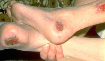
A patient came in to see me today with a sore on his heel that’s been bothering him for the past few months. He’s diabetic and has lost feeling in the bottom of his feet. He’s had these pressure sores in the past but has trouble getting them to heal up.

Pressure sore: An area of skin damaged by pressure, such as sitting or lying in one position for a long period. They are also called bedsores. They are most common where bone sits near the surface of the skin: the hips, elbows, ankles, and back or buttocks. The skin and soft tissue become damaged because not enough oxygenated blood reaches the area to support healing, usually because the soft tissue is compressed between the surface and the hard bone beneath.

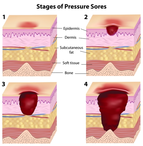
Appearance: The sores change in appearance depending on how long they have been present and how much damage has been done. At the start, the sore looks like a small red patch of skin. If it is not treated, the skin breaks down and a hole or crater forms, which is what we call an ulcer.

Stage 1: The skin is intact without ulceration, but when you press on it the color does not change the way it should. Healthy tissue is pink, and pressing on it with a finger blanches it, with the pinkness returning within a couple of seconds. That does not happen in damaged skin at this stage, and the area may look darkly pigmented.

Stage 2: There is an open, shallow ulcer with a red-pink base. Blisters may be present, either intact or ruptured.

Stage 3: Structures beneath the skin such as fat may be exposed, but at this stage you should not see bone, tendon or muscle.

Stage 4: Bone, tendon and muscle may be visible in the base of the ulcer.

People at risk: Some patients are far more likely than others to develop pressure sores.

  1. Patients who cannot move well because of a medical problem. They may sit or lie in one position for a long time and need help repositioning so the skin does not break down.
  2. Older people, who often move less and whose skin is thinner and more fragile.
  3. Patients with diabetes or nerve problems in their feet, who may not feel a pebble in a shoe or pressure building against the foot.
  4. Patients in the hospital or a nursing home, who are at especially high risk because several of these factors stack: increased age, reduced mobility, and other complicated medical problems.

Prevention: Several things lower the chance of developing pressure sores.

  1. Repositioning the patient’s body every two hours so no one area of skin stays crushed, pinched or under pressure.
  2. Putting pillows between the ankles and knees to reduce pressure on the skin over those bony areas.
  3. Raising the head of the bed when the patient is lying on their side to reduce pressure on the hip bone.
  4. Using special foam or soft mattresses that reduce pressure over the areas that bear the most load.

For patients in wheelchairs:

  1. Use a cushioned seat if possible to prevent pressure on the sacrum.
  2. Tilt forward or to the side every hour to release pressure on the seat.
  3. If the ankles or heels press against the chair, use foam padding to protect against sores.
  4. Check the skin regularly for signs of pressure or ulceration.

Treatment: Pressure sores are treated differently depending on the stage and how severe the skin damage is.

  1. If there is mild erythema, treatment is generally offloading the area by reducing the time it spends compressed, through repositioning and pillows to cushion it. We also use transparent films over the ulcers to protect them.
  2. In patients with diabetes, keeping blood sugars under good control is very important. Elevated blood sugars impede wound healing.
  3. If there is dead or dying skin or soft tissue, it often needs to be removed to help prevent infection.
  4. Special bandages may be needed to keep the healing tissue moist while preventing maceration from being too moist. The dressings we use can be expensive.
  5. Antibiotics may be prescribed if there is a wound infection.
  6. Medication for pain may also be prescribed.

There are scoring tools to grade pressure and track healing. These help for patients who return for repeat visits to a wound care clinic or their primary care provider and need their progress graded. Clinical features examined include:

  1. Amount of exudate
  2. Skin color surrounding the wound
  3. Peripheral tissue swelling
  4. Peripheral tissue firmness around the wound
  5. Amount of granulation (healing) tissue
  6. How much epithelialization is present

It’s important to optimize the nutritional status of patients with wounds. Patients with stage 3 and stage 4 ulcers in particular need enough protein and calories to heal.

Scott Rennie, D.O.

Board Certified in Obesity Medicine and Family Medicine

This blog is for educational purposes only and does not constitute individual medical advice. Always consult your own physician before making changes to your health, medications, or treatment plan.


Blog: https://doctorrennie.wordpress.com

Diagram showing proper footwear, sock selection, foot inspection, and skin moisturizing for diabetic foot care.

Diabetic Foot Care: How to Check and Protect Your Feet

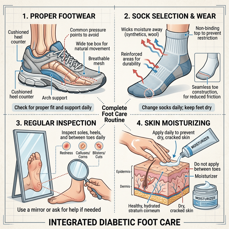
shutterstock_110360354Patients who have diabetes need to pay extra attention to their foot care to help prevent infections.  I’ve had numerous patients with diabetes need foot or toe amputations that could have been prevented with excellent foot hygiene.  Small scrapes in the skin or ingrown nails can become extremely bad very quickly with diabetes because patients who have diabetes often don’t have as much sensation (due to damage to the nerve endings and blood vessels in your feet).  This can make it difficult to detect sores and once an infection is present it can be very difficult to treat.  I thought I’d put together some tips to help you keep your feet healthy and decrease the risk for infections.

  1. Stop smoking:  If you smoke, this can decrease the blood flowing to your feet and make foot problems worse.
  2. Inspect your feet everyday:  Look for blisters, cuts, cracks or sores.  If you cannot see your feet well then use a mirror or have a family member help you.
  3. Wash your feet everyday:  Use warm (not hot) water – be sure to check the temperature with your hands rather than your feet.
  4. Dry your feet well:  Pat them dry and do not rub the skin on your feet too hard.  Dry between each toe.  If the skin on your feet stays moist, bacteria or fungus can grow and that might lead to a foot infection.
  5. Keep your feet soft:  Use a skin moisturizer such as Aveeno, Dove or Cetaphil on your feet to keep your skin soft and prevent calluses and cracks.  Don’t put the cream between your toes unless you are treating athlete’s foot with a fungal cream.  Make sure to wear socks or traction on your feet after applying the cream so you don’t slip and fall.
  6. Clean under your toenails carefully:  Don’t use sharp objects under your toenails.  Instead use the blunt end of a nail file or other rounded tool to decrease the chance of piercing the skin.
  7. Trim and file your toenails straight across:  This helps prevent ingrown nails.  Use a nail clipper instead of scissors.  Then use an emery board to smooth the edges.  If you need help trimming your nails, schedule an appointment with your medical provider.
  8. Change your socks everyday:  Socks should have a thick cushion and fit loosely around your feet.  Socks without seams are best because seams often rub the feet.  Do not wear stockings, socks, or garters that come up to the thigh or knees unless your medical provider advises you to do so because they can decrease the blood flow to your feet.
  9. Look inside your shoes before putting them on:  Check them every day for gravel, torn linings, or thorns that can cause blisters or sores.
  10. Do not go barefoot:  Don’t wear sandals or shoes with thin soles because these types of shoes are easy to puncture.  They also do not protect your feet from hot pavement or cold weather.
  11. Have your medical provider check your feet during each visit:  If you notice a problem with your feet, see your medical provider right away rather than trying to treat it with a home remedy.  Some home remedies or treatments that you can buy without a prescription (such as corn removers) can be harmful.
  12. Keep your blood sugar down:  Watch what and how you eat, monitor your blood sugar, take your medications and get regular exercise.

When to seek medical help:

A)      If you cannot do proper foot care

B)     If you have a foot sore or ulcer that is not healing after 3 days (including corns, calluses or ingrown nails)

C)     If you have black and blue areas in your toes or feet

D)    If you have peeling skin or blisters between your toes

E)     If you have a fever for more than 24 hours and a foot sore

F)     If you have new numbness or tingling in your feet that does not go away after you move your feet or change positions

G)    If you have unexplained or unusual swelling of your foot or ankle

H)    Anytime you have questions about your feet or concerns it is best to contact your medical provider


I hope that you have found this information useful.  Wishing you the best of health,

Scott Rennie, D.O.
Board Certified in Obesity Medicine and Family Medicine

This blog is for educational purposes only and does not constitute individual medical advice. Always consult your own physician before making changes to your health, medications, or treatment plan.
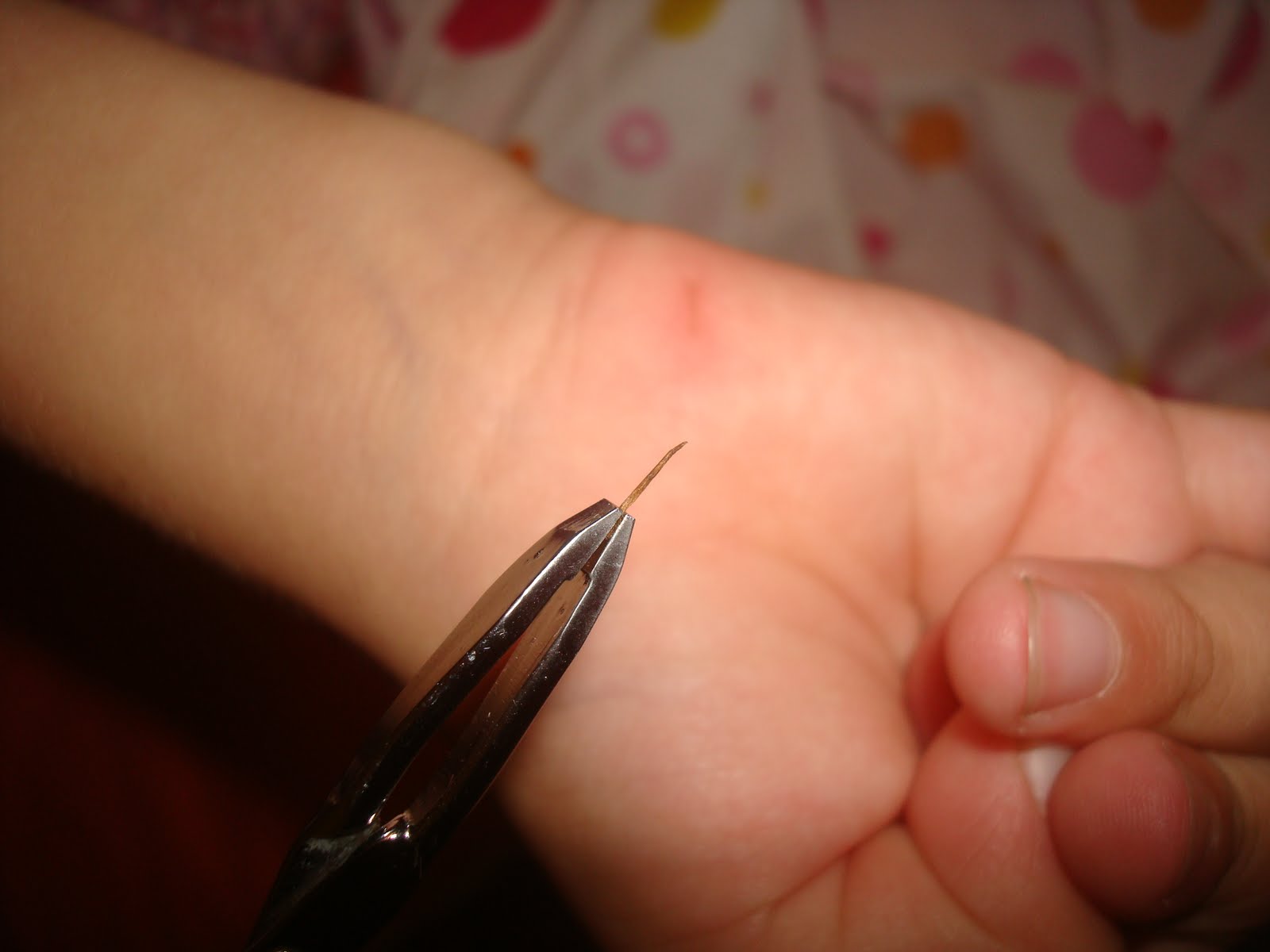
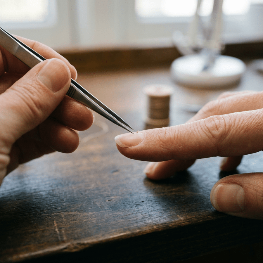
Tweezers removing debris from beneath a person’s fingernail

Splinter (Sliver) Removal


Photo credits: http://www.lemamme.it/sos-cosa-fare-quando-il-bambino-si-fa-male/

http://www.aafp.org/afp/2003/0615/p2557.html

Splinters look like the simplest possible problem, right up until one of them is not.  I thought I’d discuss some different types of splinters and some techniques that you can use at home to remove them.  In addition, it’s important to know when to have it taken out in person for help with that process.

Splinters are actually very common, and children and adults need it taken out in person usually with a small foreign body embedded into the superficial or subcutaneous soft tissues of the hands or feet.  Wood, glass and metallic splinters are the most common types.  Many splinters can be removed at home by our patients, which leaves the physicians only the deeper and larger splinters or retained splinters that have been broken down during an attempt at removal.  If splinters are not removed completely, they may cause complications such as infection and inflammation.

When to see a medical provider:  I recommend that you see a medical provider instead of trying to remove the splinter yourself if there is lots of redness around the splinter, increased swelling or pus draining from the wound.

The severity of the reaction and type of reaction that the body has to a particular splinter.  Some examples of reactions to retained foreign materials include:

1)   Glass – Mild reaction and the body attempts encapsulation

2)   Blackthorns – Severe reaction with inflammation from the plant alkaloids

3)   Wood – Severe reaction with inflammation from the oils and resins

4)   Cactus Spines – Moderate to severe reaction from the fungal coating on the plant and possibly an allergic reaction

5)   Rose thorns – Moderate to severe reaction with inflammation from the fungal coating on the plant

6)   Sea urchins – Moderate to severe reaction with inflammation and possible infection

7)   Metal – Mild reaction and the body attempts encapsulation

8)   Plant spines – Mild to severe reaction from the toxins in the plant

9)   Animal spines – Mild to severe reaction from the toxins in the animal spines

10)  Plastic – Mild reaction and the body attempts encapsulation

Sometimes splinters can be difficult to detect, especially if they are deep or very small.  Clues that a splinter is present might be swelling, tenderness, a mass, or soft tissue infection such as cellulitis, or an abscess.  Sometimes we might use a diagnostic test such as a x-ray, ultrasound, CT scan or MRI to detect a splinter if it’s deep and difficult to see.

Removal:  We try to remove splinters quickly before inflammation or infection occurs.  There are several ways to remove a splinter, some of those might include:

1)   Using tweezers: Make sure you have the right kind of tweezers.  The sharp tipped splinter tweezers work best most of the time.  If the sliver is sticking out of the skin, clean the pair of tweezers with alcohol and carefully pinch the sliver as close to the skin as possible and gently slide it out.

2)   Use a small needle:  If the sliver isn’t sticking out of the skin, often a needle can help push the sliver out.  First clean the needle with alcohol and then push the sliver from the bottom (the pointed end that entered the skin first) and angle upward toward the puncture hold in the skin’s surface.  Try to start at the bottom of the sliver so you don’t break it and leave sliver shreds in the skin.

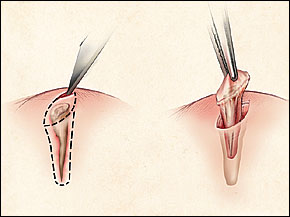
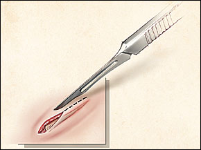
3)   Use a razor blade to make a small slice in the skin over the sliver:  If the sliver is deep and using tweezers or a needle isn’t working well, you can clean a razor blade in alcohol and make a shallow incision parallel to the sliver, just above it.  I know this sounds scary, but the top layer of skin is made of dead skin cells that do not have nerves, so this is not likely to hurt unless you cut too deeply.  Once the incision is made, gently part the skin and pick out the sliver.

4)   Clean the wound:   Whatever method you use to remove the sliver, make sure you clean the wound with soap and water after the sliver is removed.  Puncture wounds are often laced with bacteria and are very likely to become infected.

5)   Antibiotics:  Apply a small amount of antibiotic ointment such as bacitracin over the puncture wound.

6)   Examine the area at least once a day for the next several days to look for increasing redness, swelling or pain.  If any of these occur, I recommend that you see a medical provider because this might be the start of an infection.


I hope that you have found this information useful.  Wishing you the best of health,

A splinter you cannot reach is one of the few things a video visit genuinely cannot fix. I wrote about the conditions that still need an in person visit.

Related Reading

Abscesses and MRSA: What To Do About a Skin Infection

When to Use Telemedicine vs. Urgent Care: How I Spot the Sick One

Scott Rennie, D.O.
Board Certified in Obesity Medicine and Family Medicine

This blog is for educational purposes only and does not constitute individual medical advice. Always consult your own physician before making changes to your health, medications, or treatment plan.
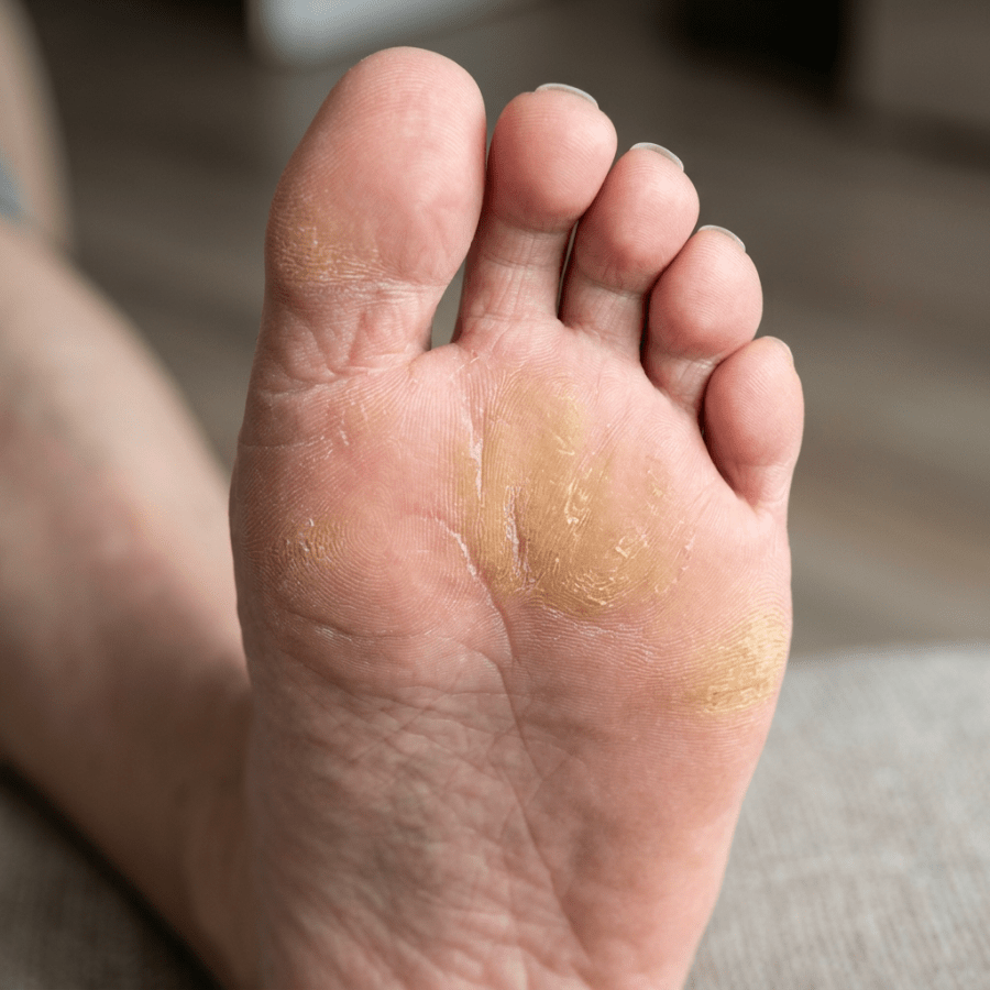
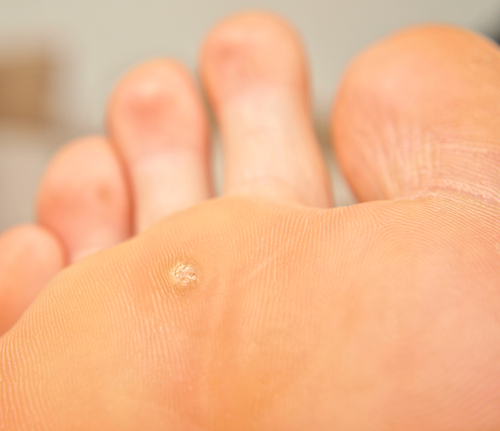
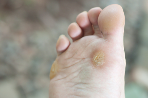
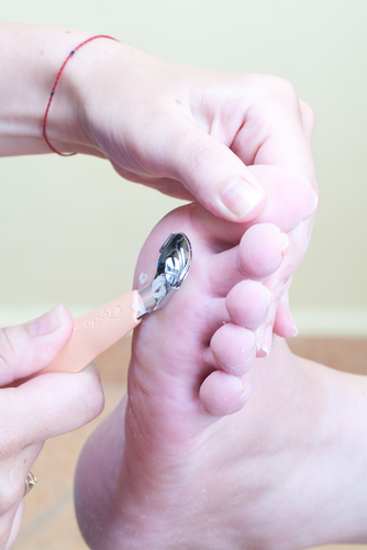
Sole of foot with dry, peeling skin and calluses

Calluses and Corns: Causes, Prevention, and Treatment

shutterstock_113610118shutterstock_93910513I can’t tell you how many patients come in to the clinic because of a callus or corn that’s bothering them – too many to count!  Corns and calluses can cause significant pain, especially when they’re on the feet because we often get them in areas over pressure areas.  When they get thickened they can re-distribute your weight onto other areas of your feet and that may lead to foot instability or worsening pain. Calluses are usually on the hands and feet and basically just thickened areas of skin that form when something rubs or presses on these areas over prolonged periods of time. Corns are thickened areas of skin that are often on the soles of the feet or sides of the toes and look like a small nodule that has a hard center.  Corns are usually more painful because they are often over a smaller area and press harder on a more specific area of the foot. Causes:  Possible reasons that calluses or corns develop include: 1)   Wearing shoes that don’t fit properly for your feet – either too loose or too tight 2)   Walking barefoot 3)   Wearing shoes without socks 4)   Calluses on the hands can be caused by repetitive sports such as rowing, golfing, tennis or biking without gloves Prevention: 1)   Get shoes that fit properly!  It’s important to be proactive and to prevent calluses or corns if they bother you.  There are special shoe stores that work with foot doctors to help you select shoes that fit your feet properly.  Some people are born with narrow or wide feet and that makes it more difficult to find shoes that fit properly.  If you are getting painful corns or calluses, take time and visit a store where someone can help you to pick out shoes that fit you well 2)   Avoid going barefoot or wearing shoes without socks 3)   If you have spots on your feet that rub inside your shoes, you can get special pads that prevent rubbing Treatment:  I often work with patients who come into the clinic to trim the corn or callus down so that it reduces the pressure in the affected area.  I use a scalpel to carefully trim away the thickened skin.  A foot doctor (podiatrist or orthopedic surgeon who specializes in treating conditions of the foot/ankle) can make special orthotic devices that can help reduce the pain and help prevent recurrence of corns/calluses.  Some people treat calluses themselves by purchasing special pads that contain medications to burn or dissolve the thick skin.  I highly recommend that patients with diabetes be seen and treated by a medical provider to reduce the risk of infection.  Diabetic patients are at increased risk of foot infections and should be seen for yearly foot examinations even if they don’t have calluses or corns.   I hope that you have found this information useful.  Wishing you the best of health,

Scott Rennie, D.O.
Board Certified in Obesity Medicine and Family Medicine

This blog is for educational purposes only and does not constitute individual medical advice. Always consult your own physician before making changes to your health, medications, or treatment plan.
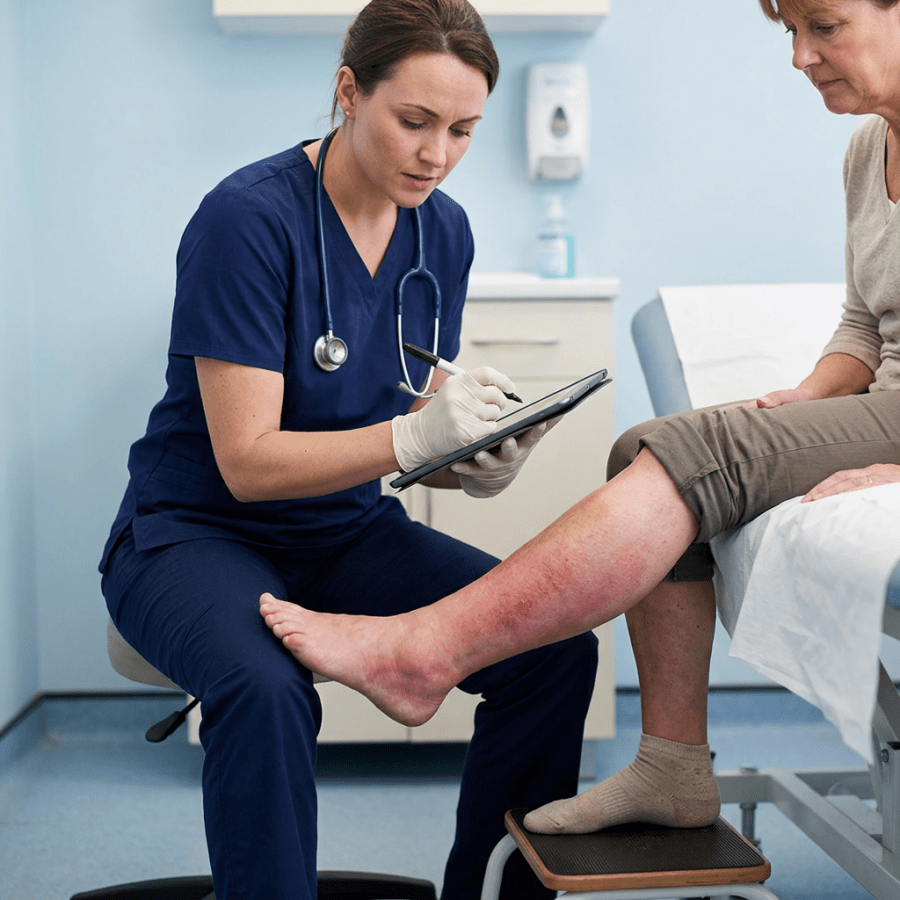
Nurse documenting examination of patient's reddened lower leg

Cellulitis – a soft tissue/skin infection – is it MRSA?

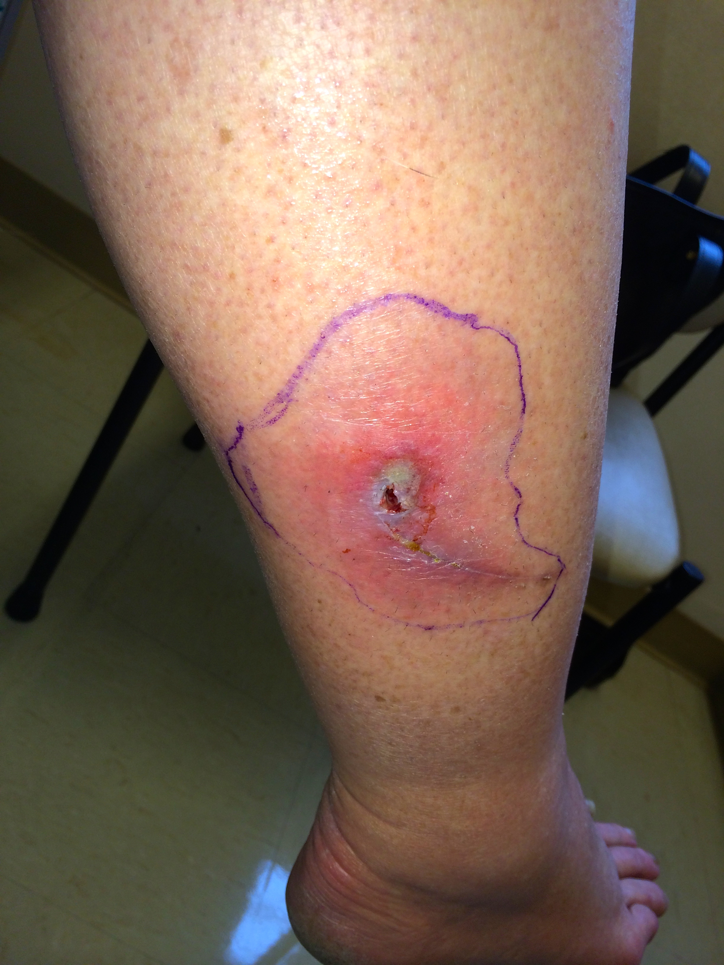
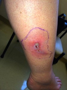
IMG_5864Cellulitis is an infection of the skin and soft tissues.  It is common and most often it’s caused by bacteria that normally live on the skin.  These bacteria don’t normally cause any problems unless the skin is scratched, torn or punctured.  The most common bacteria that cause cellulitis are streptococci “Strep” or staphylococci “Staph.” Most of the time cellulitis is mild and heals with the use of antibiotics, however it can become severe and cause an infection that spreads throughout the body if left untreated.  It can also lead to deep infections called abscesses. Risk factors: 1)   Chronic skin condition such as eczema or psoriasis 2)   Accumulation of fluid (edema) due to poor circulation possibly from heart failure, liver disease, or removal of lymph nodes 3)   Recent injury, wound, cut or laceration to the skin 4)   Current skin infection such as athlete’s foot or impetigo 5)   Being overweight Symptoms:  Pain, tenderness, increased warmth, redness and swelling in a distinct area of skin.  The skin can be smooth and shiny in this area.  Fever and chills are not usually present. Other skin infections:  different kinds of infections that can be confused with cellulitis include an abscess or boil.  These are different because they are usually bumpy, raised and filled with pus.  Abscesses are usually caused by a staph bacteria which may be MRSA.  Cellulitis is more commonly caused by “strep” bacteria. Treatment:  We commonly use antibiotics and also treat the underlying problem that caused the skin infection (such as athlete’s food or increased edema, eczema or psoriasis) if present. Keeping the area elevated to reduce inflammation is also helpful.  It is important to keep the infected area clean and dry.  Showering or bathing can be done normally, but pat the area dry with a clean towel afterwards to dry the area completely.  You can also use a bandage or gauze to protect the skin if needed. Antibiotics are often used and will be tailored to the individual patient.  The patient’s specific area of infection, medical history, history of allergies and examination will be important in determining the proper antibiotics to use.  Sometimes the antibiotics will need to be given by IV in more serious cases of cellulitis. Treatment time:  In most cases, the swelling, warmth and redness should improve within 1-3 days after starting the antibiotics.  We often use a Sharpie marker and draw a circle around the area of redness so that we can examine the red area over a few days to see if it’s increasing, decreasing or staying the same.  Symptoms of redness and swelling can persist for up to 2 weeks but it should be gradually decreasing.  If it’s not improving, you should call or return to your health care provider for re-evaluation.   I hope that you have found this information useful.  Wishing you the best of health,

Cellulitis of the lower leg often starts at a crack or a small cut on the foot, and with diabetic neuropathy that break can go unnoticed until the whole leg is red. It is the reason I keep after people about the diabetic foot check and what to look for.

Updated for 2026: Pus or No Pus Decides the Antibiotic

The most useful thing I can add to this post is a distinction that was fuzzy in 2012 and is now the thing the whole treatment decision turns on. Is there pus, or is there not. If there is an abscess, a pocket of pus you can feel giving under your finger, that is usually staph, MRSA is on the table, and it needs drainage plus an antibiotic that covers MRSA. I have written about that separately. If it is plain cellulitis, meaning spreading red, warm, tender skin with no drainable pocket, that is usually strep, and a cephalosporin like cephalexin is the right drug. Adding MRSA coverage on top does not help, and this has been tested directly. Moran and colleagues randomized patients with uncomplicated cellulitis to cephalexin plus trimethoprim-sulfamethoxazole or cephalexin alone. Cure was 83.5 percent against 85.5 percent (1). No benefit, two drugs, more side effects. So if you have been handed two antibiotics for a red leg with no abscess, that is worth a conversation. The 2014 infectious disease guidelines remain the operative document here and have not been superseded (2).

Draw a Line Around It

This is the single most useful thing you can do, and it is more valuable to me on a video visit than almost anything else. Take a pen and trace the edge of the redness. Write the time next to it. Then photograph it. When we talk tomorrow, or if you end up in an urgent care instead, that line answers the only question that really matters, which is whether this is advancing, holding, or retreating. Redness that has crossed well past the line in a few hours is a different problem from redness that has not moved. Without the line, everyone is guessing from memory, and memory is bad at this.

What I Can and Cannot Tell From a Screen

I can see color, extent, and whether there is an obvious pocket. I can ask about fever, chills, how fast it came on, whether there was a break in the skin, and how your blood sugars have been. What I cannot do is feel it. Warmth and firmness are things I am taking your word for, and the difference between cellulitis and something deeper and far more dangerous is partly a matter of how the tissue feels and how much pain there is relative to how it looks. That last one matters. Pain out of proportion to the appearance is a red flag I take seriously, and it is a reason for me to send you in rather than treat from here. Good photographs help more than people expect. Natural light, the same angle each time, and something for scale.

When To Be Seen, Today

Fever or chills. Redness advancing visibly past your marked line. Pain that seems far worse than the skin looks, or skin that is dusky, blistering, or numb. Cellulitis on the face. Any of this in someone diabetic, immunosuppressed, or with poor circulation. And no improvement after forty eight hours on the right antibiotic. The rapidly advancing, severely painful ones are surgical emergencies, not prescriptions, and they are the reason I would rather over-refer than under-refer on this particular condition.

The Bottom Line

No pus means a cephalosporin, and adding a second antibiotic for MRSA does not improve anything. Pus means drainage. Draw a line around the redness and note the time, because that line is the best data anyone will have.

Sources

1. Moran GJ, et al. Effect of Cephalexin Plus Trimethoprim-Sulfamethoxazole vs Cephalexin Alone on Clinical Cure of Uncomplicated Cellulitis. JAMA. 2017;317(20):2088-2096. https://jamanetwork.com/journals/jama/fullarticle/2627970 2. Stevens DL, et al. Practice Guidelines for the Diagnosis and Management of Skin and Soft Tissue Infections: 2014 Update. Clin Infect Dis. 2014;59(2):e10-e52. https://www.idsociety.org/practice-guideline/skin-and-soft-tissue-infections/

Related Reading

Abscesses and MRSA: What To Do About a Skin Infection Am I Truly Allergic to Penicillin? When to Use Telemedicine vs. Urgent Care: How I Spot the Sick One Scott Rennie, D.O.

Board Certified in Obesity Medicine and Family Medicine

This blog is for educational purposes only and does not constitute individual medical advice. Always consult your own physician before making changes to your health, medications, or treatment plan.
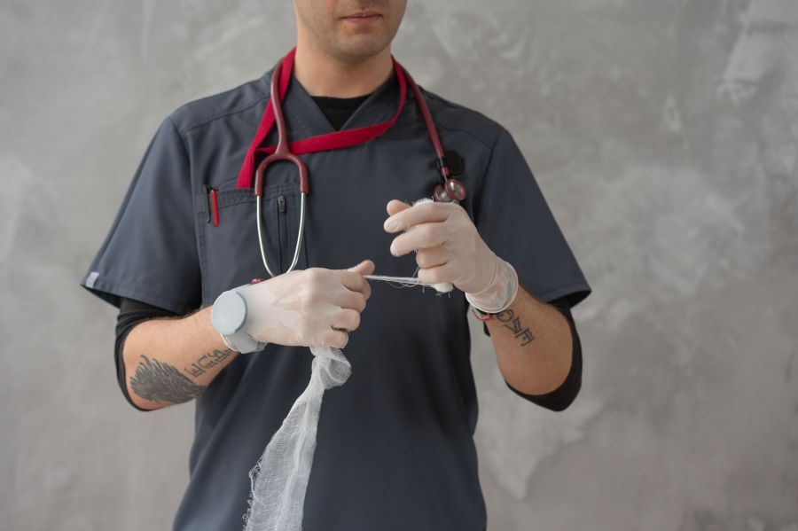
A clinician in scrubs and gloves preparing a length of sterile gauze for a dressing.

Abscesses – What to do about MRSA (Methicillin-resistant Staphylococcus aureus)

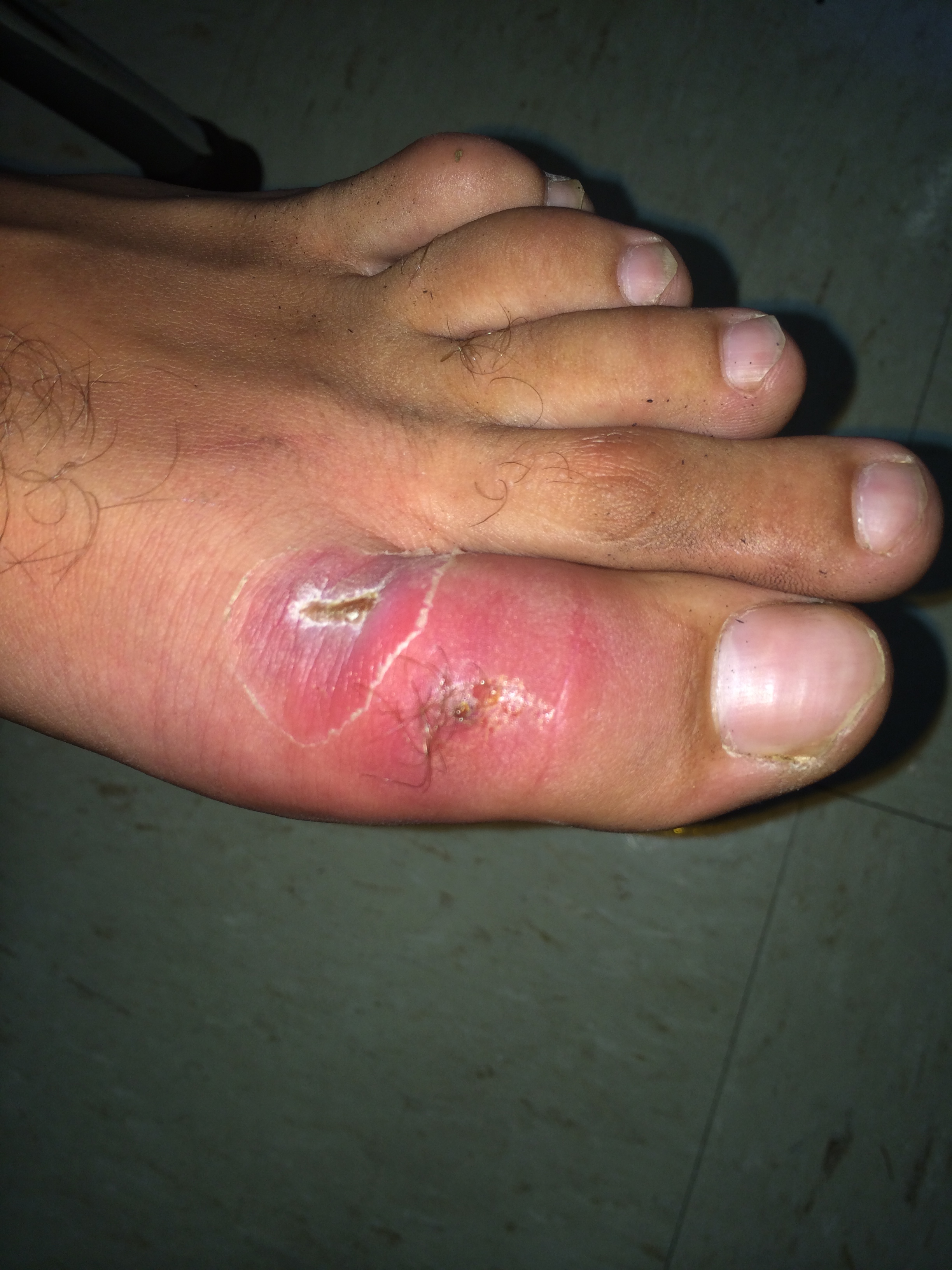
IMG_5872“Staph” infections are most often caused by the organism Staphylococcus aureus, a bacterium that is carried on the skin of about 30% of healthy individuals.  Most of the time, these bacteria do not cause any symptoms. They just hang out on the skin because the skin is a natural layer of defense against infections.  If the skin is damaged, even from a small scratch however, Staph can cause anything from a mild skin infection to a severe, life-threatening illness, especially in young children or older adults. The difference between normal Staph infections and MRSA Infections:  Staph infections in the past were treated with antibiotics such as penicillin.  Some strains of Staph have unfortunately become resistant to penicillin and related antibiotics.  These strains of MRSA cannot be cured with traditional penicillin-related medications.  MRSA is now found in up to 70% of people in the community with a diagnosed staph infection. The spread of MRSA:  Many people become “colonized” with MRSA and that means that they carry the bacteria on their skin or in their nose.  It doesn’t cause any problems unless there is an opening in the skin from a scratch or cut.  You can become colonized with MRSA bacteria in several ways: 1)   Touching the skin of someone else who has been colonized with MRSA 2)   By touching or inhaling small droplets from breathing, coughing or sneezing 3)   By touching a contaminated surface such as a phone headset, countertop or door handle Community MRSA:  You can more easily pick up MRSA by: 1)   Being overweight or obese 2)   Shaving body hair 3)   Sharing personal items or equipment that is not cleaned 4)   Skin trauma such as turf burns, cuts or sores 5)   Physical contact with someone with a draining cut or sore that has MRSA Symptoms:  A skin infection is the most common symptom and it may be mistaken for a spider bite.  A raised, red lump that is tender or cluster of  “pimples,” or large tender lump that drains pus may also be present.  If the bacteria enter the bloodstream, it is possible to develop an infection in areas other than the skin.  A staph infection can occur in a heart valve, inside a bone, a joint or in an implanted device such as an IV line, pacemaker or replacement joint.  Symptoms of these types may include fever, fatigue as well as swelling in the infected area. Diagnosis:  To actually diagnose a true MRSA infection, a culture of the infected area must be performed.  A small sample of bacteria or pus collected from the skin or bloodstream is collected and allowed to grow in an environment that the bacteria thrive in, and then we test these bacteria for resistance to certain antibiotics, including penicillin. Treatment:  in patients with community acquired MRSA we use antibiotics that may be different than patients who develop MRSA in the hospital.  This is because community acquired MRSA is usually sensitive to antibiotics such as Bactrim, clindamycin, or doxycycline.  MRSA that is acquired in the hospital often has to be treated with antibiotics given through an IV such as Vancomycin. A surgical procedure called an I&D (incision and drainage) can be done to drain the pus and heal the infection. If you’ve had an incision and drainage done at your doctor’s office:  After having a surgical procedure done at your doctors office, the wound is often packed with sterile gauze to allow the wound to heal from the bottom upwards.  This way, the wound doesn’t close up at the top and leave a space underneath the top of the skin where bacteria can start growing and cause an infection.  This type of wound needs special attention and observation each day. Basics of wound care after an I&D (incision and drainage): 1)   Keep the bandage/wound clean and dry. 2)   Only remove the bandage to clean around the wound and follow the advice of your medical provider on how to do this. 3)   The packing gauze should be removed little by little until the wound heals completely.  The decision on how much to remove (if any) should be made by a medical provider but this can be explained to the patient on an individual basis (ie. Wounds heal differently depending on the situation). 4)   You may need to wear a splint to decrease movement to the area and allow wound healing. 5)   Antibiotics and pain medication may be prescribed. Prevention:  The CDC has made several recommendations about how to prevent and control MRSA in our communities: 1)   Wash hands with soap and water paying special attention to the fingernails, wrists and between the fingers. 2)   If a sink is not available, alcohol-based hand sanitizers are a good alternative. 3)   Cover all cuts and scrapes and keep them clean until healed. 4)   Don’t touch wounds or bandages from other people 5)   Avoid sharing towels, razors, clothing, uniforms, towels, brushes, combs, make-up and towels. 6)   Athletes should shower after every sports activity and use soap and clean towels. 7)   Exercise equipment at sports clubs and schools should be wiped down with an alcohol-based solution after using it. Care for family members:  Our guidelines do not recommend that family members with MRSA be treated with antibiotics.  Proper preventative measures as described above, should be used. Should I be tested?:  Experts do not recommend widespread testing for MRSA because of the small risk of infection.  Currently only 4 out of 10,000 people in our communities develop a MRSA infection per year.   I hope that you have found this information useful.  Wishing you the best of health,

Recurrent boils and abscesses are one of the reasons I end up asking about someone’s blood sugar, since diabetes both invites skin infections and slows the healing after they are drained. If that is where you are, start with what a new diagnosis of type 2 diabetes actually means. Most of my writing now is obesity and metabolic medicine, including what Wegovy and Zepbound cost without insurance.

Updated for 2026: What Has Changed Since I Wrote This

I first published this in 2012. Most of it held up. One part did not, and the change matters enough that I would rather correct it here than leave it sitting. Back then the teaching was that a small abscess, once drained, did not need antibiotics. Drainage was the treatment. Antibiotics were for people who looked sick, ran a fever, or had redness spreading past the lump. That is what I wrote. It is what most of us were taught. Two randomized trials changed it. In 2016, Talan and colleagues published a placebo-controlled trial in the New England Journal of Medicine. Everyone got drainage. Half then got trimethoprim-sulfamethoxazole and half got placebo. Cure was 92.9 percent with the antibiotic against 85.7 percent with placebo (1). The following year Daum and colleagues ran a similar trial in smaller abscesses, adults and children together, comparing clindamycin, TMP-SMX, and placebo after drainage. Cure came in at 83.1 percent, 81.7 percent, and 68.9 percent. Clindamycin had fewer recurrences, 6.8 percent against 13.5 percent, and more diarrhea (2). Drainage still does the heavy lifting. But adding an antibiotic afterward buys a real, measurable improvement, and I now recommend it for most drained abscesses instead of holding it back for the ones who look ill. One limit worth knowing, because it cuts the other way. The benefit is specific to pus. In 2017 Moran and colleagues tested cephalexin plus TMP-SMX against cephalexin alone in uncomplicated cellulitis with no abscess. No difference, 83.5 percent against 85.5 percent (3). No pocket of pus, no reason to add MRSA coverage.

What This Actually Means on a Video Visit

I practice entirely by video, so let me be straight about what I can and cannot do with an abscess on a screen. I can look at it. I can ask you to press on it and tell me whether it feels like a firm knot or a soft pocket that gives under your finger. I can ask how fast it came up, whether you have had fever, how your blood sugars have been running. What I cannot do is drain it. If there is pus under there, somebody has to open it, and that somebody has to be in the room with you. So here is the honest version. Send me a photo of a red, painful, fluctuant lump and I am going to tell you it needs to be drained, and help you work out where to go. Once it has been drained, the antibiotic afterward is something I can handle from here without any trouble.

When You Need To Be Seen Today

Fever or chills. Redness spreading well past the edge of the lump. An abscess on your face, particularly near your eyes or nose. Poorly controlled diabetes, or a suppressed immune system. No improvement forty eight hours after drainage and antibiotics. Any of those and you belong in front of someone in person, not on a screen (4).

The Bottom Line

Cutting it open is still what fixes an abscess. The 2012 version of this post told you that was usually enough by itself. The evidence since then says an antibiotic afterward makes a real difference, and that is how I practice now.

Sources

1. Talan DA, et al. Trimethoprim-Sulfamethoxazole versus Placebo for Uncomplicated Skin Abscess. N Engl J Med. 2016;374(9):823-832. https://pmc.ncbi.nlm.nih.gov/articles/PMC4851110/ 2. Daum RS, et al. A Placebo-Controlled Trial of Antibiotics for Smaller Skin Abscesses. N Engl J Med. 2017. https://pmc.ncbi.nlm.nih.gov/articles/PMC6886470/ 3. Moran GJ, et al. Effect of Cephalexin Plus Trimethoprim-Sulfamethoxazole vs Cephalexin Alone on Clinical Cure of Uncomplicated Cellulitis. JAMA. 2017;317(20):2088-2096. https://jamanetwork.com/journals/jama/fullarticle/2627970 4. Stevens DL, et al. Practice Guidelines for the Diagnosis and Management of Skin and Soft Tissue Infections: 2014 Update. Clin Infect Dis. 2014;59(2):e10-e52. https://www.idsociety.org/practice-guideline/skin-and-soft-tissue-infections/

Related Reading

When to Use Telemedicine vs. Urgent Care: How I Spot the Sick One Allergic Antibiotic Drug Reactions: Am I Truly Allergic to Penicillin? Scott Rennie, D.O.

Board Certified in Obesity Medicine and Family Medicine

This blog is for educational purposes only and does not constitute individual medical advice. Always consult your own physician before making changes to your health, medications, or treatment plan.
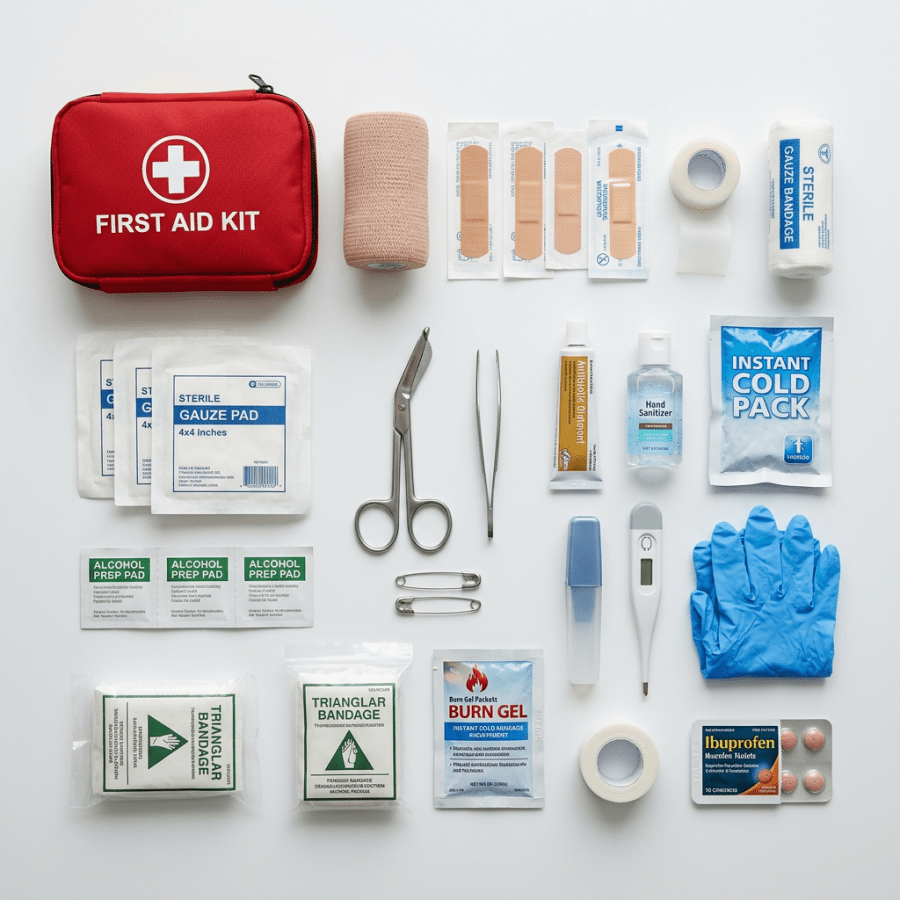
Red first aid kit with assorted bandages, scissors, gloves, thermometer, antiseptic, and medication arranged on white surface

Basic Wound Care Tips for Non-Medical Professionals

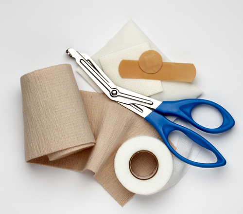
shutterstock_43127152I find that many patients who come into the clinic with a wound have questions about how to take care of it.  The wound might be a post-surgical incision or even a minor cut.  Hopefully this will help with some of those questions. Do I need stitches?:  The best way to know for sure is to be examined by a medical provider, but some basic things we look at: 1)   Depth of the skin wound – if it does not go all the way through the skin, it usually doesn’t need stiches 2)   The size of the wound, whether it’s wide, or jagged.  If it’s large, wide or jagged – there is a higher likelihood of needing stiches 3)   Where on the body the wound is – in areas of the body with lots of stress on the wound, the stitches help hold the skin together better than tape, or a butterfly bandage. 4)   How long ago the injury occurred:  If you got cut 5 days ago, there is less chance sutures would be appropriate because wound healing has already begun. 5)   If it was an animal bite or dirty wound:  We will sometimes put stitches in a wound that was created by an animal bite, but these are generally dirty wounds and we are very careful not to bring the skin together too tight so that the wound can drain. What to do at home – basic guidelines: 1)   Clean the cut or scrape with soap and water.  If there is a piece of glass, or foreign object in the wound that will not come out, you will need to see a medical provider 2)   Stop the bleeding with direct pressure on the wound.  Press a clean cloth or bandage on the wound for 20 minutes minimum without releasing.  If the bleeding is not stopping, contact a medical provider. 3)   Put a thin layer of antibiotic ointment on the wound edges – not in the wound.  I recommend bacitracin (mupirocin) and not Neosporin. 4)   Cover the wound with bandage/gauze to keep the wound clean and dry.  Change the bandage 1-2 times every day until the wound heals 5)   Examine the wound for signs of infection – see below.  Healing time for most wounds is 7-10 days but this can vary. When to see a medical provider: 1)   The wound is deep and/or you can see fatty tissue, muscle, bone or dirty/debris within the wound 2)   You are having severe pain 3)   There are signs of infection (usually begin on about 2-4 days after the wound injury) such as:  Fever, redness, swelling, increased warmth around the wound, pus draining from the wound or red streaks on the skin around the wound. 4)   The wound is from a bite 5)   The wound is a puncture wound caused when a sharp object goes through the skin into the tissue underneath.  There is a higher chance of infection with these kinds of wounds. Do I need a tetanus shot?:  You may need a tetanus booster depending on when your last one was.  If you have deep wound, it is best to contact a medical provider if you’re unsure of whether you need a tetanus vaccine. Abscess:  An abscess is an infection under the skin where bacteria (pus) collect.  A surgical procedure called an I&D (incision and drainage) can be done to drain the pus and heal the infection. If you’ve had an incision and drainage done at your doctor’s office:  After having a surgical procedure done at your doctors office, the wound is often packed with sterile gauze to allow the wound to heal from the bottom upwards.  This way, the wound doesn’t close up at the top and leave a space underneath the top of the skin where bacteria can start growing and cause an infection.  This type of wound needs special attention and observation each day. Basics of wound care after an I&D (incision and drainage): 1)   Keep the bandage/wound clean and dry. 2)   Only remove the bandage to clean around the wound and follow the advice of your medical provider on how to do this. 3)   The packing gauze should be removed little by little until the wound heals completely.  The decision on how much to remove (if any) should be made by a medical provider but this can be explained to the patient on an individual basis (ie. Wounds heal differently depending on the situation). 4)   You may need to wear a splint to decrease movement to the area and allow wound healing. 5)   Antibiotics and pain medication may be prescribed.   I hope that you have found this information useful.  Wishing you the best of health,

Scott Rennie, D.O.
Board Certified in Obesity Medicine and Family Medicine

This blog is for educational purposes only and does not constitute individual medical advice. Always consult your own physician before making changes to your health, medications, or treatment plan.