Woman seated on a sofa holding her knee with both hands

All About Knee Pain: What Every Patient Should Know

shutterstock_94626553I often have people come to the Urgent Care who have problems due to knee pain. Pain that originates in the knee can be complex because there are quite a few areas within the knee that can cause pain. The knee is a “hinge” type joint that is at the greatest risk of injury, age-related wear and tear, arthritis as well as infection related arthritis. Causes: As I mentioned above, there are many causes of knee pain. Several are listed below: 1) Patellofemoral pain syndrome – Group of symptoms that is sometimes caused from overuse of the knee. It can affect running athletes, and is more common in women. The pain is usually in the front of the knee and is made worse with squatting, running, prolonged sitting or when climbing or going down stairs. We also call this Patellar tracking syndrome because it is related to the knee cap (patella) sliding out of the groove that it normally sits in. 2) Meniscal tears – The meniscus is a specialized shock absorber that provides cushion on both sides of the knee. They can become damaged or torn from an acute knee injury or from overuse that comes with age. They have a very poor blood supply, so they don’t heal very quickly 3) Bursitis – The knee is lubricated by joint fluid that is produced by a lubricating bursa (or sac). These bursa sacs can become irritated as a result of injury or even overuse. Inflammation of the bursa is called bursitis 4) Arthritis – This refers to inflammation of the cartilage covering the ends of the bones and undersurface of the knee cap. When it gets worn down, irritated or irregular, it can become painful and is known as arthritis. 5) Tearing of a ligament – The knee is held together by a combination of ligaments including the anterior and posterior cruciate ligaments as well as the medial and collateral lateral ligaments. These ligaments function to hold the bones together and prevent side-to-side or back and forth motion. 6) Muscle strain – If the muscles on the front or back of the knee area become injured, or in spasm they can cause pain around the knee joint. These muscle groups are the quadriceps (located on the front of the top of the knee) and the hamstrings (located on the back part of the leg). These muscle groups work to give support to and move the knee joint 7) Fractures – Broken bones around the knee can obviously cause pain 8) Infection – If bacteria get into the knee joint, this can cause pain, swelling and decreased range of motion 9) Gout – This is caused by the formation of uric acid crystals that build up within the knee joint and is less common in the knee than some other joints 10) Baker’s Cyst – a cyst in the back of the knee can cause pain Diagnosis: Since there are so many different possible causes of knee pain, it is important to make an accurate diagnosis to treat the underlying problem. In addition to getting a good history from the patient about their pain, a physical evaluation will be performed and sometimes knee x-rays, an ultrasound, or an MRI will be ordered. Additionally, a small sample of synovial fluid is sometimes removed from the knee using a needle. This fluid can be examined under a microscope and/or sent for culture to look for bacteria, crystals or signs of inflammation. Treatment: The individual patients injury will dictate the kind of treatment that they need to recover the fastest. Physical therapy is often used because it speeds recovery and regain motion. We also often recommend ice, elevation of the leg and muscle-toning exercises when appropriate. Sometimes a knee brace or immobilizer may be appropriate. Medications such as ibuprofen, Aleve or Tylenol may be appropriate for knee injuries. If infection is present, antibiotics may also be prescribed. If injury to the bone or ligaments is the cause, surgery can be helpful in some patients. Limiting certain activities: Speeding the recovery and helping prevent further injury sometimes involve limiting activities temporarily. Excessive pressure on the knee joint by the following activities should be avoided to help recovery: 1) Kneeling 2) Jogging 3) Squatting 4) Twisting and pivoting 5) Aerobics/Dancing 6) Playing stop and go sports such as basketball or racquet sports 7) Swimming with frog leg or whip kick techniques 8) Rowing machine 9) Stationary bike 10) Stair stepper 11) Leg extensions with weights   I hope that you have found this information useful.  Wishing you the best of health,

Related Reading

How to Stretch Out Properly How to Manage Chronic Pain Without Relying on Medication Diagnosed With Gout? What It Means and How to Prevent Flares Hip Pain on the Side: Greater Trochanteric Bursitis Explained Why Exercise Matters for Obesity Beyond Weight Loss

Scott Rennie, D.O.
Board Certified in Obesity Medicine and Family Medicine

This blog is for educational purposes only and does not constitute individual medical advice. Always consult your own physician before making changes to your health, medications, or treatment plan.

Healthcare worker bandaging a patient’s injured hand

I Hurt My Finger or Toe: How Do I Know if It’s Fractured?

shutterstock_136665947Patients frequently come into the urgent care after injuring a finger or toe and are concerned about the possibility of a fracture. A finger or toe fracture is when a person breaks a bone within their digit. There are different types of fractures, and they are described based on how the bone breaks and whether the broken bone actually sticks out of the skin or not. Symptoms of fracture: 1) Pain 2) Swelling 3) Bruising 4) Stiffness 5) Weakness of the finger/hand or toe 6) The finger or toe looks bent in an abnormal position Testing: You healthcare provider will examine your finger or toe and obtain an X-ray Treatment: The treatment depends on the type of fracture and where it is. Some possible treatments may include: 1) Medication for pain such as Ibuprofen or Tylenol 2) Splinting or “buddy taping” 3) If the fracture is severe with a bone sticking out of the skin, you will need antibiotics and referral to a bone specialist (orthopedic physician). 3) After the splint can come off, your healthcare provider may recommend you work with a physical or occupational therapist. The therapist can show you exercises to strengthen your finger muscles and keep the fingers from getting stiff. Length of time to health: Finger/Toe fractures can take weeks to months to heal, depending on the type of fracture. Home therapy: to help the bone heal rapidly we generally recommend the following: 1) No smoking 2) Keep the cast or splint from getting wet, and keep it clean 3) Keep the finger splinted and immobile to decrease movement   I hope that you have found this information useful.  Wishing you the best of health,

Related Reading

Wrist Fracture or Colles Fracture: What Is a FOOSH Injury? What Should Every Patient Know About Ankle Sprains? Osteoporosis: How to Prevent It and How It’s Treated Diagnosed With Gout? What It Means and How to Prevent Flares Trigger Finger: What Is Stenosing Tenosynovitis Exactly?

Scott Rennie, D.O.
Board Certified in Obesity Medicine and Family Medicine

This blog is for educational purposes only and does not constitute individual medical advice. Always consult your own physician before making changes to your health, medications, or treatment plan.

Woman holding her wrist beside a laptop at a home office desk

Finger and Hand Pain: Is It Carpal Tunnel Syndrome?

shutterstock_86604217Many patients come to the Urgent or their primary care office with complaints of hand/wrist pain, numbness, tingling or a combination of these symptoms.  They often wonder if it could be due to carpal tunnel syndrome. Carpal Tunnel Syndrome:  characterized by pain and numbness in the fingers and hands, and sometimes in the arms.  It happens when the median nerve in the wrist becomes pinched or squeezed.  The median nerve travels through a small “tunnel” in the wrist that is formed by bones and a ligament.  It’s a setup for this nerve to become pinched due to our normal anatomy, but may be worse in some people or due to certain conditions.  There is some thought that the nerve gets pinched possibly due to one or more of the following: 1)   Tissues that surround the surrounding tends in the tunnel harden 2)   Tendons that go through the tunnel get swollen 3)   People hold their hands in a position that causes the tunnel to get smaller. Parts of the hand affected by the median nerve: 1)   Thumb 2)   Index finger 3)   Middle finger 4)   Half of the ring finger 5)   The parts of the palm closest to the thumb Symptoms:  Pain, and tingling in the thumb, index, middle and ring fingers.  These symptoms may be present in one or both hands.  Rarely, the pain can travel up the wrist and forearm and even cause tingling past the elbow to the shoulder. The symptoms are usually worse at night.  Activities that may trigger carpal tunnel syndrome include: 1)   Typing 2)   Reading 3)   Driving 4)   Holding a phone 5)   Sleeping at night – many people bend their wrist while sleeping Testing:  Nerve conduction studies or Electromyography can measure the speed of the electrical nerve conduction of the median nerve or show whether muscles of the hand and wrist are responding appropriately to the electrical signals.  Most of the time the surgeons want these tests to be performed before they will consider surgical treatments for carpal tunnel syndrome. Treatment:  they are tailored to the individual patient and may include: 1)   Wrist splints keep the hands in a neutral position, where the wrists are not bent forward to backward 2)   Surgery is offered to patients who have severe symptoms and that involves cutting the ligament that stretches across the wrist to form the carpal tunnel. 3)   Steroid shots or pills:  The steroid medications that we use short-term are a group of medications that control inflammation and swelling.  Sometimes we will inject a steroid directly into the carpal tunnel, but this is usually done by a hand specialist because of the risks of getting the steroid directly into the median nerve. 4)   Osteopathic manipulation: There are techniques that an Osteopathic Physician (D.O.) may perform that can actually help increase the space inside the carpal tunnel ie. “carpal bone mobilization.”   I hope that you have found this information useful.  Wishing you the best of health,

Related Reading

Trigger Finger: What Is Stenosing Tenosynovitis Exactly? Elbow Pain: Several Common Causes and Treatment Options Wrist Fracture or Colles Fracture: What Is a FOOSH Injury? How to Stretch Out Properly

Scott Rennie, D.O.
Board Certified in Obesity Medicine and Family Medicine

This blog is for educational purposes only and does not constitute individual medical advice. Always consult your own physician before making changes to your health, medications, or treatment plan.

Swollen, reddened big toe resting on a beige ottoman

Diagnosed With Gout? What It Means and How to Prevent Flares

shutterstock_114066385At least once a week, I have patient who presents to the clinic due to pain from gout.  It is a painful condition that develops in some patients who have elevated levels of uric acid in their blood (hyperuricemia).  Up to 66% of patients who have elevated uric acid levels never develop symptoms.  We’re not sure why some people develop symptoms and others don’t have symptoms at all. Uric acid crystals most commonly affect one or multiple joints in the body, but may also cause uric acid crystals to develop within the kidney or urinary tract.  A stone within the kidney may cause impaired kidney function or pain.  Only 15% of people with gout develop kidney stones due to uric acid. Gout risk factors:  Gout is estimated to affect about 2% of the population in the USA.  It is most common in men between 30-45 and women between 55-70.  There is an increased risk of gout with the following: 1)   Taking medications that affect uric acid levels, such as diuretics 2)   Obesity 3)   Ingesting large amounts of meat or seafood 4)   High blood pressure 5)   Consuming high quantities of alcohol 6)   Overeating 7)   Recent surgery or injury Symptoms: 1)   Sudden severe joint pain (usually one joint), sometimes with redness and swelling 2)   Pain and inflammation are worst within several hours and improve over a few days to weeks completely even if untreated Gout phases: 1)   Acute gouty arthritis:  attacks usually involve the big toe or knee which become painful and inflamed 2)   Intercritical period:  The time between attacks.  Usually a second attack occurs within 2 years.  If uric acid levels are uncreated over several years the time between attacks may shorten or the symptoms may become more severe when attacks occur. 3)   Chronic tophaceous gout:  Large numbers of uric acid crystals may collect in joints, bones or cartilage and cause a nodule called a tophus to form.  This tophus is not usually tender, but cause erosion of bone and permanent deformity.  The tophi when the occur on the knuckles can cause an unfortunate cosmetic problem. Diagnosis:  Gout may be suspected if the person has the sudden onset of joint pain in the commonly affected joints such as the big toe.  If the patient has a period when there are no symptoms, and then symptoms return this is very typical of the disease.  The best way to diagnose gout is to examine fluid from the affected joint to look for uric acid crystals.  A medical provider uses a needle and syringe to withdraw a small sample of synovial fluid from the joint and this fluid is sent to the lab for microscopic analysis.  Other criteria for diagnosis gout may be: 1)   Blood testing showing high uric acid levels 2)   Rapid improvement of symptoms after the patient is treated with colchicine 3)   History of pain and inflammation involving one joint at a time, especially at the base of the large toe 4)   The symptoms totally go away between attacks Treatment:  The goals of gout treatment are to decrease the pain of a gout attack and also to reduce the frequency of their attacks.  Some of the treatment options are listed below: 1)   Nonsteroidal anti-inflammatory drugs (NSAIDs) reduce the swelling in the joint.  Examples of these are Ibuprofen or indomethacin.  They are most effective if started as soon as possible after the gout attack starts. 2)   Colchicine is a medication that may decrease the pain of a gout attack very rapidly. 3)   Steroids such as prednisone may be used if an NSAID is not used.  This type of medication can be injected directly into the affected joint or can be given as pills or intramuscular injection.  There is a risk of “rebound gout attack” as the steroids are stopped however, so usually steroids are slowly tapered over 7-10 days. Prevention therapy:  Some medications as well as dietary changes can be helpful to reduce the frequency of gout attacks. 1)   Uric acid lowering medications are used indefinitely because there is not a benefit from taking a break from the medications.  Examples of this are Probenacid, Losartan, and Allopurinol.  Rapid lowering of uric acid can cause a gout flare . 2)   Increased water intake – at least 2 liters/day are recommended Dietary changes:  Obesity is a major risk factor for gout so changing diet may decrease the frequency of gout attacks.  Current recommendations also include eating less of the following: 1)   Red meat 2)   Seafood 3)   Beer and hard alcohol (gin, vodka, etc.) 4)   Foods and drinks that contain high-fructose corn syrup (fond in some non-diet sodas for example) Some foods that may be good to eat or drink if you have gout may include: 1)   Low fat dairy 2)   Foods with complex carbohydrates such as whole grains, brown rice, oats and beans 3)   Vitamin C (500mg/day has been found to have a mild uric acid lowering affect). 4)   Coffee in moderation 5)   Wine in moderation – (1-2 5oz servings/day me be helpful) 6)   Pineapples – see my blog article about preventing gout by eating pineapples 7)   Drink plenty of water – twelve 8oz glasses of water during every day.   I hope that you have found this information useful.  Wishing you the best of health,

Related Reading

Can Eating Pineapple Help Treat Gout Pain Naturally? How to Manage Chronic Pain Without Relying on Medication All About Knee Pain: What Every Patient Should Know I Hurt My Finger or Toe: How Do I Know if It’s Fractured? How Do You Actually Lose Weight? A Doctor Explains

Scott Rennie, D.O.
Board Certified in Obesity Medicine and Family Medicine

This blog is for educational purposes only and does not constitute individual medical advice. Always consult your own physician before making changes to your health, medications, or treatment plan.

A rolled elastic compression bandage and an ice pack wrapped in a towel beside a pair of sneakers on a wooden floor

What Should Every Patient Know About Ankle Sprains?

Editor’s note, August 2026. This post recommends RICE with two to three days of rest. Sports medicine has moved away from that. Prolonged rest and routine icing are no longer advised, and getting a gentle amount of weight and movement through the ankle sooner produces better outcomes. Read what follows as the older protocol.

shutterstock_134396747Another common condition that we see in the urgent care and clinic is the ankle sprain.  Sometimes there can be some worry about whether there is an ankle fracture by the patient and the doctor.  Here is some general information about ankle pain that might be helpful if you have or know someone who has ankle pain. Ankle sprains include stretch injuries, partial ruptures or even a complete ruptures of at least on ligament of the ankle.  Ligaments provide stability and help direct the motion of the ankle.  If the ankle starts to move in a direction that causes too much stretch and puts the ligaments at risk of injury, there are sensors built into the ligament that help provide a reflex to move the ankle back to a safe position.  This happens without conscious effort of the patient.  These stretch receptors within the ligament can become damaged with a prior ankle sprain.  If this happens, they might not work properly to protect the ankle from future sprains. How bad is the sprain?/Grading:  There are three classes of ankle sprains that help describe how bad the sprain is by loss of function and clinical signs.  Grade 1 is less severe and results from mild stretching of the ligament with microscopic tears.  There is no joint instability on exam.  Grade 2 sprains are more severe and involve the complete tear of a ligament.  There is usually mild to moderate joint instability on exam with reduced range of motion and loss of function.  Walking with a grade 2 ankle sprain is painful.  Grade 3 ankle sprains involve a complete tear of a ligament, and there is significant mechanical instability.  Patients are usually unable to walk with these sprains. Lateral ankle sprain:  The most common injury to the ankle is when the ankle is plantar-flexed (toes pointed down) and the ankle rolls inward.  This causes damage to the lateral ligament complex of the ankle (anterior talofibular ligament [ATF], calcaneofibular ligament [CFL] and posterior talofibular ligament [PTF]).  The ATF is the first or only ligament that is injured in almost all ankle sprains.  If the ankle injury is more severe, the TF and CFL might become injured, which can result in ankle joint instability.  Very strong forces to the ankle may result in injuries to all three ligaments.  These are less common injuries, but  are more debilitating and are often associated with ankle nerve injury. Medial ankle sprain:  The strongest ligament of the ankle is the medial deltoid ligament complex.  If the ankle rolls outward in a forced motion, it can cause damage to this structure but usually this type of motion causes an avulsion fracture of the medial malleolus. High Ankle Sprain:  1-11% of all ankle sprains are this type and are more common with contact sports.  This sprain is caused by the toes pointing upwards and rolling the ankle outwards.  This can cause a sprain that includes the interosseous membrane as well as the ATF, PTF and transverse tibiofibular ligaments.  These ligaments are critical to ankle stability. Physical exam of the ankle:  We look at the ankle and look for several signs on examination which may include:  Swelling, bruising, palpating the fibula, the Achilles tendon, the edge or tip of the lateral malleolus, the posterior edge or tip of the medial malleolus, the base of the 5th metatarsal and the navicular bone.  We use a set of rules called the “Ottawa ankle Rules” to determine the need for an ankle x-ray. Treatment:  For lateral ankle sprains, the treatment goal is to limit inflammation and swelling to maintain range of motion to the ankle.  This is usually done via early treatment with RICE (rest, ice, compression and elevation) for the first 2-3 days. 1)   Rest is done by limiting any standing or walking and using crutches until the patient can walk normally. 2)    Ice is applied (or cold water emersion) for 15-20 minutes every 2-3 hours during the first 2 days or until the swelling has improved. 3)   The sprained ankle should be elevated above the level of the heart to reduce swelling. 4)   Compression with a gel splint or elastic bandage such as an ACE wrap or SAM splint is helpful to minimize swelling and reduce further injury. 5)   Ibuprofen,  Aleve or Tylenol can be used to reduce pain. Rehabilitation/Physical Therapy: For ankle sprains, I feel that early rehabilitation with range of motion exercises can be very helpful for retuning to activity and preventing chronic instability.  In most patients with mild to moderate sprains, rehab should begin as soon as the initial pain and swelling have decreased to allow them to perform simple exercises.  The program will take several weeks in order for the ankle to become stronger and limit the chance of re-injury. Indications for referral to an ankle surgeon include: 1)   Ankle fracture 2)   Dislocation or subluxation 3)   Syndesmosis injury 4)   Tendon rupture 5)   Wound that goes into the ankle joint 6)   Uncertain diagnosis Prevention of ankle sprains:  Some possible options for prevention of ankle sprains include: 1)   External ankle supports (lace up, high-top shoes, taping, etc) 2)   Stretching and strengthening 3)   Proprioceptive ankle training using a wobble board 4)   Appropriate shoes/boots for the planned activity – ie. Hiking boots for a hike on uneven terrain.   I hope that you have found this information useful.  Wishing you the best of health,

Related Reading

I Hurt My Finger or Toe: How Do I Know if It’s Fractured? Plantar Fasciitis Explained: A Real Pain in the Foot How to Stretch Out Properly Wrist Fracture or Colles Fracture: What Is a FOOSH Injury?

Scott Rennie, D.O.
Board Certified in Obesity Medicine and Family Medicine

This blog is for educational purposes only and does not constitute individual medical advice. Always consult your own physician before making changes to your health, medications, or treatment plan.

Man in navy suit holding his lower back in pain in office hallway

Low Back Pain: What Can Actually Help You Find Relief?

shutterstock_115219366

Low back pain is one of the most common reasons people see a doctor. About 8 out of 10 adults will have at least one episode at some point in their lives. The pain is usually felt along the lumbar spine and can worsen with bending, twisting, or even just sitting down. In many cases the muscles that support the back are the main source of the pain. Sometimes arthritis or a bulging disk irritates the nerve roots and that’s when patients notice symptoms like numbness, tingling, or weakness in the legs.

Sciatica is a well-known example. That’s when one of the nerve roots of the sciatic nerve is irritated, sending sharp, burning pain down the back or side of the thigh and often all the way to the ankle or foot. It may also come with numbness or tingling.

Most back pain gets better with conservative care, but there are times when patients should see a doctor right away. If someone over 70 suddenly develops back pain, if the pain persists at night even while lying down, or if it comes with weakness in the legs, loss of bladder or bowel control, unexplained fever, or weight loss, those are red flags. A history of cancer, osteoporosis, or a significant fall also raises concern. And if pain hasn’t improved at all after four weeks, it’s worth reevaluating.

Doctors usually start with a history and physical exam. Imaging tests like x-rays can pick up things such as compression fractures or alignment issues. CT or MRI scans are used when more detail is needed, especially to look at soft tissues, disks, or nerves. A bulging disk will often improve on its own over time as the body reabsorbs it, so surgery isn’t the first step unless there are severe symptoms.

Activity is important. Studies show patients recover faster when they keep moving. Staying in bed too long can actually make pain worse. Heat can ease stiffness in the first couple of weeks, and alternating ice and heat sometimes helps too. I usually encourage people to keep working if possible, but to avoid heavy lifting, prolonged standing, or twisting motions.

Medications have a role. Over-the-counter options like acetaminophen, ibuprofen, or naproxen can reduce pain and inflammation. Muscle relaxants such as cyclobenzaprine or baclofen may help in the short term, but they can cause drowsiness, so patients need to be cautious with driving or operating machinery. Narcotics are occasionally used but carry more risks and aren’t typically needed for most back pain.

Exercise is helpful once the pain begins to settle. Early stretching can sometimes aggravate symptoms, but as recovery progresses, a structured program that builds flexibility and strengthens core muscles supports long-term improvement. If pain lingers beyond four to six weeks, physical therapy is often recommended. Therapists focus on strengthening, posture, and mobility to reduce recurrences.

As an osteopathic physician, I sometimes use hands-on manipulation. By improving range of motion in restricted areas, it’s possible to break the cycle of stiffness and pain. Other approaches that can help certain patients include massage, yoga, acupuncture, injections, or even traction. Braces may be useful in select cases. Surgery is an option, but usually only when there’s severe nerve involvement or persistent symptoms that don’t respond to other treatments.

Prevention is key. Staying active, exercising regularly, and keeping the core muscles strong all help protect the back. Avoiding repetitive heavy lifting or twisting motions reduces strain. When lifting can’t be avoided, bending at the knees rather than the waist protects the spine. Stretching the hamstrings, quadriceps, piriformis, and gluteal muscles also supports spinal health.

Related Reading

How to Stretch Out Properly

How to Manage Chronic Pain Without Relying on Medication

Cervicalgia – A Pain in the Neck

Hip Pain on the Side: Greater Trochanteric Bursitis Explained

Scott Rennie, D.O.
Board Certified in Obesity Medicine and Family Medicine

This blog is for educational purposes only and does not constitute individual medical advice. Always consult your own physician before making changes to your health, medications, or treatment plan.

Person stretching leg on rock near ocean at sunset

How to Stretch Out Properly

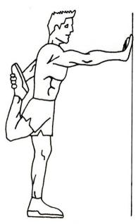
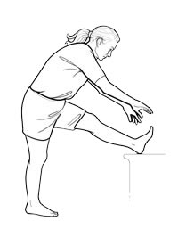
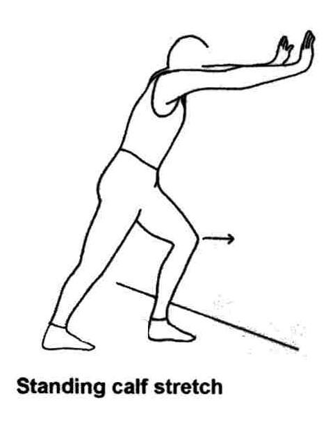
I often get questions from patients about how they should warm up or prepare before starting to exercise.  I have a few suggestions that I’ve learned over the years from physical therapists, personal trainers and patients who have come in from sports related injuries.  A good stretching warm up raises the temperature of your muscles and promotes optimal flexibility with the goal of preventing muscle strain or spasms. 1)  Understand the muscles you will be using during your particular sport.  It’s important to know what your workout will involve and then stretch those muscles.  For example, if you are a runner then warming up the hamstrings, quads and gluts are more important than the muscles of the upper body. 2)  Begin slowly.  Learn to gradually lengthen the muscles to prevent injury during exercise.  You don’t need to touch your toes immediately.  If fact if you over-stretch your muscles too quickly, it can put them into a spasm. 3)  Hold the stretch.  Begin by holding your position once your muscles reach their limit for 10-15 seconds, then relax and perform that stretch again.  Repeat on the other side of the body or extremity as necessary. 4)  Once you’ve finished your workout, stretch again.  You will increase your flexibility and it’s a great way to cool down after your exercise routine. 5)  Don’t bounce.  If you bounce when you stretch, you may increase your risk of injury and over-stretch.   You should not feel like you’re hurting yourself with stretching.  If it hurts, stop the stretch immediately. Every medical professional has their favorite stretches.  Some of my favorites include the following: A)  Quadricep Stretches:  The quadriceps are four muscles located in the anterior thigh (the Vastus lateralis, Vatus medialis, Vastus intermedius and Rectus femoris.)  This muscle group acts to extend the leg while straightening the knee.  Running, biking, jumping, hopping, and jogging all involve the quads and dysfunction in these muscles may lead to injury.  The way I stretch this muscle group is by: 1)  While standing, grab  a stable structure such as the table or counter for balance with one hand. 2)  Bend your knee back by grasping your ankle with the hand on the same side of your body 3)  Maintain the position for 15-30 seconds and if no stretch is felt, you may bend forward at the hips, then relax to a standing position. 4)  Repeat the procedure for the other leg and continue repeating for 15 minutes. B)  Hamstring stretch:  Your hamstring  is the muscle group that runs along the back of your upper leg.  Three muscles that make up the hamstrings are the biceps femoris, semimembranosus and semitendonosus.  They are mostly used as a hip extensor and 1)  Place your heel on an object approximately 18″ high, and stand as erectly as possible. 2)  Extend your low back, tilting your pelvis forward, and bend forward from the hips, maintaining the curve of your low back pain. 3)  For added emphasis, tilt your toes back toward you.  You should feel the stretch in the back of your leg.  Hold for 15-30 seconds and then repeat for the other leg for a total of 15 minutes. C)  Glute Stretch:  The glute muscles are defined as the buttocks.  They encompass the Gluteus Maximus, gluteus medius and gluteus minimus.  They play a role in movement and are used in walking, running, jumping, bicycle riding, and more.  They extend and rotate the leg. 1)  Lie on the floor or mat.  Bend knees with feet on the floor. 2)  Cross lower leg over thigh and grasp back of thigh of the lower leg with both hands 3)  Pull leg toward torso and hold stretch for 15-30 seconds.  Repeat for the opposite leg and continue for a total of 15 minutes. D)  Calf Stretch:   The calf muscles consist of the gastrocnemius and the soleus. The gastrocnemius is the big muscle at the back of the lower leg.  It helps us plantar flex (point the food down).  It also helps with knee extension.  The Soleus muscle’s action is ankle plantar flexion. 1)  Place the toes of one foot up onto the wall so that your heel is still on the ground 2)  Lean forward until a stretch is felt in your calf, keeping your knee straight. 3)  Hold for 15-30 seconds and then repeat with the other foot.  Continue for a total of 15 minutes.   I hope that you have found this information useful.  Wishing you the best of health,

Related Reading

Low Back Pain: What Can Actually Help You Find Relief? Cervicalgia – A Pain in the Neck Plantar Fasciitis Explained: A Real Pain in the Foot Shoulder Pain: Common Causes and Treatment Options How to Manage Chronic Pain Without Relying on Medication

Scott Rennie, D.O.
Board Certified in Obesity Medicine and Family Medicine

This blog is for educational purposes only and does not constitute individual medical advice. Always consult your own physician before making changes to your health, medications, or treatment plan.

Ripe pineapples resting on a green surface

Can Eating Pineapple Help Treat Gout Pain Naturally?

Gout is a common condition that happens when uric acid crystals build up inside the joints. The result is sudden, severe pain with swelling, redness, and loss of function. The joint at the base of the big toe is the one most often affected. Doctors call this podagra. But gout doesn’t stop there. I’ve seen it flare in the ankle, the wrist, the knee, and even the shoulder or hip. It can also involve the kidneys if crystals begin to form there, which can complicate the picture.

When someone has been diagnosed with gout, one of the first conversations is about diet. Certain foods can raise uric acid levels and set off an attack. Red meat, some seafood, beer, and drinks with high fructose corn syrup are well-known triggers (National Institute of Arthritis and Musculoskeletal and Skin Diseases, 2011). Patients often notice the pattern themselves after a few flares.

During an acute attack, the goal is relief. In the office, I’ll usually talk through standard treatment options. Nonsteroidal anti-inflammatory drugs like ibuprofen or naproxen are often first line. Colchicine is another option, and in severe cases, a corticosteroid injection directly into the joint can bring fast relief (American College of Rheumatology, 2010). These treatments can be effective, but they aren’t without risk. Side effects, cost, and access are real considerations for many patients.

Some people ask about natural approaches. I’ve had patients report success with pineapple, which contains bromelain, an enzyme with anti-inflammatory properties. One patient told me that eating pineapple right after the onset of pain worked better for them than any pill. While that’s anecdotal, bromelain has been studied as a supplement and is available in capsule form as well (University of Maryland Medical Center, 2010). As with any supplement, I advise patients to check with their doctor before starting it.

Hydration is another piece of advice I return to often. Uric acid crystals form more easily in concentrated urine. Drinking water dilutes the urine and helps the kidneys flush uric acid more efficiently. The Arthritis Foundation has long recommended at least eight to twelve glasses of water a day for patients with gout (Arthritis Foundation, 2011). It’s a simple step but one that can make a difference.

Gout is painful, but with attention to triggers, thoughtful use of medications, and supportive measures like hydration, patients can reduce the frequency and severity of attacks.

Related Reading

Diagnosed With Gout? What It Means and How to Prevent Flares

How to Manage Chronic Pain Without Relying on Medication

I Hurt My Finger or Toe: How Do I Know if It’s Fractured?

Scott Rennie, D.O.
Board Certified in Obesity Medicine and Family Medicine

This blog is for educational purposes only and does not constitute individual medical advice. Always consult your own physician before making changes to your health, medications, or treatment plan.


Sources:

  • National Institute of Arthritis and Musculoskeletal and Skin Diseases. “Questions and Answers about Gout.” NIH, 2011.

  • American College of Rheumatology. “Gout Clinical Practice Guidelines.” ACR, 2010.

  • Arthritis Foundation. “Living with Gout: Lifestyle and Diet.” Arthritis Foundation, 2011.

  • University of Maryland Medical Center. “Bromelain.” UMMC Complementary and Alternative Medicine Guide, 2010.

Yoga mat with resistance bands, heating pad, and bowl of water in bright room

How to Manage Chronic Pain Without Relying on Medication

We all deal with physical pain at some point. When it lingers for more than three to six months, it’s considered chronic pain. The causes are many. It might be diabetic neuropathy. It might be arthritis in the joints. It could be long-term back pain tied to muscle spasms. The list is long, and so are the options for treatment.

Patients often ask how to manage pain without relying on narcotics (also called opioids). The concern is real. These medications can cause drowsiness, constipation, and for some people, dependence or addiction. Before prescribing them, it’s important to talk through those risks and explore other options first.

Finding the right treatment usually starts with finding the cause. Sometimes that means getting tests. Depending on the symptoms, this might be an MRI, CT scan, ultrasound, nerve conduction study, or X-rays. Pinpointing the source of the pain helps guide treatment more precisely.

Non-medication options can make a big difference. Exercise, weight reduction, and a balanced diet all play a role. Sleep matters too. Some patients get relief from meditation or acupuncture. In my own practice, I’ve seen osteopathic manipulative medicine help certain patients. Others do well with stress reduction strategies like biofeedback, sometimes with support from a counselor. Avoiding alcohol, tobacco, and recreational drugs also helps the body recover and stay healthier. For some, surgery is the right path. And yes, sometimes medications are still part of the plan, but usually not the only piece.

One example that sticks with me is a patient with chronic low back pain. He had been through multiple prescriptions without much relief. We worked on a mix of stretching, weight loss, and stress management, and added in osteopathic manipulation. It wasn’t a cure, but his pain decreased enough that he could get back to walking his dog each morning, which mattered more to him than the number on a pain scale.

Managing chronic pain is rarely simple. It can be frustrating for both the patient and the doctor. But using a combination of treatments usually works better than leaning on just one approach. There are many ways to manage pain without heavy reliance on narcotics, and that gives patients real choices.

This information is meant for general education and shouldn’t be taken as medical advice for an individual patient. If you’re dealing with chronic pain, it’s best to talk with your own medical provider.

Wishing you good health,

Related Reading

How to Stretch Out Properly

Low Back Pain: What Can Actually Help You Find Relief?

Cervicalgia – A Pain in the Neck

All About Knee Pain: What Every Patient Should Know

Can Eating Pineapple Help Treat Gout Pain Naturally?

Scott Rennie, D.O.
Board Certified in Obesity Medicine and Family Medicine

This blog is for educational purposes only and does not constitute individual medical advice. Always consult your own physician before making changes to your health, medications, or treatment plan.


Sources: