
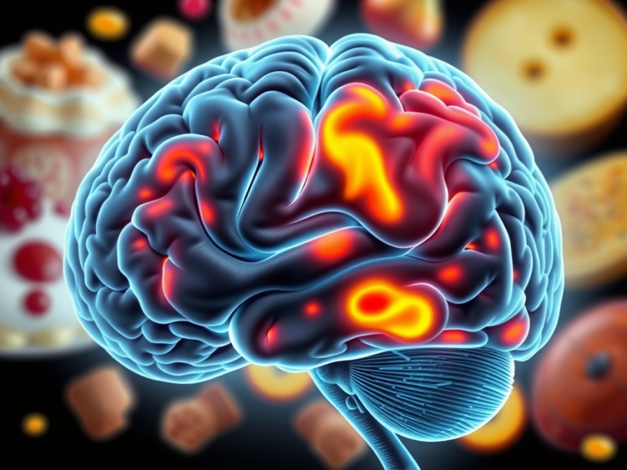
Patients say a version of the same thing constantly: “I know what I should eat, but I still crave the wrong things.” That gap between knowledge and behavior is what pushed researchers toward the brain. Functional MRI has shown that appetite runs on circuits that defend fat mass and respond to food cues, and that willpower is a small part of the story.
For years we leaned on BMI as the working definition of obesity. A BMI over 30 got the label, and the number explained nothing about why weight gain happened or why some patients struggle far more than others. Schwartz and colleagues reframed it in 2017 as “a disorder of energy homeostasis, characterized by the defense of an elevated body fat mass” (Schwartz et al., Endocr Rev, 2017). That definition earns its keep. It says the body is working to hold fat stores high, and that when weight comes off, biology answers with stronger hunger signaling, slower metabolism, and shifted hormones.
The gut-fat-brain conversation sits at the center. Leptin, ghrelin, insulin, GLP-1, and PYY all shape hunger and satiety, and fMRI shows how those signals land. High-calorie food cues light up the amygdala, striatum, medial orbitofrontal cortex, and ventral tegmental area, all reward and craving territory (Schur et al., Int J Obes, 2009; Melhorn et al., Am J Clin Nutr, 2018). After weight loss, that reward response doesn’t fade, which is a large part of why relapse is the rule. Interventions do move it. Leptin replacement, intranasal insulin, GLP-1 agonists, and bariatric surgery all reduce this activation (Holsen et al., Int J Obes, 2018; van Bloemendaal et al., Diabetes, 2014).
One finding deserves more attention than it gets: looking at pictures of calorie-dense food predicts what people actually eat. In studies where participants later chose from a buffet, those with higher reward activation to food images selected more high-fat, high-calorie items. The brain response translated into behavior at the table.
That has treatment implications. Patients with persistent reward-driven responses may get the most from GLP-1 agonists like semaglutide. For others, agents acting on central insulin or leptin signaling may fit better. Bupropion-naltrexone targets reward pathways directly and may suit patients where hedonic eating is the main driver. Obesity is a brain-based condition, and it needs brain-aware treatment.
Inflammation belongs in this picture too. Valdearcos and colleagues showed that rodents on a high-fat diet developed hypothalamic gliosis, an inflammatory response in the brain, before they gained significant weight (Valdearcos et al., Cell Metab, 2017). Human MRI findings line up. Individuals with obesity are more likely to show signs of hypothalamic gliosis (Schur et al., Obesity, 2015; Kreutzer et al., Diabetes, 2017). Inflammation may disrupt appetite regulation early, helping drive the defense of elevated fat mass.
For clinicians, this changes the posture. Blaming patients for “failing” when weight returns misreads the physiology. Their biology is built to resist fat loss. Medications acting on appetite centers belong in long-term care rather than short courses. Diet quality may matter for brain inflammation as well as calorie balance. And as with any other chronic disease, the expectation should be continuous management rather than a one-time fix.
Framing obesity as a chronic brain and inflammatory disease does something useful for the room. It takes stigma out of it. Patients are living with a condition in which the brain defends fat mass through powerful signals, and that framing replaces shame with something we can actually treat.
References:
1. Schwartz MW, Seeley RJ, Zeltser LM, et al. Obesity Pathogenesis: An Endocrine Society Scientific Statement. Endocr Rev. 2017;38(4):267-296. https://pubmed.ncbi.nlm.nih.gov/28898979/
2. Schur EA, et al. Activation in brain energy regulation and reward centers by food cues varies with choice of visual stimulus. Int J Obes (Lond). 2009;33(6):653-661. https://pubmed.ncbi.nlm.nih.gov/19365394/
3. Melhorn SJ, et al. Am J Clin Nutr. 2018;107(4):574-582.
4. Holsen LM, et al. Int J Obes (Lond). 2018;42(4):785-793.
5. van Bloemendaal L, et al. GLP-1 receptor activation modulates appetite- and reward-related brain areas in humans. Diabetes. 2014;63(12):4186-4196. https://pubmed.ncbi.nlm.nih.gov/25071023/
6. Valdearcos M, et al. Microglial Inflammatory Signaling Orchestrates the Hypothalamic Immune Response to Dietary Excess and Mediates Obesity Susceptibility. Cell Metab. 2017;26(1):185-197.e3. https://pubmed.ncbi.nlm.nih.gov/28683286/
7. Kreutzer C, et al. Hypothalamic Inflammation in Human Obesity Is Mediated by Environmental and Genetic Factors. Diabetes. 2017;66(9):2407-2415. https://pubmed.ncbi.nlm.nih.gov/28576837/
8. Schur EA, et al. Radiologic evidence that hypothalamic gliosis is associated with obesity and insulin resistance in humans. Obesity (Silver Spring). 2015;23(11):2142-2148. https://pubmed.ncbi.nlm.nih.gov/26530930/
Board Certified in Obesity Medicine and Family Medicine
This blog is for educational purposes only and does not constitute individual medical advice. Always consult your own physician before making changes to your health, medications, or treatment plan.
 Doctor Rennie's Blog