

 http://www.dvtforum.com/index.asp?action=start
A few days ago a patient came into the clinic with complaints of pain in the back of his knee after spending hours driving his car back to Washington from the east coast. He didn’t recall injuring his leg or knee and told me that the pain was worse when he starts walking. He also noted some swelling in the leg and ankle. I became concerned about the possibility of a blood clot (also called a deep vein thrombosis or DVT) in his leg after hearing about his long journey because sitting for extended periods of time in a car or on an airplane are big risk factors for a deep vein thrombosis. After the ultrasound confirmed a blood clot in his left leg, we started him on the treatment and he is doing great today.
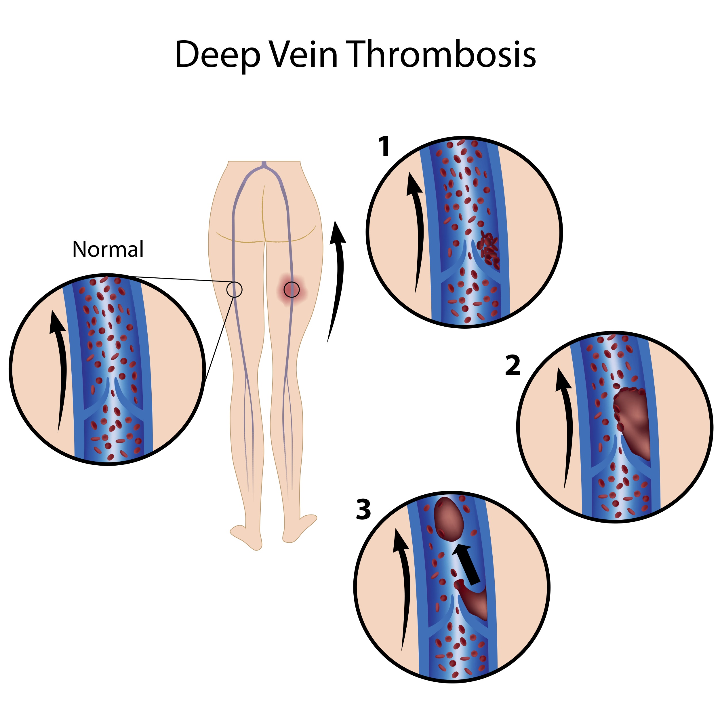
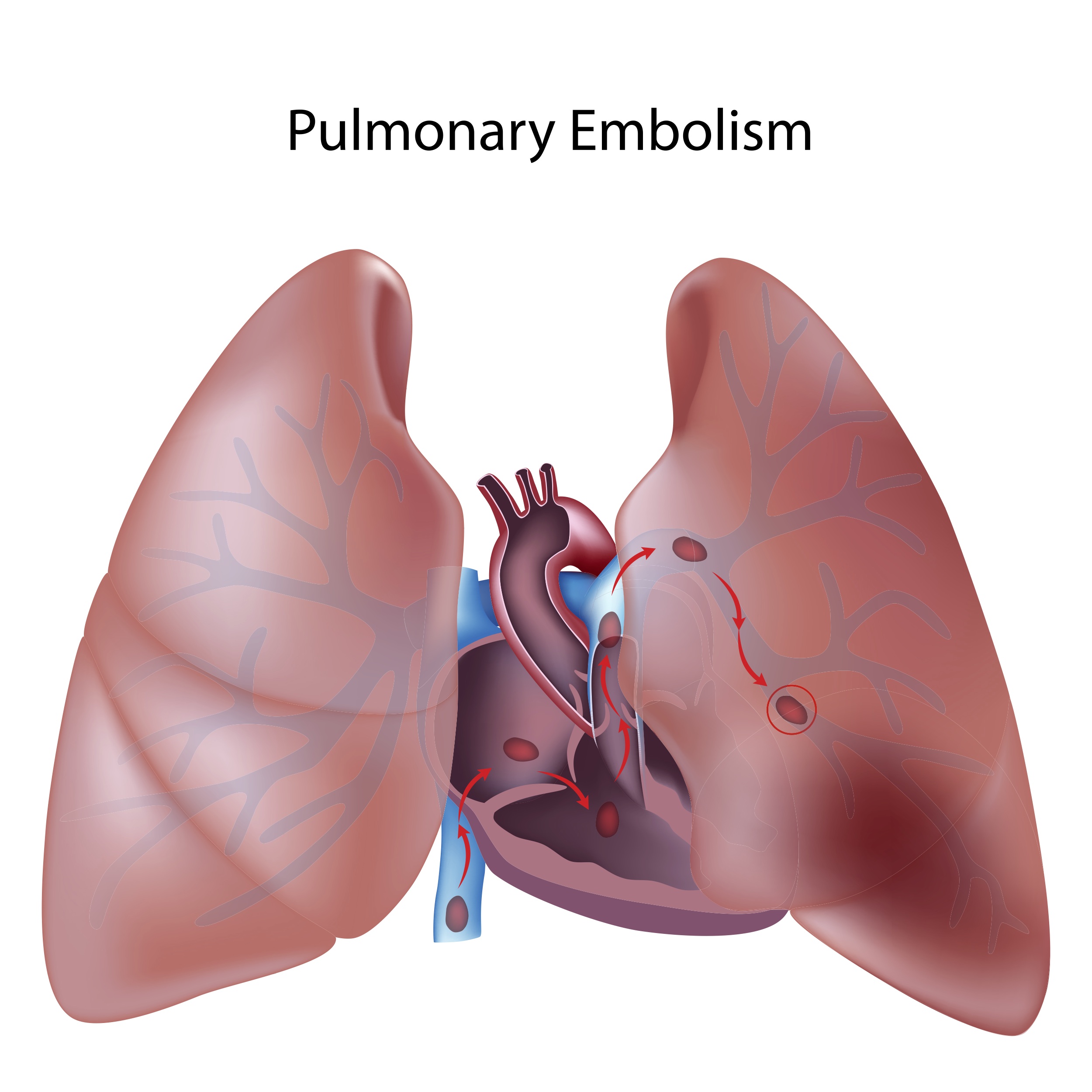
A blood clot (also called a thrombus) can be very serious and even life threatening if it breaks and travels through the circulatory system. If that happens, the blot clot (now called an embolis to indicate that it broke off from the main clot and is traveling through the veins of the body) can lodge in the lung and cause a pulmonary embolism (PE) which is a serious condition that causes over 50,000 deaths a year in the U.S.A.
A deep vein thrombosis (DVT) is a problem where a blood clot is created inappropriately within the vein. Most commonly these veins are in the “deep veins” of the leg, pelvis, or thigh. The thrombus can block or partially block the blood flowing through the vein and cause pain and swelling.
Symptoms: These may or may not all be present:
1) Pain – usually in one leg, or knee
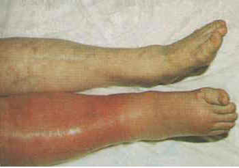
2) Swelling in that leg or knee
3) Warmth and redness in the leg or knee area
Diagnosis: If a medical provider suspects that you might have a DVT, there are certain tests that can be done to look for this including:
1) Compression ultrasonography (ultrasound) which uses sound waves to allow the visualization of the blood flow through the veins of the leg.
2) D-dimer is a blood test that is often elevated in people who have a blood clot. It can also be elevated for other reasons, so an elevated D-dimer does not tell us that a patient has a blood clot, but if the D-dimer is not elevated and the risk factors are low, there is less chance of the patient having a blood clot.
3) MRI uses a very strong magnet to create photos inside the leg and is expensive so it’s not the first choice the diagnose blood clots.
4) Contrast venography is when a small tube (called a catheter) is inserted into the vein and a liquid that shows up on x-ray (we call this contrast) is injected through the tube while xray is used to visualize the blood flow of the contrast through the vein. We usually prefer to use ultrasound, but this test can be helpful when ultrasound is not available.
Risk factors for DVT:
1) Some people have a genetic problem called thrombophilia which causes blood to clot more easily than it should
2) Cancer
3) Smoking
4) Heart failure
5) Pregnancy
6) Being overweight or obese
7) Increased age
8) Having had a DVT or PE in the past
9) Certain kidney problems
10) Medications such as birth control pills or hormone replacement therapy
11) Recent surgery – especially involving the knee, hip or pelvis
12) Trauma – such as a fall or deep bruise where the blood vessels may have become injured
13) Sitting for long periods of time such on a long flight or a long drive in a vehicle. We also think about this for elderly patients who don’t move around very much
Treatment: The goal in treating a DVT is to prevent the blood clot from dislodging or breaking off and becoming an embolism that could lodge into the lung (pulmonary embolism). We also want to prevent the clot from getting larger or new clots from forming.
Medications can be used to prevent new blood clots from forming(anticoagulants), and other medications can be used to dissolve a blood clot (thrombolytic). Most people might think that the primary treatment would be to dissolve the clot using thrombolytic therapy, however this is usually not the case. The body has natural substances to dissolve blood clots that are formed inappropriately in blood vessels and we usually prefer the body to do this naturally while assisting by giving medications to prevent new blood clots from forming. Anticoagulant medications such as warfarin or heparin are the most common agents used to treat patients with DVTs at this time. For patients who continually develop blood clots despite being on medications or who cannot take the recommended medications, a filter might be inserted into one of the largest veins in the body (the inferior vena cava) to block the circulation of blood clots. We strategically place the filter so that if a blood clot forms, it is not allowed to reach the lungs.
Prevention: For patients at high risk of developing a blood clot such as a patient who just had surgery, or someone who has cancer, they might be started on anticoagulant medications to decrease the risk of a blood clot. Some patients are fitted with inflatable compression devices that are worn on the legs and periodically fill with air and put compression on the legs and work similar to how an electronic blood pressure cuff inflates.
If you or someone you know is having symptoms that you think might be related to a blood clot or DVT, I recommend that you have them evaluated immediately.
I hope that you have found this information useful. Wishing you the best of health,
http://www.dvtforum.com/index.asp?action=start
A few days ago a patient came into the clinic with complaints of pain in the back of his knee after spending hours driving his car back to Washington from the east coast. He didn’t recall injuring his leg or knee and told me that the pain was worse when he starts walking. He also noted some swelling in the leg and ankle. I became concerned about the possibility of a blood clot (also called a deep vein thrombosis or DVT) in his leg after hearing about his long journey because sitting for extended periods of time in a car or on an airplane are big risk factors for a deep vein thrombosis. After the ultrasound confirmed a blood clot in his left leg, we started him on the treatment and he is doing great today.
A blood clot (also called a thrombus) can be very serious and even life threatening if it breaks and travels through the circulatory system. If that happens, the blot clot (now called an embolis to indicate that it broke off from the main clot and is traveling through the veins of the body) can lodge in the lung and cause a pulmonary embolism (PE) which is a serious condition that causes over 50,000 deaths a year in the U.S.A.
A deep vein thrombosis (DVT) is a problem where a blood clot is created inappropriately within the vein. Most commonly these veins are in the “deep veins” of the leg, pelvis, or thigh. The thrombus can block or partially block the blood flowing through the vein and cause pain and swelling.
Symptoms: These may or may not all be present:
1) Pain – usually in one leg, or knee
2) Swelling in that leg or knee
3) Warmth and redness in the leg or knee area
Diagnosis: If a medical provider suspects that you might have a DVT, there are certain tests that can be done to look for this including:
1) Compression ultrasonography (ultrasound) which uses sound waves to allow the visualization of the blood flow through the veins of the leg.
2) D-dimer is a blood test that is often elevated in people who have a blood clot. It can also be elevated for other reasons, so an elevated D-dimer does not tell us that a patient has a blood clot, but if the D-dimer is not elevated and the risk factors are low, there is less chance of the patient having a blood clot.
3) MRI uses a very strong magnet to create photos inside the leg and is expensive so it’s not the first choice the diagnose blood clots.
4) Contrast venography is when a small tube (called a catheter) is inserted into the vein and a liquid that shows up on x-ray (we call this contrast) is injected through the tube while xray is used to visualize the blood flow of the contrast through the vein. We usually prefer to use ultrasound, but this test can be helpful when ultrasound is not available.
Risk factors for DVT:
1) Some people have a genetic problem called thrombophilia which causes blood to clot more easily than it should
2) Cancer
3) Smoking
4) Heart failure
5) Pregnancy
6) Being overweight or obese
7) Increased age
8) Having had a DVT or PE in the past
9) Certain kidney problems
10) Medications such as birth control pills or hormone replacement therapy
11) Recent surgery – especially involving the knee, hip or pelvis
12) Trauma – such as a fall or deep bruise where the blood vessels may have become injured
13) Sitting for long periods of time such on a long flight or a long drive in a vehicle. We also think about this for elderly patients who don’t move around very much
Treatment: The goal in treating a DVT is to prevent the blood clot from dislodging or breaking off and becoming an embolism that could lodge into the lung (pulmonary embolism). We also want to prevent the clot from getting larger or new clots from forming.
Medications can be used to prevent new blood clots from forming(anticoagulants), and other medications can be used to dissolve a blood clot (thrombolytic). Most people might think that the primary treatment would be to dissolve the clot using thrombolytic therapy, however this is usually not the case. The body has natural substances to dissolve blood clots that are formed inappropriately in blood vessels and we usually prefer the body to do this naturally while assisting by giving medications to prevent new blood clots from forming. Anticoagulant medications such as warfarin or heparin are the most common agents used to treat patients with DVTs at this time. For patients who continually develop blood clots despite being on medications or who cannot take the recommended medications, a filter might be inserted into one of the largest veins in the body (the inferior vena cava) to block the circulation of blood clots. We strategically place the filter so that if a blood clot forms, it is not allowed to reach the lungs.
Prevention: For patients at high risk of developing a blood clot such as a patient who just had surgery, or someone who has cancer, they might be started on anticoagulant medications to decrease the risk of a blood clot. Some patients are fitted with inflatable compression devices that are worn on the legs and periodically fill with air and put compression on the legs and work similar to how an electronic blood pressure cuff inflates.
If you or someone you know is having symptoms that you think might be related to a blood clot or DVT, I recommend that you have them evaluated immediately.
I hope that you have found this information useful. Wishing you the best of health,
Scott Rennie, D.O.
Board Certified in Obesity Medicine and Family Medicine
This blog is for educational purposes only and does not constitute individual medical advice. Always consult your own physician before making changes to your health, medications, or treatment plan.

 Doctor Rennie's Blog