
Hip Pain on the Side: Greater Trochanteric Bursitis Explained

Lateral hip pain that lingers and gets in the way of ordinary activity comes up constantly in my practice. One of the usual culprits is greater trochanteric bursitis, or GTB. It frustrates patients, partly because it takes a while to get named correctly. It gets called arthritis. It gets called a back problem. Catching it early changes how the whole thing goes.

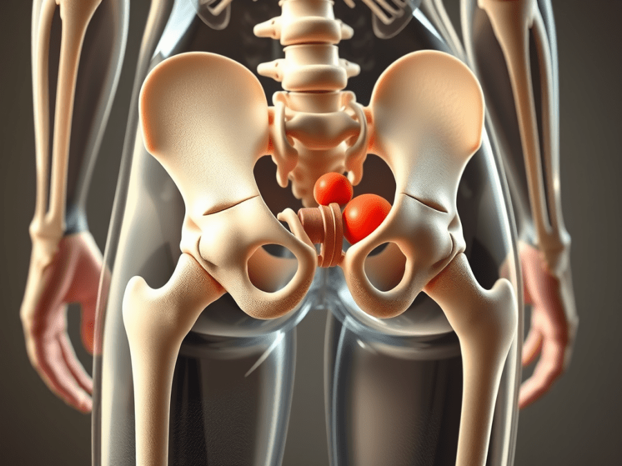
The greater trochanter is the bony bump on the outside of the hip, the one you can find with your fingers. Bursae sit around it, small fluid-filled sacs that keep bone, muscle, and tendon from grinding against each other. Inflame one and you get pain along the outer hip.

Symptoms

Patients describe an ache or a burn over the side of the hip. It can run down the outside of the thigh, though it rarely travels past the knee. Activity makes it worse: stairs, long walks, and lying on that side at night, which is the complaint that finally brings people in. The area is almost always tender to pressure. Some patients mention stiffness after sitting a while, or difficulty getting the hip moving again.

The symptom picture overlaps with lumbar radiculopathy and osteoarthritis, which is exactly why it’s so often mislabeled.

Diagnosis

History does most of the work. When did it start, what makes it worse, was there a fall, and did anything change recently in activity or training. That last question earns its keep more often than people expect.

Point tenderness directly over the greater trochanter is the classic finding. A positive Trendelenburg sign, which reflects gluteal weakness, points toward associated tendon involvement rather than isolated bursitis.

Imaging isn’t always needed. X-rays rule out arthritis. Ultrasound or MRI can show bursal inflammation or gluteal tendinopathy when the case isn’t behaving.

Treatment

Most patients get better with conservative care, and I want to be clear that conservative here doesn’t mean passive. Rest from whatever’s aggravating it, ice over the outer hip, and an NSAID like ibuprofen or naproxen for pain and swelling will settle the acute phase.

Physical therapy is the piece that actually changes the trajectory. Strengthening the gluteal muscles and improving hip stability relieves current symptoms and makes recurrence less likely. I’ve had patients who could barely sleep on their side due to the pain, and after a few weeks of targeted therapy, they were back to normal activities.

When conservative care runs out of road, a corticosteroid injection into the bursa can help, and ultrasound guidance improves accuracy enough to be worth asking for. Platelet-rich plasma and shockwave therapy are still being studied in resistant cases. Surgical bursectomy is rare and belongs to the severe cases that have failed everything else.

Prevention and Long-Term Outlook

Preventing recurrence means dealing with what caused it. Weight management reduces load through the hip. Footwear matters, particularly for anyone on their feet all day on hard floors. Regular hip strengthening and flexibility work keeps the area stable, and patients who avoid long stretches of unbroken sitting or standing tend to hold their gains.

Working Together

For colleagues: keep the differential wide on lateral hip pain, and get physical therapy involved early rather than after the third failed round of anti-inflammatories. A multidisciplinary approach is what produces durable outcomes here.

For patients: this hurts, it can drag on, and it’s genuinely treatable. Most people get their mobility back and return to normal routines.

Scott Rennie, D.O.

Sources

  • Segal NA, Felson DT, Torner JC, et al. Greater trochanteric pain syndrome: epidemiology and associated factors. Arch Phys Med Rehabil. 2007;88(8):988-992.
  • Strauss EJ, Nho SJ, Kelly BT. Greater trochanteric pain syndrome. Sports Med Arthrosc Rev. 2010;18(2):113-119.
  • Bird PA, Oakley SP, Shnier R, Kirkham BW. Prospective evaluation of magnetic resonance imaging and physical examination findings in patients with greater trochanteric pain syndrome. Arthritis Rheum. 2001;44(9):2138-2145.

Board Certified in Obesity Medicine and Family Medicine

This blog is for educational purposes only and does not constitute individual medical advice. Always consult your own physician before making changes to your health, medications, or treatment plan.
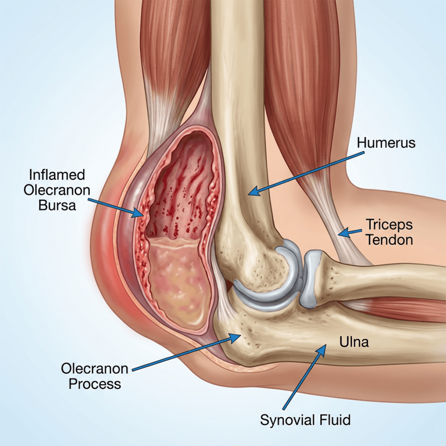
Anatomical diagram of elbow joint highlighting inflamed olecranon bursa, humerus, ulna, olecranon process, triceps tendon, and synovial fluid.

What Is Olecranon Bursitis, Also Called Elbow Bursitis?

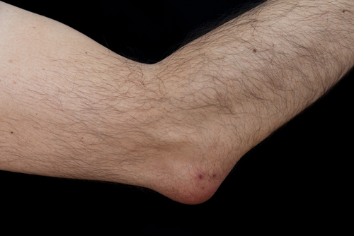
  shutterstock_159146555shutterstock_138510524A patient came in today complaining about a lump on her elbow and that is has been slightly painful for the last week.  She is a student and spends many hours reading books and resting her elbow on a table while studying.  This disorder called olecranon bursitis is a fairly common condition in which the bursa, which is a sac-like structure that protects the elbow from pressure injuries becomes inflamed. Bursas are found all over the body including the shoulders, elbows, knees, feet and hips.   They contain a lubricating fluid that fills the sac and decreases the friction between ligaments, bones and tendons. Bursitis of the elbow can be caused from:
  1. Prolonged pressure on a bursa (such as when leaning on the elbow while studying for long periods of time or resting the elbow on a car door during a long trip).
  2. Repetitive motions that cause irritation (laying carpet is an example).
  3. An injury or trauma
  4. Medical conditions such as gout or rheumatoid arthritis or pseudogout
  5. An infection within the bursa (called septic bursitis)
Usually a bursitis is not caused from an infection, but when there is redness and warmth to the skin there is a higher chance of infection.  If the skin overlying the elbow becomes punctured or injured there is a higher chance of a septic elbow bursitis. How would I know if I have bursitis or some other type of problem causing the elbow pain?  A medial provider who has experience with joint conditions will likely be able to diagnose the problem after examining you and hearing about your symptoms.  If there is redness or increased warmth to the area in addition to the swelling, a medical provider might ask to use a syringe and needle to remove a sample of fluid from the bursa to test for infection.   The fluid from the elbow can also be tested for crystals that can be caused by gout or pseudogout.  If you have uric acid crystals in your elbow, the medial provider will help you lower your uric acid levels to prevent worsening symptoms and decrease the chances of crystals forming in other joints. For more information about gout, please see my blog article on that topic. Sometimes an x-ray, ultrasound or MRI is ordered if there is still some uncertainty about the cause of the elbow problem. What can I do for the bursitis?  Treatment of elbow bursitis involved decreasing the pressure or motion that is causing the inflammation.  If there is infection, treating with antibiotics is important.  If the bursitis is not due to an infection however, then draining the fluid usually is not very helpful because the bursa will create more fluid and only enlarge again.  In addition, there is a risk of introducing infection into the joint if a needle is put poked through the skin and into the bursa.  If there was not an infection already present within the bursa or joint, there is a greater chance of introducing infection by attempting to withdraw the fluid.  It is important to rest the joint and apply ice.  We also usually prescribe an anti-inflammatory medication such as naproxen or ibuprofen.  Sometimes heat or a steroid injection is also used to help decrease the inflammation.  Heat can be applied to the elbow with a hot pack, a heating pad or hot water bottle.  Ice can be applied using a frozen gel pack or a bag of frozen peas.  I usually don’t recommend using either ice or heat for more than 20 minutes at a time and recommend caution so you don’t burn the skin. How can I prevent bursitis?  Irritation to the small bursal sacs can be minimized by decreasing repetitive motions, using cushions or pads to reduce the pressure on joints and taking periodic short breaks from tasks that increase joint pressure.  If you start having pain in a joint, this is a warning that there is too much stress and the activity should be modified or avoided.   I hope that you have found this information useful.  Wishing you the best of health,

Scott Rennie, D.O.
Board Certified in Obesity Medicine and Family Medicine

This blog is for educational purposes only and does not constitute individual medical advice. Always consult your own physician before making changes to your health, medications, or treatment plan.